. 2016 Jan 6:6:18741.
doi: 10.1038/srep18741.

Exon-centric regulation of ATM expression is population-dependent and amenable to antisense modification by pseudoexon targeting

Affiliations

Exon-centric regulation of ATM expression is population-dependent and amenable to antisense modification by pseudoexon targeting

Jana Kralovicova et al. Sci Rep. .

Erratum in

Abstract

ATM is an important cancer susceptibility gene that encodes a critical apical kinase of the DNA damage response (DDR) pathway. We show that a key nonsense-mediated RNA decay switch exon (NSE) in ATM is repressed by U2AF, PUF60 and hnRNPA1. The NSE activation was haplotype-specific and was most promoted by cytosine at rs609621 in the NSE 3' splice-site (3'ss), which is predominant in high cancer risk populations. NSE levels were deregulated in leukemias and were influenced by the identity of U2AF35 residue 34. We also identify splice-switching oligonucleotides (SSOs) that exploit competition of adjacent pseudoexons to modulate NSE levels. The U2AF-regulated exon usage in the ATM signalling pathway was centred on the MRN/ATM-CHEK2-CDC25-cdc2/cyclin-B axis and preferentially involved transcripts implicated in cancer-associated gene fusions and chromosomal translocations. These results reveal important links between 3'ss control and ATM-dependent responses to double-strand DNA breaks, demonstrate functional plasticity of intronic variants and illustrate versatility of intronic SSOs that target pseudo-3'ss to modify gene expression.


Conflict of interest statement

A part of this work is subject to a UK patent application. The authors declare no other competing financial interests.

Figures

Figure 1
Figure 1
Identification of a U2AF-repressed cryptic exon in ATM intron 28. (a) Schematics of NSE activation. NSE sequence (upper panel) is boxed, asterisk denotes rs609621, black rectangles show the indicated antisense oligonucleotides. Genome browser views of RNA-Seq data from RNAi- or SSO-mediated depletions of both U2AF35 isoforms (ab-), U2AF35a (a-), U2AF35b (b-) and controls (c) are shown in the lower panel. SSOs targeting 3′ ss of U2AF1 exons Ab and 3 and U2AF35 siRNAs were as described. Y axis, read densities. NSE inclusion/exclusion is schematically shown by dotted lines at the top. ATM exons (gray boxes) are numbered as in ref. . The 29-nt NSE introduced a stop codon in the ATM mRNA. (b) Validation of the NSE activation by RT-PCR (upper panel) in independent depletions (lower panel). RT-PCR primers (ATM-F, ATM-R, Table S1) are denoted by arrows in panel a. Spliced products are shown to the right, the percentage of transcripts with NSE is at the top. Error bars denote SDs of two transfections experiments. ***p < 0.0001, **p < 0.001. (c) NSE inclusion in mature transcripts inversely correlates with residual U2AF. r, Pearson correlation. Estimates of heterodimer levels were as described.
Figure 2
Figure 2
NSE activation and ATM expression are modified by rs609261. (a) Allelic frequencies at rs609261 in the indicated populations. (b) Minigene schematics. An XhoI/XbaI segment of ATM containing NSE and exon 29 was cloned between U2AF1 exons 2 and 4 (black boxes). RT-PCR primers to amplify exogenous transcripts (PL3 and ATM-R, Table S1) are denoted by arrows. (c) The rs609261-dependent NSE activation in exogenous pre-mRNAs. HEK293 cells depleted of U2AF35 or U2AF65 were transiently transfected with T (black) and C (gray) minigenes. Final concentration of the U2AF35 and U2AF65 siRNAs was 30 and 60 nM, respectively. (d) Identification of cell lines homozygous at rs609261 (asterisk). NSE is boxed. (e,f) Allele-specific activation of NSE in endogenous transcripts limits ATM expression in a dose-dependent manner. The source of endogenous transcripts is at the bottom, antibodies are to the right. Concentration of siRNAs in HEK293 cultures was 3, 10 and 30 nM. Concentration of siRNAs in HeLa cultures was 6.6, 20 and 60 nM. C1, C2, control siRNAs. Transfection efficiency was monitored by a GFP-plasmid and fluorescent microscopy. (g) UPF1 depletion increased NSE activation (upper panel) and upregulated isoform U2AF1c (lower panel). The U2AF1c isoform contains both exons Ab and 3 and is repressed by NMD. Final concentration of the UPF1 siRNA was 7, 20 and 60 nM. SC, a scrambled control. Error bars are SDs of independent transfections. (h) NSE inclusion levels in cells depleted of U2AF-related proteins and a subset of heterogenous nuclear RNPs. Error bars denote SDs of two transfections. Immunoblots are shown to the right. Final concentration of the U2AF35 siRNA was 25 nM; the remaining siRNAs were at 60 nM. C, controls. (i) Overexpression of PUF60 induced NSE skipping. Immunoblots are shown below, antibodies to the right.
Figure 3
Figure 3
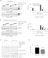
Rescue of U2AF-repressed ATM expression by SSOs targeting NSE. (a,b) Efficient SSO-mediated NSE inhibition in exogeneous (a) and endogenous (b) ATM transcripts. Mean NSE inclusion levels of two transfection experiments are shown in the right panels. (c) Restoration of ATM protein levels by SSOs that blocks access to NSE. Cells lacking U2AF35 and control cells were transfected with the SSO targeting the NSE 3’ss and a control SSOs (Fig. 1a and Table S1), as described. After 48 hrs, the cells were exposed to ionizing radiation (IR, 10 Gy) and harvested 1 hr later. Cell lysates were separated using a gradient SDS-PAGE. Western blotting was with antibodies shown to the right. (d) SSO-NSE3-mediated enhancement of ATM protein in cell lines homozygous at rs609261. Columns represent the average fold difference in ATM expression between SSO-NSE3-treated and SSO-C-treated homozygous cells depleted of U2AF35. Error bars represent SD of two biological replicates. P-values comparing SSO-C with SSO-NSE3-treated counterparts are shown at the top. Representative immunoblots are in Fig. S1a,b.
Figure 4
Figure 4
Identification of intronic cis-elements and SSOs that modulate NSE activation. (a) Schematics of two pseudoexons in ATM intron 28. Canonical exons, NSE and PE are shown as gray, white and checkered boxes, respectively. Asterisk indicates location of the IVS28-159A >G substitution. In this A-T case, both NSE and PE were included in the ATM mRNA together with the intervening sequence. Canonical and aberrant transcripts are denoted by dotted lines above and below the pre-mRNA. Middle panel shows RNA-Seq read densities for NSE in cells depleted of both U2AF35 isoforms (ab-) together with U2AF65 tags/high-confidence binding sites (horizontal lines/rectangles) identified by crosslinking and immunoprecipitation. The 100 basewise vertebrate conservation by Phylop (100 VC) is at the bottom. The lower panel shows mutations (in red and underlined) introduced in the C-minigene. (b) Splicing pattern of wildtype and mutated C minigenes. Mutations are at the bottom of panel (a); RNA products are shown schematically to the right. The largest product produced by clone PE delPPT/AG includes the shortened pseudointron. (c) Splicing pattern of C minigenes mutated in NSE (lanes 2, 3, 7 and 8) or PE (lanes 4, 5, 9 and 10) in (mock)-depleted HEK293 cells. Mutations are at the bottom; their sequences are in Table S2. Spliced products are schematically shown to the right. A hairpin symbol above PE denotes the MIR stem-loop insertion; cr3’ss, a cryptic 3’ss activation 7 nt downstream of the authentic NSE 3’ss. (d,e) SSO-induced pseudoexon switching. Transfected minigenes are shown at the top, spliced products to the right and SSOs at the bottom. SSO sequences are in Table S1. Final concentration of SSOs shown in panels (d–g) was 3, 10 and 30 nM. (f) PE SSOs induced NSE skipping. (g) SSOs targeting a sequence that activates NSE upon deletion (PEdelPPT/AG; panel a and b) inhibit PE. (h) NSE activation is haplotype-dependent. Minigene haplotypes at the indicated variants are at the bottom. Columns represent mean NSE inclusion, error bars are SDs, asterisks denote statistically significant differences as in Fig. 1b.
Figure 5
Figure 5
Exon-centric regulation of ATM signalling. (a) U2AF-regulated gene- and exon-level expression changes in MRN-ATM-CHEK2-CDC25-cdc2/cyclin B pathway (left panel). Log2fold- and q-values are shown in parentheses. Exon usage of CHEK2 and CDC25A genes is shown by RNA-Seq browser shots; PCR validation gels are in the right panels. CHEK2 exon 9 is a NMD switch exon; exon 11 encodes a portion of the kinase domain. Full spectrum of U2AF-mediated expression changes in the ATM signalling pathway is shown in Fig. S2; examples of the U2AF-mediated splicing regulation are in Figs S3–S6. (b) Impaired ATM signalling in U2AF35 depleted cells following IR. HEK293 cells were (mock)depleted of U2AF35 and subjected to IR (10 Gy) 48 hrs later. Expression was examined by immunoblotting at the indicated time points. Antibodies are shown to the right. CHEK2 exon 9 skipping levels are at the bottom; their measurements in control (U2AF35+) and depleted cells (U2AF35−) are in panel (c). (d) CHEK2 exon 9 inclusion in UPF1 depleted cells. Final concentration of the UPF1 siRNA (Table S1) was 12.5, 25, 50, and 100 nM. (e) Repression of CHEK2 exon 9 by SSO reduced CHEK2 levels and promoted NSE inclusion. Final concentration of SSO targeting CHEK2 exon 9 was 3, 10 and 30 nM. (f) CHEK2 exon 9 inclusion upon transfection of HEK293 cells with the indicated SSOs. (g) A lack of SF3B1 induced CHEK2 exon 9 skipping but did not alter NSE activation. Final concentration of each siRNA targeting SF3B1 was 20 nM.
Figure 6
Figure 6
Rescue of NSE repression by cancer-associated mutations in U2AF35. Rescue of U2AF(35)-dependent NSE splicing of the C minigene by zinc finger 1 and 2 substitutions in U2AF35 (upper panel). All substitutions were made in the U2AF1a construct (35a). Cancer-associated mutations (bottom) are boxed; splice products are to the right. Error bars are SDs of 2 transfections. Immunoblot with U2AF35 and GFP antibodies is shown in the lower panel; ex, exogenous; en, endogenous U2AF35.

Similar articles

Cited by

References

    1. Wahl M. C., Will C. L. & Luhrmann R. The spliceosome: design principles of a dynamic RNP machine. Cell 136, 701–718 (2009). - PubMed
    1. Shcherbakova I. et al. Alternative spliceosome assembly pathways revealed by single-molecule fluorescence microscopy. Cell Rep. 5, 151–165 (2013). - PMC - PubMed
    1. Zamore P. D. & Green M. R. Identification, purification, and biochemical characterization of U2 small nuclear ribonucleoprotein auxiliary factor. Proc. Natl. Acad. Sci. USA 86, 9243–9247 (1989). - PMC - PubMed
    1. Wu S., Romfo C. M., Nilsen T. W. & Green M. R. Functional recognition of the 3′ splice site AG by the splicing factor U2AF35. Nature 402, 832–835 (1999). - PubMed
    1. Sun H. & Chasin L. A. Multiple splicing defects in an intronic false exon. Mol. Cell. Biol. 20, 6414–6425 (2000). - PMC - PubMed

Publication types

MeSH terms