

TRAIL-coated leukocytes that prevent the bloodborne metastasis of prostate cancer
- PMID: 26732555
- PMCID: PMC4724502
- DOI: 10.1016/j.jconrel.2015.12.048
TRAIL-coated leukocytes that prevent the bloodborne metastasis of prostate cancer
Abstract
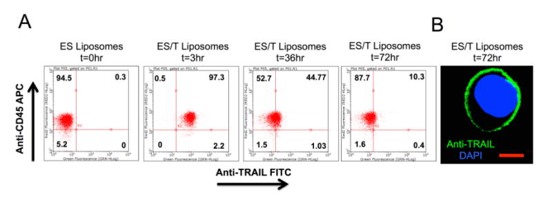
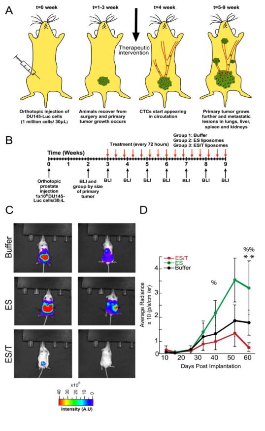
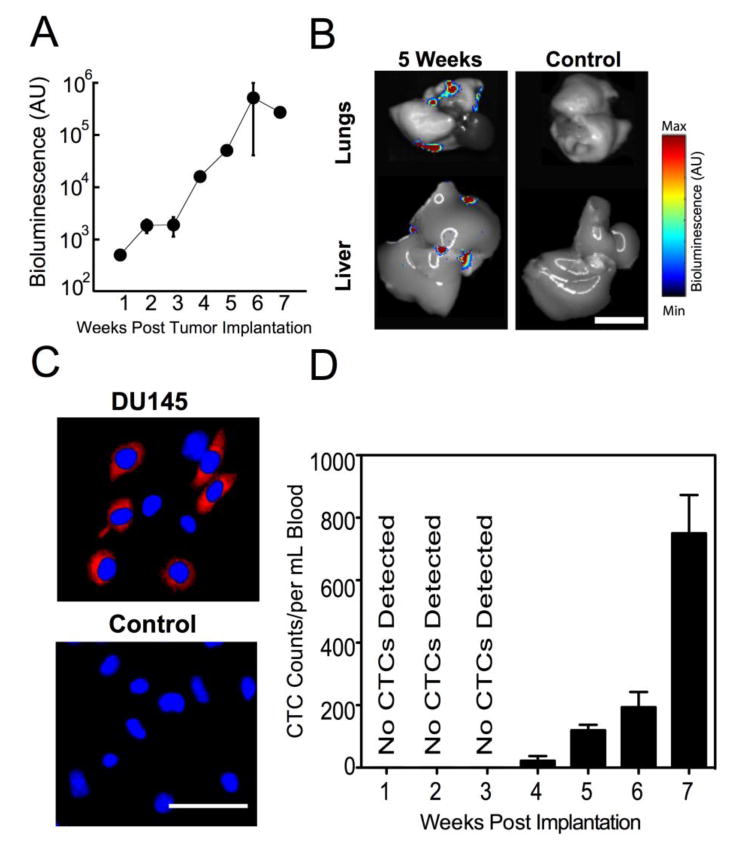
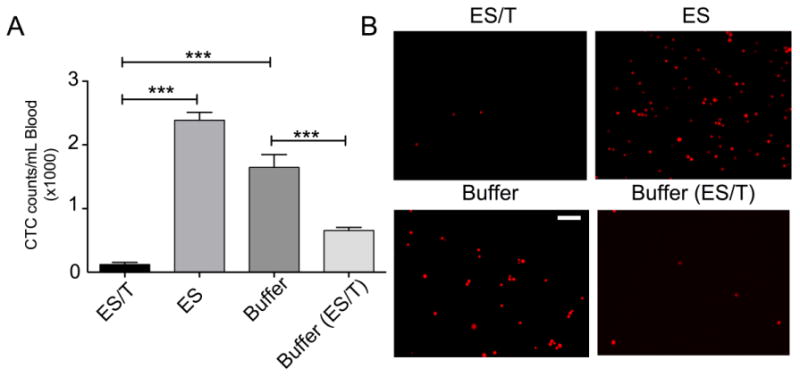
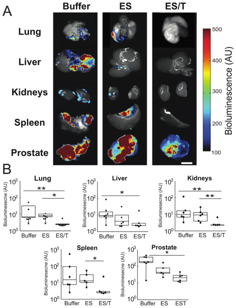
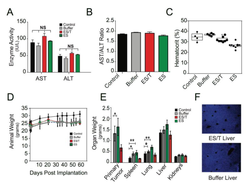
Prostate cancer, once it has progressed from its local to metastatic form, is a disease with poor prognosis and limited treatment options. Here we demonstrate an approach using nanoscale liposomes conjugated with E-selectin adhesion protein and Apo2L/TRAIL (TNF-related apoptosis-inducing ligand) apoptosis ligand that attach to the surface of leukocytes and rapidly clear viable cancer cells from circulating blood in the living mouse. For the first time, it is shown that such an approach can be used to prevent the spontaneous formation and growth of metastatic tumors in an orthotopic xenograft model of prostate cancer, by greatly reducing the number of circulating tumor cells. We conclude that the use of circulating leukocytes as a carrier for the anti-cancer protein TRAIL could be an effective tool to directly target circulating tumor cells for the prevention of prostate cancer metastasis, and potentially other cancers that spread through the bloodstream.
Keywords: Circulating tumor cells; Metastasis; Nanomedicine; TRAIL.
Copyright © 2015 Elsevier B.V. All rights reserved.
Figures
Similar articles
-
Targeting prostate cancer cells en route to dissemination.J Control Release. 2016 Feb 10;223:224. doi: 10.1016/j.jconrel.2016.01.025. J Control Release. 2016. PMID: 26802318 No abstract available.
-
Unnatural killer cells to prevent bloodborne metastasis: inspiration from biology and engineering.Expert Rev Anticancer Ther. 2014 Jun;14(6):641-4. doi: 10.1586/14737140.2014.916619. Epub 2014 May 5. Expert Rev Anticancer Ther. 2014. PMID: 24791860 Free PMC article.
-
Spatial distribution of tumor-associated macrophages in an orthotopic prostate cancer mouse model.Pathol Oncol Res. 2024 Apr 16;30:1611586. doi: 10.3389/pore.2024.1611586. eCollection 2024. Pathol Oncol Res. 2024. PMID: 38689823 Free PMC article.
-
Circulating Tumor Cells: Diagnostic and Therapeutic Applications.Annu Rev Biomed Eng. 2018 Jun 4;20:329-352. doi: 10.1146/annurev-bioeng-062117-120947. Epub 2018 Mar 14. Annu Rev Biomed Eng. 2018. PMID: 29539267 Review.
-
The "emigration, migration, and immigration"of prostate cancer.Clin Prostate Cancer. 2005 Jun;4(1):24-30. doi: 10.3816/cgc.2005.n.008. Clin Prostate Cancer. 2005. PMID: 15992458 Review.
Cited by
-
TRAIL in the Treatment of Cancer: From Soluble Cytokine to Nanosystems.Cancers (Basel). 2022 Oct 19;14(20):5125. doi: 10.3390/cancers14205125. Cancers (Basel). 2022. PMID: 36291908 Free PMC article. Review.
-
Peptide functionalized liposomes for receptor targeted cancer therapy.APL Bioeng. 2021 Jan 25;5(1):011501. doi: 10.1063/5.0029860. eCollection 2021 Mar. APL Bioeng. 2021. PMID: 33532673 Free PMC article. Review.
-
Engineering and physical sciences in oncology: challenges and opportunities.Nat Rev Cancer. 2017 Nov;17(11):659-675. doi: 10.1038/nrc.2017.83. Epub 2017 Oct 13. Nat Rev Cancer. 2017. PMID: 29026204 Free PMC article. Review.
-
Immune Cell-Mediated Biodegradable Theranostic Nanoparticles for Melanoma Targeting and Drug Delivery.Small. 2017 Mar;13(10):10.1002/smll.201603121. doi: 10.1002/smll.201603121. Epub 2016 Dec 27. Small. 2017. PMID: 28026115 Free PMC article.
-
Dual Affinity Nanoparticles for the Transport of Therapeutics from Carrier Cells to Target Cells under Physiological Flow Conditions.ACS Omega. 2023 Nov 1;8(45):42748-42761. doi: 10.1021/acsomega.3c05605. eCollection 2023 Nov 14. ACS Omega. 2023. PMID: 38024679 Free PMC article.
References
-
- Torre LA, Bray F, Siegel RL, Ferlay J. Global cancer statistics, 2012. CA Cancer J Clin. 2015;65:87–108. - PubMed
-
- Crehange G, Roach MI, Martin E, Cormier L, Peiffert D, Cochet A, et al. Salvage reirradiation for locoregional failure after radiation therapy for prostate cancer: Who, when, where and how? Cancer Radiotherapie. 2014 Oct;18(5-6):524–34. - PubMed
-
- Delacruz A. Using circulating tumor cells as a prognostic indicator in metastatic castration-resistant prostate cancer. Clin J Oncol Nurs. 2012 Apr;16(2):E44–7. - PubMed
-
- Ma X, Xiao Z, Li X, Wang F, Zhang J, Zhou R, et al. Prognostic role of circulating tumor cells and disseminated tumor cells in patients with prostate cancer: a systematic review and meta-analysis. Tumour Biol. 2014 Jun;35(6):5551–60. - PubMed
MeSH terms
Substances
Grants and funding
LinkOut - more resources
Full Text Sources
Other Literature Sources
Medical
