. 2016 Feb 1;196(3):963-77.
doi: 10.4049/jimmunol.1501531. Epub 2016 Jan 4.

Molecular Mechanisms of Airway Hyperresponsiveness in a Murine Model of Steroid-Resistant Airway Inflammation

Affiliations

Molecular Mechanisms of Airway Hyperresponsiveness in a Murine Model of Steroid-Resistant Airway Inflammation

Michelle L Manni et al. J Immunol. .

Abstract

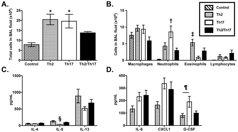
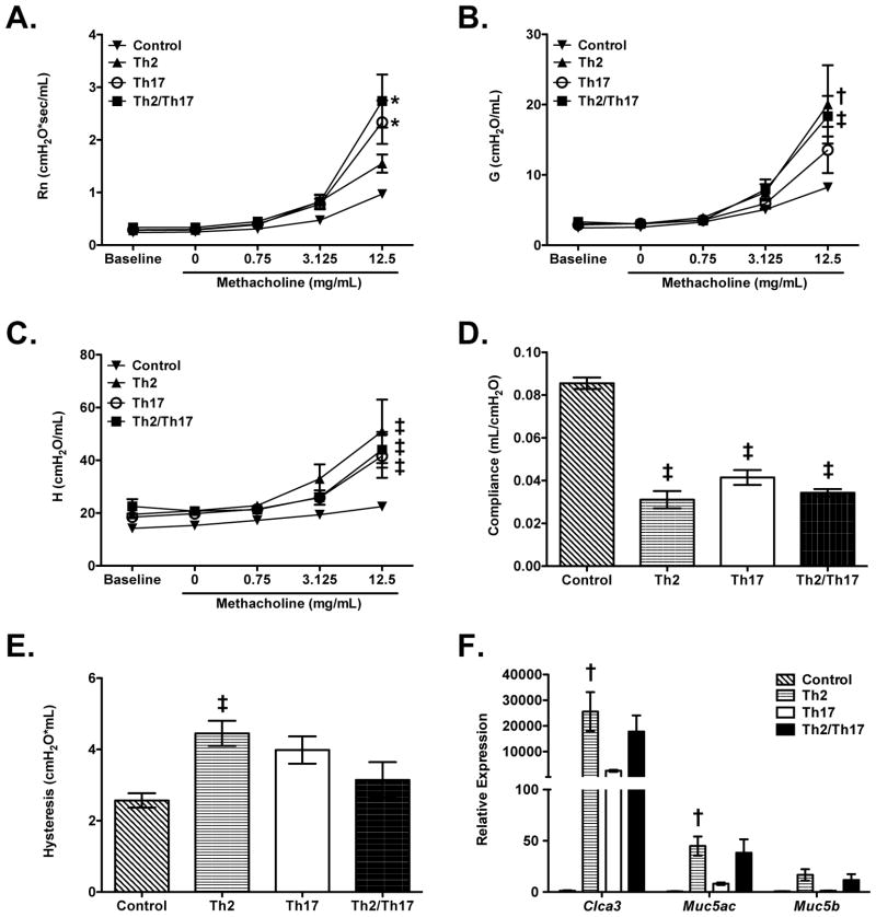
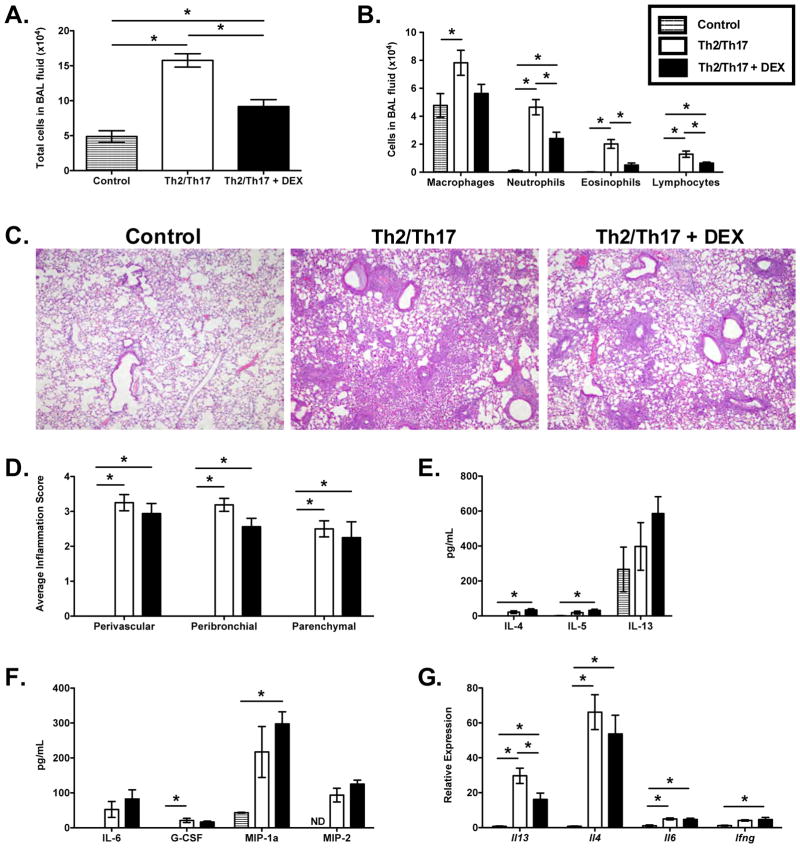
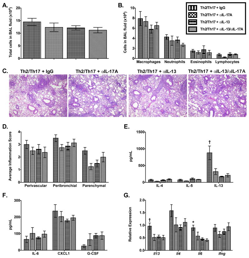
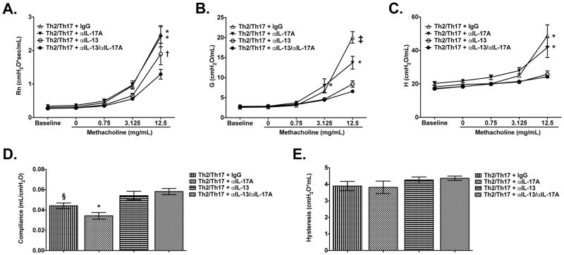
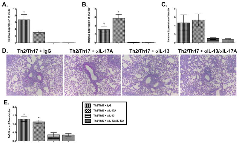
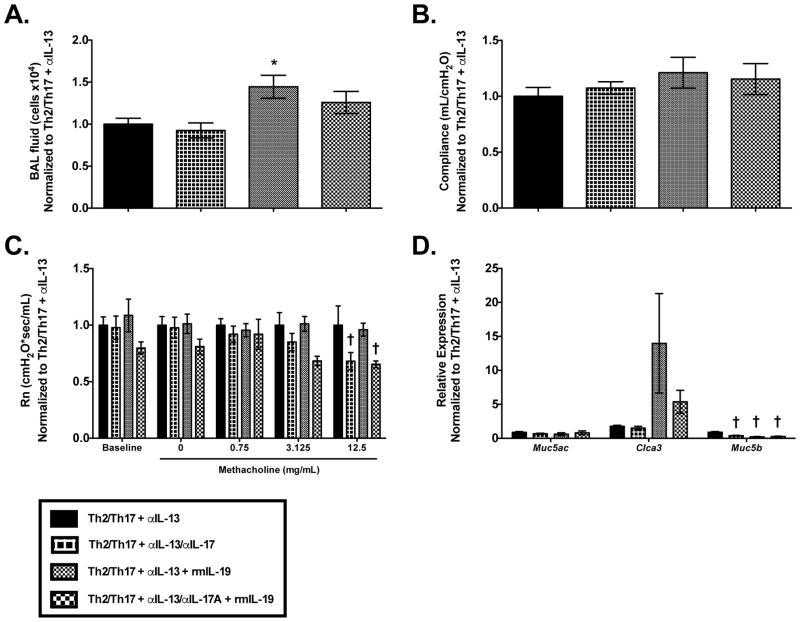
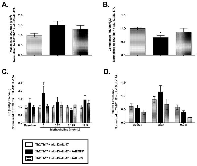
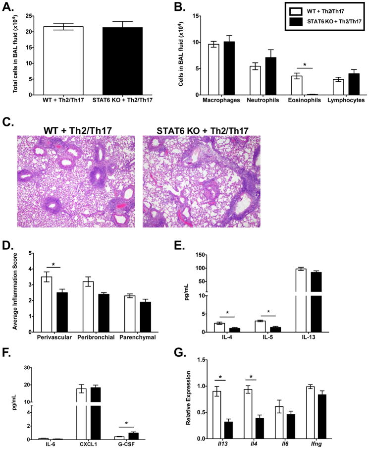
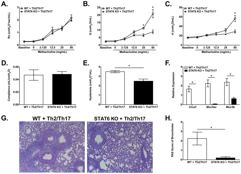
IL-13 and IL-17A, produced mainly by Th2 and Th17 cells, respectively, have an influential role in asthma pathogenesis. We examined the role of IL-13 and IL-17A in mediating airway hyperresponsiveness (AHR), lung inflammation, and mucus metaplasia in a dual Th2/Th17 model of asthma. IL-13 and/or IL-17A were neutralized using mAbs. Th2/Th17 adoptive transfer induced a mixed asthma phenotype characterized by elevated eosinophilia and neutrophilia, tissue inflammation, mucus metaplasia, and AHR that were partially reversible with steroid treatment. Pulmonary inflammation and quasi-static lung compliance were largely unaffected by neutralization of IL-13 and/or IL-17A. However, neutralization of IL-13 alone or in combination with IL-17A significantly attenuated AHR and mucus metaplasia. Further, STAT6 activation was attenuated following IL-13 and IL-13/IL-17A Ab treatment. We next assessed the role of STAT6 in Th2/Th17-mediated allergic airway disease using STAT6(-/-) mice. STAT6(-/-) mice adoptively transferred with Th2/Th17 cells had decreased AHR compared with controls. These data suggest that IL-13 drives AHR and mucus metaplasia in a STAT6-dependent manner, without directly contributing to airway or tissue inflammation. IL-17A independently contributes to AHR, but it only partially mediates inflammation and mucus metaplasia in a mixed Th2/Th17 model of steroid-resistant asthma.


Figures

Figure 1
Figure 1
Th2 and/or Th17 cell transfer and OVA challenge induces pulmonary inflammation in BALB/c SCID mice. Cellular inflammation in the airspaces as (A) total cells in the BAL fluid and (B) cell differentials. (C) Th2- and (D) Th17-related cytokine and chemokine levels in lung homogenates. Graphs show data for control (n=4), Th2 (n=4–8), Th17 (n=4–8), and Th2/Th17 (n=7–8) combined from three independent experiments. *p<0.05 when compared to control group, †p<0.05 when compared to control and Th2/Th17 groups, ‡p<0.05 when compared to control and Th17 groups, §p<0.05 when compared to all other groups, ¶p<0.05 for comparisons shown.
Figure 2
Figure 2
Th2 and/or Th17 cell transfer and OVA challenge induces allergic airway disease in BALB/c SCID mice. Lung function analyses of OVA-challenged mice adoptively transferred with Th2, Th17, or both Th2 and Th17 cells. (A) Airway resistance, Rn, (B) tissue damping, G, and (C) tissue elastance/stiffness, H, parameters to increasing doses of methacholine as well as (D) quasi-static lung compliance and (E) hysteresis. (F) Clca3, Muc5ac, and Muc5b mRNA levels in the lungs normalized to Hprt (relative to Th2/Th17 group). Graphs show data for control (n=4), Th2 (n=7–8), Th17 (n=7–8), and Th2/Th17 (n=7–8) combined from three independent experiments. *p<0.05 when compared to control and Th2 groups, †p<0.05 when compared to control and Th17 groups, ‡p<0.05 when compared to control.
Figure 3
Figure 3
Effect of dexamethasone treatment on pulmonary inflammation in Th2/Th17 cell transfer, OVA challenged mice. Cellular inflammation in the airspaces as (A) total cells in the BAL fluid and (B) cell differentials (n=6–8/group). (C) Representative H&E-stained lung sections (40x magnification) and (D) histological quantification of perivascular, peribronchial, and parenchymal inflammation in the lung (n=6–8/group). (E) Th2- and (F) Th17-related cytokine and chemokine levels in lung homogenates (n=3–4/group). (G) Relative expression of Il13, Il4, Il6, and Ifng in the lung normalized to Hprt (relative to control group). Graphs show data combined from four independent experiments. *p<0.05 for comparisons shown, ND=not determined.
Figure 4
Figure 4
Th2 and Th17 cell transfer and OVA challenge induces steroid-resistant allergic airway disease in BALB/c SCID mice. (A) Airway resistance, Rn, (B) tissue damping, G, and (C) tissue elastance/stiffness, H, parameters to increasing doses of methacholine as well as (D) quasi-static lung compliance and (E) hysteresis. (F) Clca3, Muc5ac, and Muc5b mRNA levels in the lungs normalized to Hprt (relative to control group). (G) Representative Periodic Acid-Schiff (PAS) staining of airway mucus in Th2/Th17 cell transfer, OVA-challenged mice following dexamethasone treatment (100x magnification) and (H) histological quantification of mucus production (n=6–8/group). Graphs show data combined from four independent experiments. *p<0.05 when compared to control group.
Figure 5
Figure 5
Pulmonary inflammation in Th2/Th17 transfer, OVA-challenged mice following IL-13 and/or IL-17A neutralization. Cellular inflammation in the airspaces as (A) total cells in the BAL fluid and (B) cell differentials. (C) Representative H&E-stained lung sections (40x magnification) and (D) histological quantification of perivascular, peribronchial, and parenchymal inflammation in the lung. (E) Th2- and (F) Th17-related cytokine and chemokine levels in lung homogenates. (G) Relative expression of Il13, Il4, Il6, and Ifng in the lung normalized to Hprt (relative to Th2/Th17 + IgG group). Graphs show data for Th2/Th17 + IgG (n=5–12), αIL-17A (n=4–8), αIL-13 (n=4–8), αIL-13/αIL-17A (n=4–8) combined from seven independent experiments. *p<0.05 when compared to αIL-13 and αIL-13/αIL-17A, †p<0.05 when compared to all other groups.
Figure 6
Figure 6
Lung function analyses of Th2/Th17 transfer, OVA-challenged mice following IL-13 and/or IL-17 neutralization. (A) Airway resistance, Rn, (B) tissue damping, G, and (C) tissue elastance/stiffness, H, parameters to increasing doses of methacholine as well as (D) quasi-static lung compliance and (E) hysteresis. Graphs show data for Th2/Th17 + IgG (n=11–12), αIL-17A (n=7–8), αIL-13 (n=8), αIL-13/αIL-17A (n=8), *p<0.05 when compared to αIL-13 and αIL-13/αIL-17A combined from seven independent experiments. †p<0.05 when compared to αIL-13, and ‡p<0.05 when compared to all other groups, §p<0.05 when compared to αIL-13/αIL-17A.
Figure 7
Figure 7
Mucus metaplasia in Th2/Th17 cell transfer, OVA-challenged mice following neutralization of IL-13 and/or IL-17A. (A) Clca3, (B) Muc5ac, and (C) Muc5b mRNA levels in the lung tissue normalized to Hprt (relative to Th2/Th17 + IgG group). (D) Representative Periodic Acid-Schiff (PAS) staining of airway mucus (100x magnification) and (E) histological quantification of mucus production. Graphs show data for Th2/Th17 + IgG (n=8–12), αIL-17A (n=4–8), αIL-13 (n=8), and αIL-13/αIL-17A (n=8) combined from seven independent experiments. *p<0.05 when compared to αIL-13 and αIL-13/αIL-17A, †p<0.05 when compared to all other groups.
Figure 8
Figure 8
Th2–immunoregulatory cytokines and STAT3 in the lungs of Th2/17 cell transfer, OVA-challenged mice. Relative expression of Th2-promoting cytokines (A) Il19, (B) Il25, and (C) Il33, as well as anti-inflammatory, immune suppressive cytokine (D) Il10 in lung tissue were normalized to Hprt (relative to control group). (E) Pulmonary Stat3 expression was normalized to Hprt (relative to control group). Graphs show data for control (n=6), Th2/17 + IgG (n=12), Th2/17 + αIL-17A (n=7–8), Th2/17 + αIL-13 (n=8), and Th2/17 + αIL-13/αIL-17A (n=8) combined from seven independent experiments. *p<0.05 when compared to control, Th2/17 + αIL-13, and Th2/17 + αIL-13/αIL-17A.
Figure 9
Figure 9
Exogenous IL-19 treatment of Th2/17 cell transfer, OVA-challenged mice following IL-13 and IL-17A neutralization. Cellular inflammation in the airspaces was assessed by measuring (A) total cells in the BAL fluid. (B) Quasi-static compliance and (C) airway resistance, Rn, in response to methacholine challenge were assessed. (D) Relative expression of Muc5ac, Clac3, and Muc5b in the lung was normalized to Hprt. Graphs show data for n=6–8/group combined from four independent experiments. *p<0.05 when compared to Th2/17 + αIL-13 and Th2/17 + αIL-13/αIL-17A. †p<0.05 when compared to Th2/17 + αIL-13.
Figure 10
Figure 10
Overexpression of IL-33 in the lungs of Th2/17 cell transfer, OVA-challenged mice following IL-13 and IL-17A neutralization. Cellular inflammation in the airspaces was assessed by measuring (A) total cells in the BAL fluid. (B) Quasi-static compliance and (C) airway resistance, Rn, in response to methacholine challenge were assessed. (D) Relative expression of Muc5ac, Clac3, and Muc5b in the lung was normalized to Hprt. Graphs show data for Th2/17 + αIL-13/αIL-17A (n=8), Th2/17 + αIL-13/αIL-17A + AdEGFP (n=4), Th2/17 + αIL-13/αIL-17A + AdIL-33 (n=4) combined from three independent experiments. *p<0.05 when compared to Th2/17 + αIL-13/αIL-17A, †p<0.05 when compared to Th2/17 + αIL-13/αIL-17A and Th2/17 + αIL-13/αIL-17A + AdIL-33.
Figure 11
Figure 11
STAT6 levels in the lungs of Th2/Th17 cell transfer, OVA-challenged mice following IL-13 and/or IL-17 neutralization. (A) STAT6 mRNA levels in the lung normalized to Hprt (relative to Th2/Th17 + IgG group). Graph shows data for control (n=6), Th2/Th17 + IgG (n=12), Th2/17 + αIL-17A (n=8), Th2/17 + αIL-13 (n=8), and Th2/17 + αIL-13/αIL-17A (n=6–8) combined from seven independent experiments. (B–C) Phosphorylated (p)-STAT6 and total STAT6 in the lungs normalized to β-actin. *p<0.05 when compared to Th2/Th17 + IgG.
Figure 12
Figure 12
Pulmonary inflammation in Th2/Th17 cell transfer, OVA challenged BALB/cJ and STAT6 knockout mice. Cellular inflammation in the airspaces as (A) total cells in the BAL fluid and (B) cell differentials. (C) Representative H&E-stained lung sections (40x magnification) and (D) histological quantification of perivascular, peribronchial, and parenchymal inflammation in the lung. (E) Th2- and (F) Th17-related cytokine and chemokine levels in lung homogenates. (G) Relative expression of Il13, Il4, Il6, and Ifng in the lung normalized to Hprt (relative to WT + Th2/Th17 group) n=5/group, *p<0.05 for comparisons shown.
Figure 13
Figure 13
Pulmonary function and mucus metaplasia in Th2/Th17 cell transfer, OVA challenged BALB/cJ and STAT6 knockout mice. (A) Airway resistance, Rn, (B) tissue damping, G, and (C) tissue elastance/stiffness, H, parameters to increasing doses of methacholine and (D) quasi-static lung compliance and (E) hysteresis. (F) Relative expression of Clca3, Muc5ac, and Muc5b in the lung normalized to Hprt (relative to WT + Th2/Th17 group). (G) Representative Periodic Acid-Schiff (PAS) staining of airway mucus (100x magnification) and (H) histological quantification of mucus production. n=4–5/group, *p<0.05 for comparisons shown.

Comment in

Similar articles

Cited by

References

    1. Wenzel S. Severe asthma: from characteristics to phenotypes to endotypes. Clin Exp Allergy. 2012;42:650–658. - PubMed
    1. Xie M, Wenzel SE. A global perspective in asthma: from phenotype to endotype. Chinese medical journal. 2013;126:166–174. - PubMed
    1. Manni ML, Trudeau JB, Scheller EV, Mandalapu S, Elloso MM, Kolls JK, Wenzel SE, Alcorn JF. The complex relationship between inflammation and lung function in severe asthma. Mucosal immunology. 2014;7:1186–1198. - PMC - PubMed
    1. Hellings PW, Kasran A, Liu Z, Vandekerckhove P, Wuyts A, Overbergh L, Mathieu C, Ceuppens JL. Interleukin-17 orchestrates the granulocyte influx into airways after allergen inhalation in a mouse model of allergic asthma. Am J Respir Cell Mol Biol. 2003;28:42–50. - PubMed
    1. Aujla SJ, Alcorn JF. T(H)17 cells in asthma and inflammation. Biochim Biophys Acta. 2011;1810:1066–1079. - PubMed

Publication types

MeSH terms