Expression of Sirt1 and FoxP3 in classical Hodgkin lymphoma and tumor infiltrating lymphocytes: Implications for immune dysregulation, prognosis and potential therapeutic targeting
- PMID: 26722524
- PMCID: PMC4680469
Expression of Sirt1 and FoxP3 in classical Hodgkin lymphoma and tumor infiltrating lymphocytes: Implications for immune dysregulation, prognosis and potential therapeutic targeting
Abstract
Background: Hodgkin Reed-Sternberg (HRS) cells may promote differentiation of CD4+ naïve T cells toward both FoxP3+ T regulatory (Treg) cells and TIA-1+ cytotoxic T lymphocytes (CTL). Previous studies suggest that an overabundance of cytotoxic TIA-1+ cells in relation to FoxP3+ T reg cells portends unfavorable outcomes in classical Hodgkin lymphoma (cHL), raising the possibility that its pathogenesis may be related to immune dysregulation. Sirt1 deacetylates FoxP3 and leads to decreased Treg functionality. Our objective was to compare Sirt1 and FoxP3 expressions in Hodgkin lymphoma infiltrating lymphocytes (HLIL) and confirm Sirt1 expression in HRS cells.
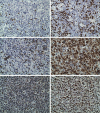
Design: Immunohistochemical staining of paraffin-embedded tissue with antibodies to Sirt1, FoxP3, TIA-1, and CD8 was performed. Expression of Sirt1 was assessed in both the HRS cells and in the HLILs in twenty-four cases. Adequate tissue was available in 13 cHL cases to permit the enumeration of FoxP3, TIA-1 and CD8 by giving their percent staining of HLILs.
Results: In HLILs, nuclear expression of Sirt1 was 32-88% (mean 67%); FoxP3 expression was 9-40% (mean 23.9%); TIA-1 expression was 15-87% (mean 32%); and CD8 expression was 10-45% (mean = 31%). Sirt1 to FoxP3 ratio was 0.96-5.5 (mean 3.2). TIA-1 to FoxP3 ratio was 0.6-5.1 (mean 1.6). CD8 to FoxP3 ratio was 0.43-3.7 (mean 1.5). There was a difference of Sirt1 to FoxP3 ratios between remission and recurrence groups, being significantly higher in the recurrence group (P = 0.005). Sirt1 demonstrated high nuclear expression in the HRS cells of 21 out of 24 (88%) cases analyzed.
Conclusion: The relative overexpression of Sirt1 to FoxP3 in HLILs may be considered possible targets for immune modulation. Histone deacetylase inhibitors may increase the efficacy of existing treatment regimens by downregulating SIRT1 gene mRNA/Sirt1 protein function and together with rapamycin could expand the T regulatory/FoxP3 population and functionality and improve prognosis for remission in cHL. Targeting Sirt1 in the HRS cells may facilitate their ability to promote naïve T cell differentiation toward Treg cells over CTL.
Keywords: FoxP3; Hodgkin lymphoma; Sirt1; immune dysregulation; morphoproteomics.
Figures


Similar articles
-
FOXP3(+) regulatory and TIA-1(+) cytotoxic T lymphocytes in HIV-associated Hodgkin lymphoma.Pathol Int. 2012 Feb;62(2):77-83. doi: 10.1111/j.1440-1827.2011.02754.x. Pathol Int. 2012. PMID: 22243776
-
Immunohistochemical expression of vitamin D receptor and forkhead box P3 in classic Hodgkin lymphoma: correlation with clinical and pathologic findings.BMC Cancer. 2020 Jun 8;20(1):535. doi: 10.1186/s12885-020-07026-6. BMC Cancer. 2020. PMID: 32513132 Free PMC article.
-
Hodgkin lymphoma: A complex metabolic ecosystem with glycolytic reprogramming of the tumor microenvironment.Semin Oncol. 2017 Jun;44(3):218-225. doi: 10.1053/j.seminoncol.2017.10.003. Epub 2017 Oct 10. Semin Oncol. 2017. PMID: 29248133 Free PMC article.
-
Evidence of abortive plasma cell differentiation in Hodgkin and Reed-Sternberg cells of classical Hodgkin lymphoma.Hematol Oncol. 2005 Sep-Dec;23(3-4):127-32. doi: 10.1002/hon.764. Hematol Oncol. 2005. PMID: 16342298 Review.
-
Apoptosis of Hodgkin-Reed-Sternberg cells in classical Hodgkin lymphoma revisited.APMIS. 2010 May;118(5):339-45. doi: 10.1111/j.1600-0463.2010.02600.x. APMIS. 2010. PMID: 20477808 Review.
Cited by
-
CCR7 in Blood Cancers - Review of Its Pathophysiological Roles and the Potential as a Therapeutic Target.Front Oncol. 2021 Oct 29;11:736758. doi: 10.3389/fonc.2021.736758. eCollection 2021. Front Oncol. 2021. PMID: 34778050 Free PMC article. Review.
-
Role of Sirtuins in the Pathobiology of Onco-Hematological Diseases: A PROSPERO-Registered Study and In Silico Analysis.Cancers (Basel). 2022 Sep 23;14(19):4611. doi: 10.3390/cancers14194611. Cancers (Basel). 2022. PMID: 36230534 Free PMC article. Review.
-
Regulatory Mechanisms and Reversal of CD8+T Cell Exhaustion: A Literature Review.Biology (Basel). 2023 Apr 1;12(4):541. doi: 10.3390/biology12040541. Biology (Basel). 2023. PMID: 37106742 Free PMC article. Review.
-
Characterization of the Microenvironment of Nodular Lymphocyte Predominant Hodgkin Lymphoma.Int J Mol Sci. 2016 Dec 16;17(12):2127. doi: 10.3390/ijms17122127. Int J Mol Sci. 2016. PMID: 27999289 Free PMC article.
-
Identifying highly active anti-CCR4 CAR T cells for the treatment of T-cell lymphoma.Blood Adv. 2023 Jul 25;7(14):3416-3430. doi: 10.1182/bloodadvances.2022008327. Blood Adv. 2023. PMID: 37058474 Free PMC article.
References
-
- Kuppers R. Molecular biology of Hodgkin’s lymphoma. Adv Cancer Res. 2002;84:277–312. - PubMed
-
- Kuppers R, Hansmann ML. The Hodgkin and Reed/Sternberg cell. Int J Biochem Cell Biol. 2005;37:511–517. - PubMed
-
- Hummel M, Ziemann K, Lammert H, Pileri S, Sabattini E, Stein H. Hodgkin’s disease with monoclonal and polyclonal populations of Reed-Sternberg cells. N Engl J Med. 1995;333:901–906. - PubMed
-
- Stein H, Hummel M. Cellular origin and clonality of classic Hodgkin’s lymphoma: immunophenotypic and molecular studies. Semin Hematol. 1999;36:233–241. - PubMed
-
- Kuppers R. Identifying the precursors of Hodgkin and Reed-Sternberg cells in Hodgkin’s disease: role of the germinal center in B-cell lymphomagenesis. J Acquir Immune Defic Syndr. 1999;21(Suppl 1):S74–79. - PubMed
MeSH terms
Substances
LinkOut - more resources
Full Text Sources
Medical
Research Materials
Miscellaneous
