

Free-water imaging in Parkinson's disease and atypical parkinsonism
- PMID: 26705348
- PMCID: PMC5790142
- DOI: 10.1093/brain/awv361
Free-water imaging in Parkinson's disease and atypical parkinsonism
Abstract
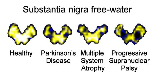
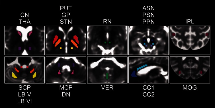
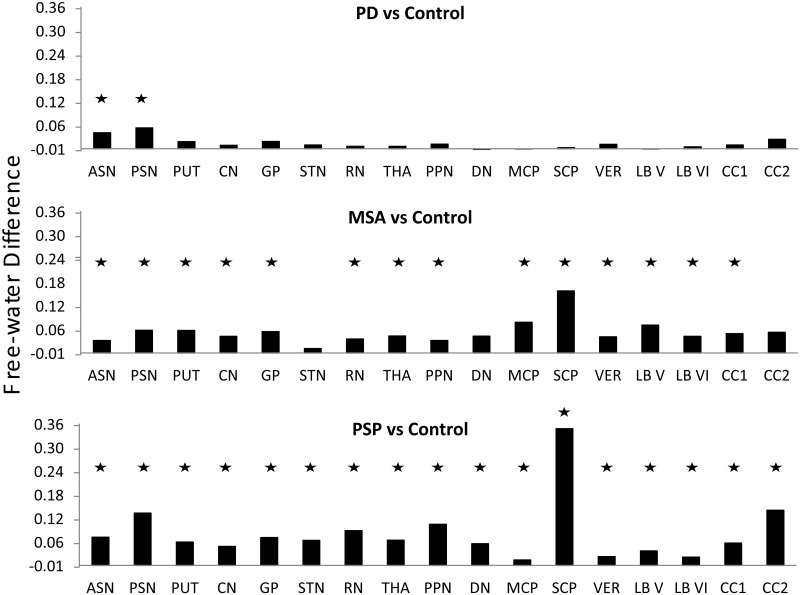
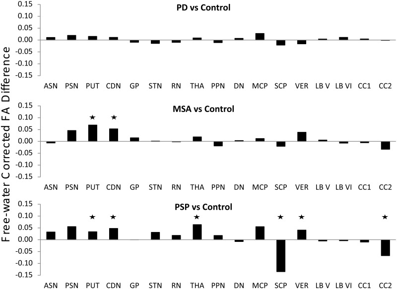
Conventional single tensor diffusion analysis models have provided mixed findings in the substantia nigra of Parkinson's disease, but recent work using a bi-tensor analysis model has shown more promising results. Using a bi-tensor model, free-water values were found to be increased in the posterior substantia nigra of Parkinson's disease compared with controls at a single site and in a multi-site cohort. Further, free-water increased longitudinally over 1 year in the posterior substantia nigra of Parkinson's disease. Here, we test the hypothesis that other parkinsonian disorders such as multiple system atrophy and progressive supranuclear palsy have elevated free-water in the substantia nigra. Equally important, however, is whether the bi-tensor diffusion model is able to detect alterations in other brain regions beyond the substantia nigra in Parkinson's disease, multiple system atrophy, and progressive supranuclear palsy and to accurately distinguish between these diseases. Free-water and free-water-corrected fractional anisotropy maps were compared across 72 individuals in the basal ganglia, midbrain, thalamus, dentate nucleus, cerebellar peduncles, cerebellar vermis and lobules V and VI, and corpus callosum. Compared with controls, free-water was increased in the anterior and posterior substantia nigra of Parkinson's disease, multiple system atrophy, and progressive supranuclear palsy. Despite no other changes in Parkinson's disease, we observed elevated free-water in all regions except the dentate nucleus, subthalamic nucleus, and corpus callosum of multiple system atrophy, and in all regions examined for progressive supranuclear palsy. Compared with controls, free-water-corrected fractional anisotropy values were increased for multiple system atrophy in the putamen and caudate, and increased for progressive supranuclear palsy in the putamen, caudate, thalamus, and vermis, and decreased in the superior cerebellar peduncle and corpus callosum. For all disease group comparisons, the support vector machine 10-fold cross-validation area under the curve was between 0.93-1.00 and there was high sensitivity and specificity. The regions and diffusion measures selected by the model varied across comparisons and are consistent with pathological studies. In conclusion, the current study used a novel bi-tensor diffusion analysis model to indicate that all forms of parkinsonism had elevated free-water in the substantia nigra. Beyond the substantia nigra, both multiple system atrophy and progressive supranuclear palsy, but not Parkinson's disease, showed a broad network of elevated free-water and altered free-water corrected fractional anisotropy that included the basal ganglia, thalamus, and cerebellum. These findings may be helpful in the differential diagnosis of parkinsonian disorders, and thereby facilitate the development and assessment of targeted therapies.
Keywords: Parkinson’s disease; diffusion MRI; extracellular space; multiple system atrophy; progressive supranuclear palsy.
© The Author (2015). Published by Oxford University Press on behalf of the Guarantors of Brain. All rights reserved. For Permissions, please email: journals.permissions@oup.com.
Figures
Similar articles
-
Development and validation of the automated imaging differentiation in parkinsonism (AID-P): a multicentre machine learning study.Lancet Digit Health. 2019 Sep;1(5):e222-e231. doi: 10.1016/S2589-7500(19)30105-0. Epub 2019 Aug 27. Lancet Digit Health. 2019. PMID: 33323270
-
Diffusion tensor imaging of Parkinson's disease, atypical parkinsonism, and essential tremor.Mov Disord. 2013 Nov;28(13):1816-22. doi: 10.1002/mds.25491. Epub 2013 May 14. Mov Disord. 2013. PMID: 23674400 Free PMC article.
-
Brain alpha-synuclein accumulation in multiple system atrophy, Parkinson's disease and progressive supranuclear palsy: a comparative investigation.Brain. 2010 Jan;133(Pt 1):172-88. doi: 10.1093/brain/awp282. Epub 2009 Nov 10. Brain. 2010. PMID: 19903734
-
Differential diagnosis of Parkinson's disease and atypical parkinsonian disorders by magnetic resonance imaging.Neurol Sci. 2003 May;24 Suppl 1:S35-7. doi: 10.1007/s100720300036. Neurol Sci. 2003. PMID: 12774211 Review.
-
Magnetic resonance imaging in progressive supranuclear palsy and other parkinsonian disorders.J Neural Transm Suppl. 1994;42:93-110. doi: 10.1007/978-3-7091-6641-3_8. J Neural Transm Suppl. 1994. PMID: 7964700 Review.
Cited by
-
The Role of Magnetic Resonance Imaging for the Diagnosis of Atypical Parkinsonism.Front Neurol. 2020 Jul 17;11:665. doi: 10.3389/fneur.2020.00665. eCollection 2020. Front Neurol. 2020. PMID: 32765399 Free PMC article. Review.
-
Association between neuromelanin-sensitive MRI signal and psychomotor slowing in late-life depression.Neuropsychopharmacology. 2021 Jun;46(7):1233-1239. doi: 10.1038/s41386-020-00860-z. Epub 2020 Sep 12. Neuropsychopharmacology. 2021. Retraction in: Neuropsychopharmacology. 2024 Jun;49(7):1202. doi: 10.1038/s41386-024-01851-0. PMID: 32919398 Free PMC article. Retracted.
-
Neuromelanin-MRI to Quantify and Track Nigral Depigmentation in Parkinson's Disease: A Multicenter Longitudinal Study Using Template-Based Standardized Analysis.Mov Disord. 2022 May;37(5):1028-1039. doi: 10.1002/mds.28934. Epub 2022 Feb 15. Mov Disord. 2022. PMID: 35165920 Free PMC article.
-
Multimodal brain and retinal imaging of dopaminergic degeneration in Parkinson disease.Nat Rev Neurol. 2022 Apr;18(4):203-220. doi: 10.1038/s41582-022-00618-9. Epub 2022 Feb 17. Nat Rev Neurol. 2022. PMID: 35177849 Review.
-
Free-water imaging reveals unique brain microstructural deficits in hispanic individuals with Dementia.Brain Imaging Behav. 2024 Feb;18(1):106-116. doi: 10.1007/s11682-023-00819-w. Epub 2023 Oct 31. Brain Imaging Behav. 2024. PMID: 37903991 Free PMC article.
References
-
- Agosta F, Galantucci S, Svetel M, Lukić MJ, Copetti M, Davidovic K, et al. Clinical, cognitive, and behavioural correlates of white matter damage in progressive supranuclear palsy. J Neurol 2014; 261: 913–24. - PubMed
-
- Agosta F, Pievani M, Svetel M, Ječmenica Lukić M, Copetti M, Tomić A, et al. Diffusion tensor MRI contributes to differentiate Richardson's syndrome from PSP-parkinsonism. Neurobiol Aging 2012; 33: 2817–26. - PubMed
-
- Ahmed Z, Asi YT, Sailer A, Lees AJ, Houlden H, Revesz T, et al. The neuropathology, pathophysiology and genetics of multiple system atrophy [Review]. Neuropathol Appl Neurobiol 2012; 38: 4–24. - PubMed
-
- Ahmed Z, Josephs KA, Gonzalez J, DelleDonne A, Dickson DW. Clinical and neuropathologic features of progressive supranuclear palsy with severe pallido-nigro-luysial degeneration and axonal dystrophy. Brain 2008; 131: 460–72. - PubMed
-
- Arai K, Braak E, De Vos RA, Jansen Steur EN, Braak H. Mossy fiber involvement in progressive supranuclear palsy. Acta Neuropathol 1999; 98: 341–4. - PubMed
Publication types
MeSH terms
Substances
Grants and funding
LinkOut - more resources
Full Text Sources
Other Literature Sources
Medical
