. 2016 Jan;22(1):46-53.
doi: 10.1038/nm.4011. Epub 2015 Dec 21.

Tau-driven 26S proteasome impairment and cognitive dysfunction can be prevented early in disease by activating cAMP-PKA signaling

Affiliations

Tau-driven 26S proteasome impairment and cognitive dysfunction can be prevented early in disease by activating cAMP-PKA signaling

Natura Myeku et al. Nat Med. 2016 Jan.

Abstract

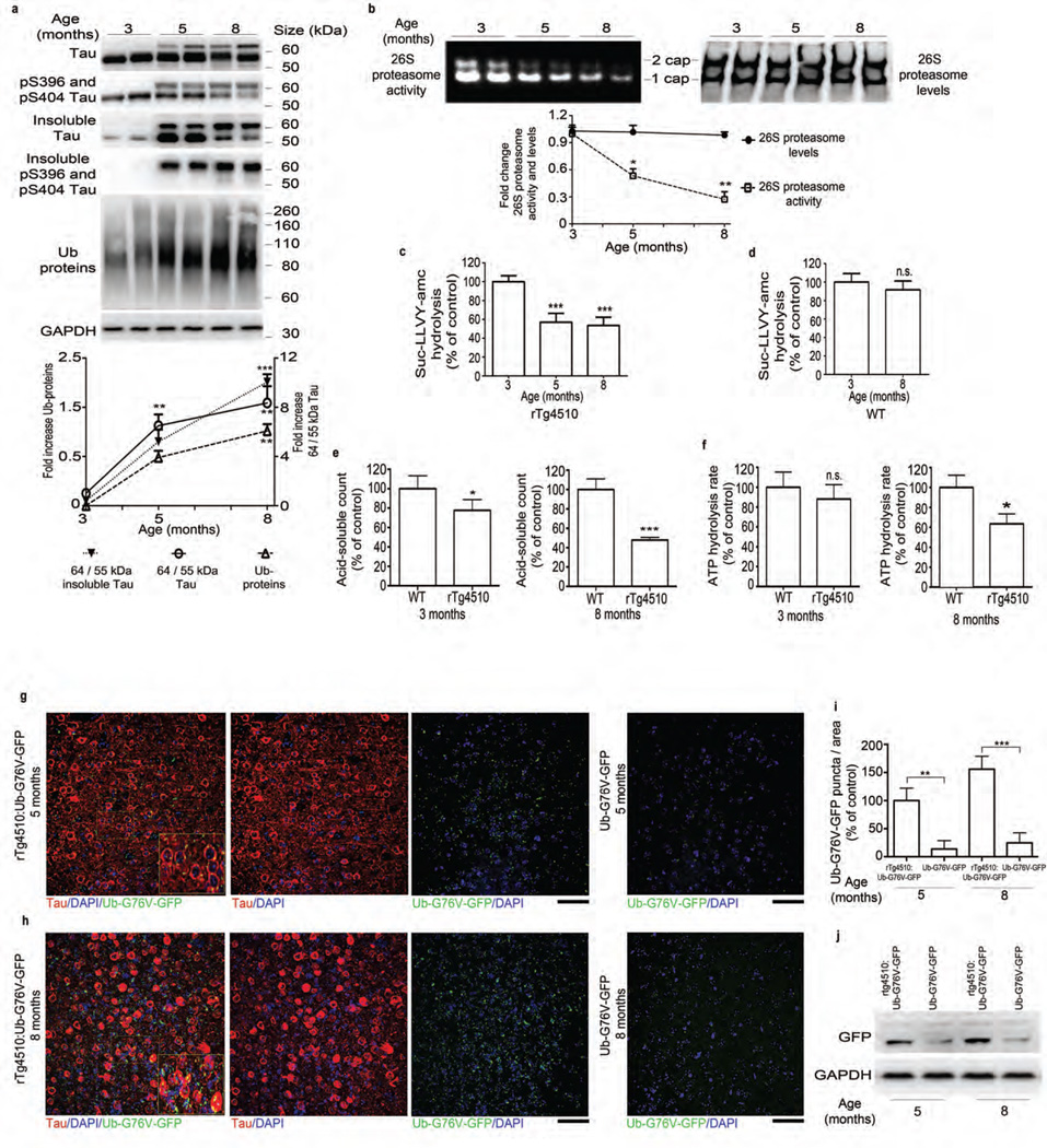
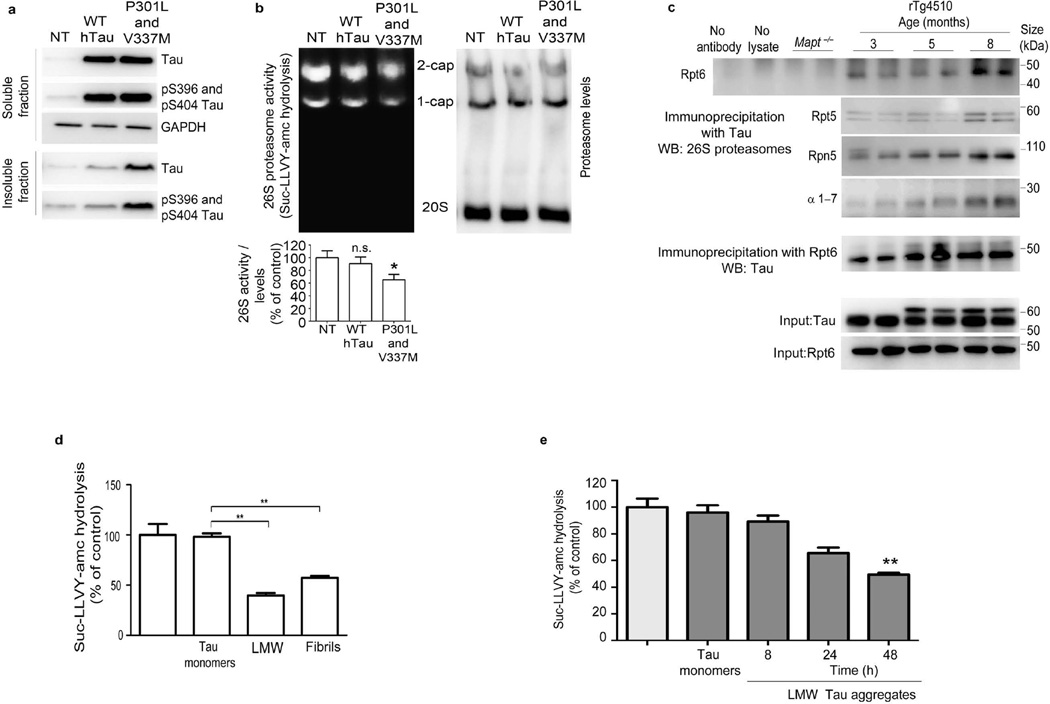
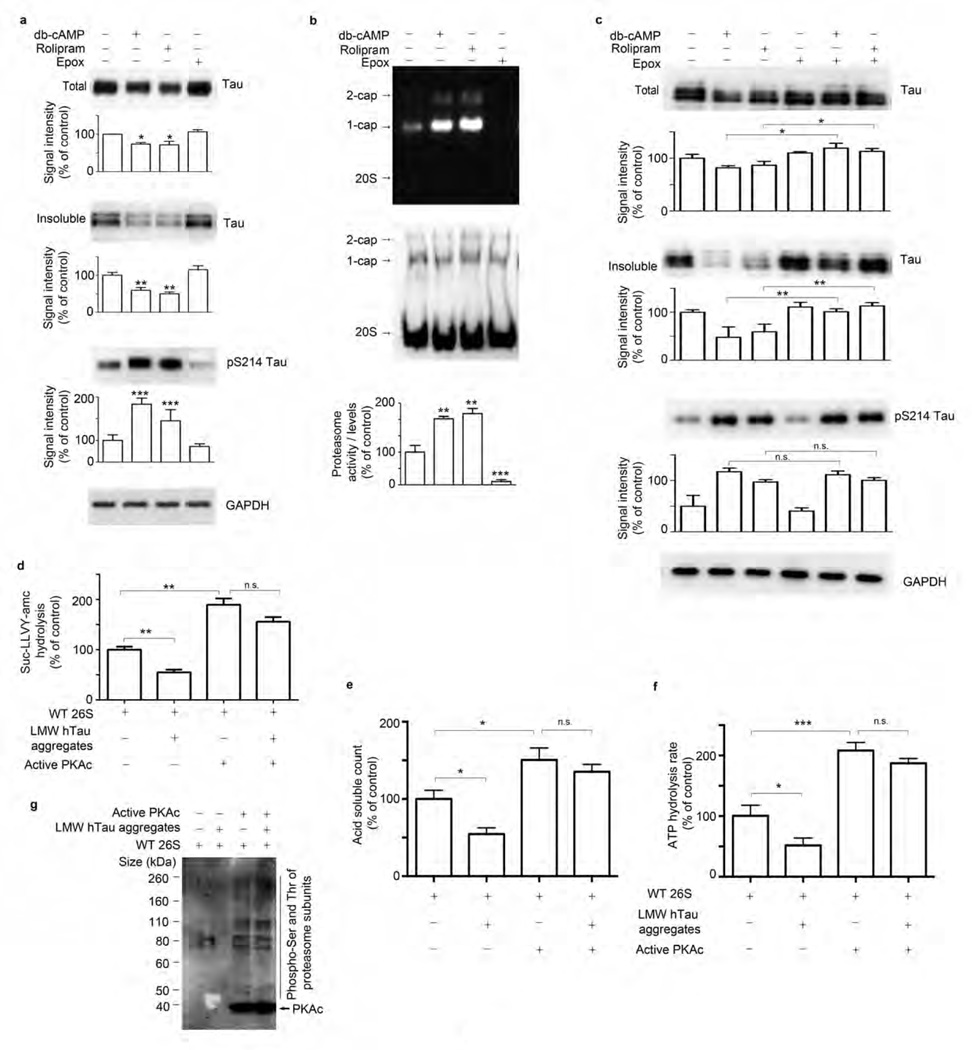
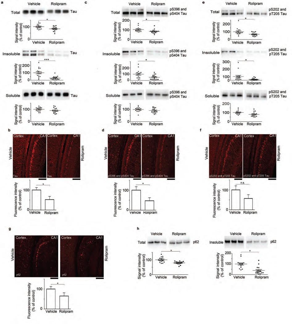
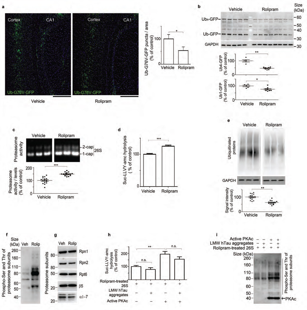
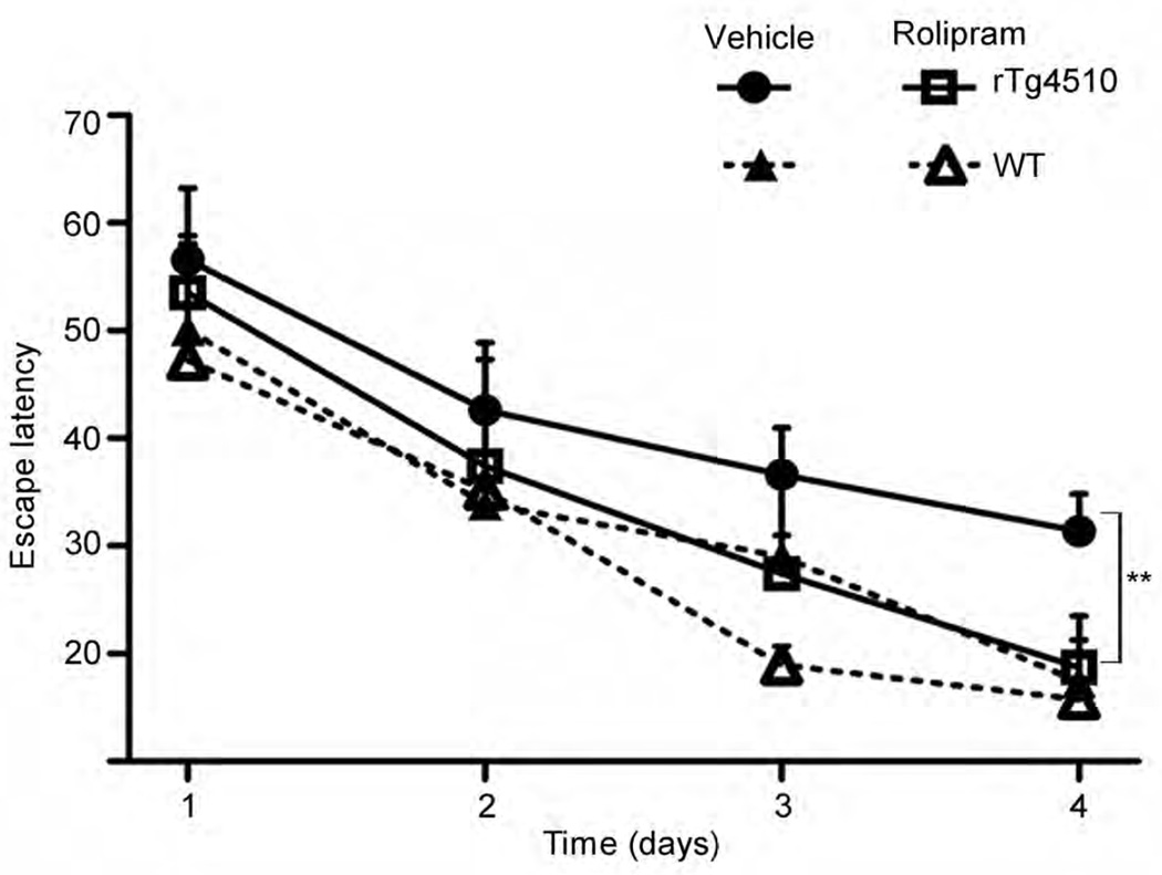
The ubiquitin proteasome system (UPS) degrades misfolded proteins including those implicated in neurodegenerative diseases. We investigated the effects of tau accumulation on proteasome function in a mouse model of tauopathy and in a cross to a UPS reporter mouse (line Ub-G76V-GFP). Accumulation of insoluble tau was associated with a decrease in the peptidase activity of brain 26S proteasomes, higher levels of ubiquitinated proteins and undegraded Ub-G76V-GFP. 26S proteasomes from mice with tauopathy were physically associated with tau and were less active in hydrolyzing ubiquitinated proteins, small peptides and ATP. 26S proteasomes from normal mice incubated with recombinant oligomers or fibrils also showed lower hydrolyzing capacity in the same assays, implicating tau as a proteotoxin. Administration of an agent that activates cAMP-protein kinase A (PKA) signaling led to attenuation of proteasome dysfunction, probably through proteasome subunit phosphorylation. In vivo, this led to lower levels of aggregated tau and improvements in cognitive performance.


Figures

Figure 1
Figure 1
Tauopathy is associated with a progressive decrease in proteasome function. (a) Top, immunoblot analysis of tau and pS396 and pS404 tau, Ub (ubiquitin) and GAPDH (for normalization) in total and sarkosyl-insoluble extracts from rTg4510 mice. Bottom, quantified densitometry of 64/55-kDa tau ratio in total and insoluble tau and ubiquitin, expressed as fold change relative to 3 months of age. (b) Native PAGE of 26S proteasome activity and levels (immunoprobing for Rpt6) and quantified densitometry (bottom). (c,d) Chymotrypsin-like activity of purified 26S proteasomes from rTg4510 (c) and WT (d) mice at indicated ages. (e) Degradation rate of 32P-labeled Ub5-DHFR by purified 26S proteasomes from rTg4510 and WT mice at indicated ages. (f) ATPase activity of purified 26S proteasomes from rTg4510 and WT (control) mice at indicated ages (g,h) Immunofluorescence labeling of human tau (red) and GFP signal (green, detected without antibody) in the frontal cortex of rTg4510:Ub-G76V-GFP and Ub-G76V-GFP mice at 5 (g) and 8 (h) months of age. Insets, high-magnification views of outlined areas. Scale bars, 50 µm. (i) Quantification of GFP puncta from analyses in g and h. Control, 5-month-old rTg4510:Ub-G76V-GFP. (j) Immunoblot analysis of GFP expression in rTg4510:Ub-G76V-GFP and Ub-G76V-GFP mice. For a,b and j at least three biological experiments (two mice per experiment, n = 6 mice) were performed. For c–f, n = 6 cortical brains per age group were used to elute 26S proteasomes, and at least three independent experiments were performed. Quantification of GFP signal for g and h was performed on slices from 6 mice per group. Error bars, mean ± s.e.m.; n.s., not significant; *P < 0.05, **P < 0.01, ***P < 0.001 (one-way ANOVA followed by Tukey’s multiple comparison post hoc test).
Figure 2
Figure 2
Aggregated tau directly inhibits 26S proteasomes and associates with proteasomes in brains of mice with tauopathy. (a) Immunoblot analysis for tau and phosphorylated (pS396 and pS404) tau epitopes from soluble and insoluble fractions in HEK293 cells nontransfected (NT) or stably transfected with WT human tau (hTau) or double-mutant P301L and V337M tau. (b) Native PAGE assay (top) and quantification (bottom) of 26S proteasome activity and levels (assessed by immunoblotting for β5 subunit) in NT HEK293 cells and HEK293 cells transfected with WT hTau or P301L and V337M tau. The densitometric quantification of 26S proteasome activity normalized to 26S proteasome levels (c) Immunoprecipitation with antibody to human tau of cortical brain lysates from Mapt−/− (negative control) and rTg4510 mice at indicated ages and immunoblot analysis of Rpt6, Rpt5, Rpn5 and 20S α-subunits 1–7 (α1–7) expression and a reciprocal experiment using anti-Rpt6 for immunoprecipitation and anti-hTau for immunoblotting. The total extracts (input) for tau and Rpt6 are shown as loading controls. (d,e) Rate of succinyl-Leu-Leu-Val-Tyr-amc (Suc-LLVY-amc) hydrolysis by 26S proteasomes, incubated with tau monomers, LMW aggregates and fibrils generated from recombinant tau (d), or incubated with tau monomers and LMW aggregates generated by incubating recombinant tau at indicated time points (e). Untreated WT 26S proteasomes were used as a control for d and e. At least three biological replicates were performed with stably transfected clones (a,b). For immunoprecipitation (c), two animals per experiment were analyzed in three biological replicates (n = 6 mice). Purified proteasomes from n = 6 brains from 3-month-old WT were used for d and e, and at least three independent experiments were performed (d,e). Error bars, means ± s.e.m. *P < 0.05, **P < 0.01 (one-way ANOVA followed by Tukey’s multiple comparison post hoc test).
Figure 3
Figure 3
Activation of PKA stimulates hydrolyzing activity of the proteasome in slices-ex vivo and in vitro. (a–c) Immunoblot analysis and corresponding densitometric quantification of total and insoluble extracts for tau, pS214 tau epitope and GAPDH (a,c) and native PAGE analysis and quantification of 26S proteasome activity and level using antibody to the β5 subunit (b) in acute organotypic cortical slices from 3- to 4-month-old rTg4510 mice treated with vehicle, db-cAMP, rolipram or epoxomicin (Epox) alone or a combination of epoxomicin and db-cAMP or rolipram. (d–f) Hydrolysis rate of succinyl-Leu-Leu-Val-Tyr-amc (Suc-LLVY-amc) (d), 32P-labeled Ub5-DHFR substrate (e) and ATP (f) by purified 26S proteasomes from WT mice untreated (WT 26S), incubated with LMW tau aggregates and/or pre-incubated with PKA. (g) Immunoblot analysis of phosphorylated serine and threonine (pSer and pThr) epitopes of proteasome subunits by PKA in purified 26S proteasomes treated as in d–f. For a–c, two animals per experiment were analyzed in three biological replicates (n = 6 mice). For d–g, purified proteasomes from n = 6 brains from 3-month-old WT mice were used for each hydrolyzing assay and at least three independent experiments were performed. Error bars, mean ± s.e.m.; n.s., not significant; *P < 0.05, **P < 0.01, ***P < 0.001 (one-way ANOVA followed by Tukey’s multiple comparison post hoc test).
Figure 4
Figure 4
Rolipram administration reduces accumulation of tau species and p62 in vivo. (a,c,e) Immunoblot analysis and corresponding densitometric quantification of total, insoluble and soluble extracts of tau (a) and pS396 and pS404 (c) and pS202 and pT205 (e) tau epitopes from cortical tissue of rTg4510 mice treated with vehicle or rolipram (b,d,f,g). Immunofluorescence labeling and quantification of fluorescence intensity for tau (b), pS396 and pS404 (d) and pS202 and pT205 (f) tau epitopes, and p62 (g) in the CA1 region of the hippocampus of rTg4510:Ub-G76V-GFP mice treated with vehicle or rolipram. Scale bars, 200 µm. (h) Immunoblot analysis and densitometric quantification of p62 in total and insoluble extracts from cortical tissue. Scatter plots represent quantification of immunoreactivity normalized to GAPDH. Statistical analyses of rolipram (n = 16) and vehicle-treated (n = 16) mice for a,c and e were performed in two sets. For quantification of immunofluorescence signal (b,d,f,g), slices from 6 mice per treatment group were analyzed. Error bars, mean ± s.e.m.; n.s., not significant; *P < 0.05, ***P < 0.001 (unpaired two-tailed Student’s t-test). Vehicle treatment served as control for all experiments.
Figure 5
Figure 5
Rolipram treatment increases proteasome function and reduces ubiquitinated protein accumulation in vivo. (a) GFP signal and quantification of GFP puncta from hippocampus CA1 and the cortex of rTg4510:Ub-G76V-GFP mice. Scale bars, 200 µm. (b) Immunoblot analysis and densitometric quantification of GFP and GAPDH in vehicle- and rolipram-treated rTg4510:Ub-G76V-GFP mice. (c) Native PAGE of 26S proteasome and densitometric quantification of activity normalized to 26S proteasome levels in vehicle- and rolipram-treated rTg4510 mice. (d) Rate of hydrolysis by 26S proteasomes from vehicle- and rolipram-treated mice, incubated with succinyl-Leu-Leu-Val-Tyr-amc (Suc-LLVY-amc). (e) Immunoblot analysis and corresponding densitometric quantification of ubiquitinated protein and GAPDH in rTg4510 mice treated with vehicle or rolipram. (f,g) Immunoblot analysis of phosphorylated serine and threonine (pSer and pThr) (f) and proteasome subunits Rpn1, Rpn2, Rpt6, β5 and α-subunits 1–7 (α1–7) (g) in purified 26S proteasomes from vehicle- and rolipram-treated rTg4510 mice. (h) Rate of Suc-LLVY-amc hydrolysis by 26S proteasomes purified from rolipram-treated rTg4510 mice (control) or incubated with LMW tau aggregates and/or pre-incubated with PKA. (i) Immunoblot analysis of serine and threonine phosphorylation in rolipram-treated 26S proteasomes treated as in h. Statistical analyses for rTg4510 (rolipram n = 16 and vehicle n = 16) (c and e) and for rTg4510:Ub-G76V-GFP (rolipram n = 8 and vehicle n = 5) mice (b) and for quantification of GFP signal (n = 3 mice per treatment group) (a) were carried out using unpaired two-tailed Student’s t-test between groups. Purified proteasomes were pooled from n = 6 brains (d, f–i), and at least three independent experiments were performed. Statistical analyses employed two-tailed Student’s t-test between groups (d) and one-way ANOVA followed by Tukey’s multiple comparison post hoc test (h). Error bars, mean ± s.e.m.; n.s., not significant; *P < 0.05, **P < 0.01, ***P < 0.001. Veh, vehicle; rolip, rolipram.
Figure 6
Figure 6
Rolipram treatment improves cognition in rTg4510 mice with early-stage disease. Cognitive performance, assessed by Morris water maze escape latency, of rTg4510 and WT mice treated with vehicle or rolipram. n = 16 (vehicle) or 15 (rolipram) in rTg4510 mice group; and n = 7 (vehicle) or n =7 (rolipram) in WT mice group. Error bars, mean ± s.d. **P < 0.01 (repeated-measures ANOVA test (days 1–3) plus one-way ANOVA with Bonferroni correction (day 4)).

Comment in

Similar articles

Cited by

References

    1. Goldberg AL. Protein degradation and protection against misfolded or damaged proteins. Nature. 2003;426:895–899. - PubMed
    1. Finley D. Recognition and processing of ubiquitin-protein conjugates by the proteasome. Annual review of biochemistry. 2009;78:477–513. - PMC - PubMed
    1. Lander GC, et al. Complete subunit architecture of the proteasome regulatory particle. Nature. 2012;482:186–191. - PMC - PubMed
    1. Beck F, et al. Near-atomic resolution structural model of the yeast 26S proteasome. Proceedings of the National Academy of Sciences of the United States of America. 2012;109:14870–14875. - PMC - PubMed
    1. Asai M, et al. PKA rapidly enhances proteasome assembly and activity in in vivo canine hearts. Journal of molecular and cellular cardiology. 2009;46:452–462. - PubMed

Publication types

MeSH terms