Comparative Study
. 2016 Jan 26;7(4):4724-34.
doi: 10.18632/oncotarget.6619.

Identification of a novel RNA giant nuclear body in cancer cells

Affiliations
Comparative Study

Identification of a novel RNA giant nuclear body in cancer cells

Hong Zhou et al. Oncotarget. .

Abstract

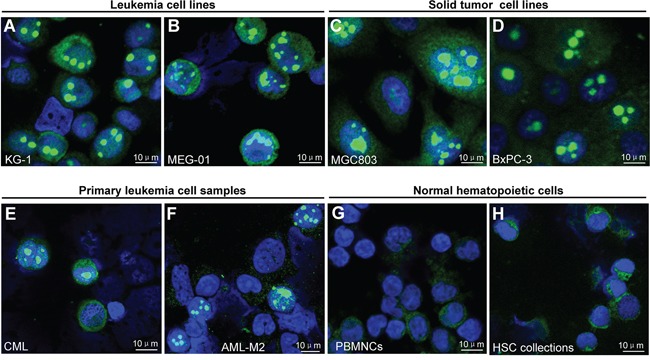
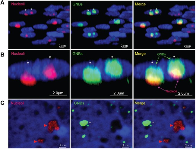
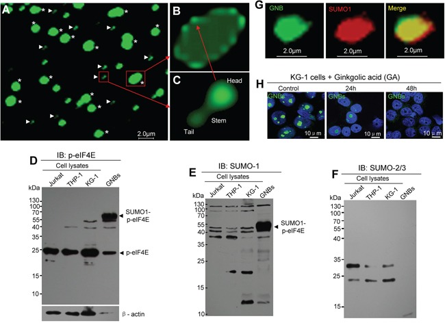
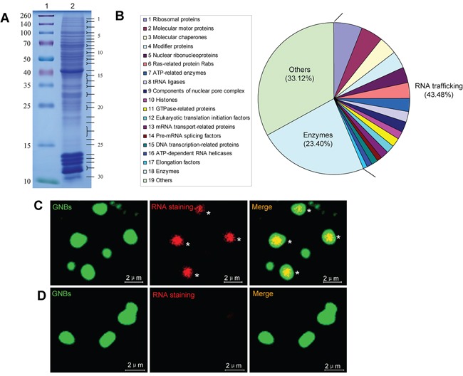
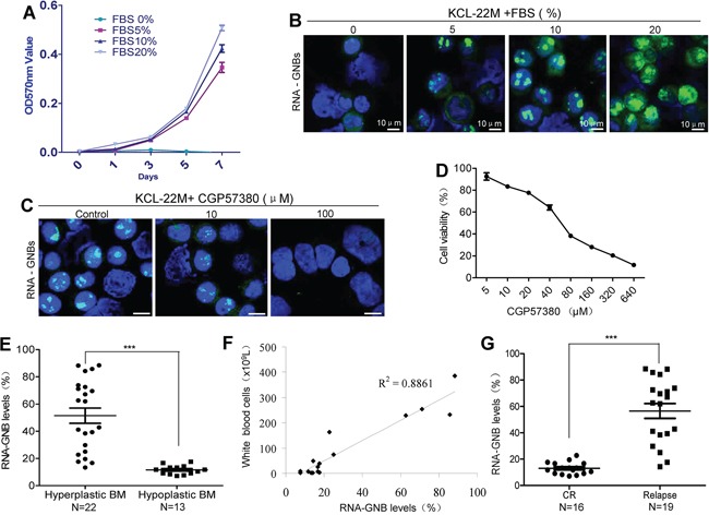
Constitutive synthesis of oncogenic mRNAs is essential for maintaining the uncontrolled growth of cancer cells. However, little is known about how these mRNAs are exported from the nucleus to the cytoplasm. Here, we report the identification of a RNA giant nuclear body (RNA-GNB) that is abundant in cancer cells but rare in normal cells. The RNA-GNB contains a RNA core surrounded by a protein shell. We identify 782 proteins from cancer-associated RNA-GNBs, 40% of which are involved in the nuclear mRNA trafficking. RNA-GNB is required for cell proliferation, and its abundance is positively associated with tumor burden and outcome of therapies. Our findings suggest that the RNA-GNB is a novel nuclear RNA trafficking organelle that may contribute to the nuclear mRNA exporting and proliferation of cancer cells.

Keywords: RNA giant nuclear body; cancer cell; nuclear RNA trafficking.


Conflict of interest statement

CONFLICTS OF INTEREST

The authors declare no competing financial interests.

Figures

Figure 1
Figure 1. Novel giant nuclear bodies (GNBs) in cancer cells and normal blood cells
A–H. Immunofluorescent staining images of novel giant nuclear bodies (green nuclear bodies) in the indicated leukemia (A–B) and solid tumor cell lines (C–D) and primary leukemia cell sample (E–F) and normal hematopoietic cell samples (G–H) with p-eIF4E antibody. Cell nuclei (blue) were stained using 4′,6-diamidino-2-phenylindole (DAPI).
Figure 2
Figure 2. Giant nuclear bodies (GNBs) are different from nucleoli in cancer cells
A. 3-D images of nuclei (blue), nucleoli (red bodies) and GNBs (green bodies) in cancer cells. GNBs attach on the surface of nucleoli. B. Magnified image of 3-D images of nuclei (blue), nucleoli (red bodies) and GNBs (green bodies) in cancer cells. Two GNBs attach on the surface of two nucleoli in the nucleus of cancer cells. C. GNBs (green bodies) were removed completely from nucleoli (red nuclear bodies) in nuclei of KG-1 cells after treatment with 0.05% NP40 for 5 min. Cell nuclei (blue) were stained using 4′,6-diamidino-2-phenylindole (DAPI).
Figure 3
Figure 3. GNB contains a shell composed of drumstick-shaped subunits and inner structure
A. Immunofluorescent staining images of purified GNBs (asterisks) and the subunits of GNB shells (arrowheads) from KCL-22M leukemia cells. B. Structure image of an intact GNB with a shell composed with several subunits. C. Structure image of a single subunit of the shell contains a head, a stem and a tail. D–F. Profile analyses of p-eIF4E protein (D), SUMO1-modified proteins (E), and SUMO2/3-modified proteins (F) of total cell lysates and purified GNB lysates using Western blotting with antibodies against p-eIF4E, SUMO1 or SUMO2/3. G. Two-color confocal images of p-eIF4E (green) and SUMO1(red) in GNB by immunofluorescence staining. GNBs were incubated with antibodies against p-eIF4E and SUMO1 followed by a secondary antibody labeled with FITC (p-eIF4E, green color) and Alexa Fluor 568(SUMO1, red color). A colocalization of p-eIF4E and SUMO1 in the GNB was evident (merge, yellow). H. SUMOylation inhibition decreased GNBs in KG-1 leukemia cells. Leukemia cells were treated with GA at 200 μM for different time points and then collected for analyses of GNBs using immunofluorescence staining. Cell nuclei (blue) were stained using DAPI.
Figure 4
Figure 4. GNB is a highly complex nuclear protein structure with a RNA core
A. Analysis of GNB proteins separated by SDS-PAGE. GNB proteins were separated by SDS-PAGE on a 12% polyacrylamide gel and stained with Coomassie blue (lane 2). Molecular masses of known proteins separated in the same gel are indicated on the left of the image (lane 1). B. Functional classes of 782 different proteins identified in proteomic analyses of purified GNBs from leukemia cells. C. RNA staining of GNBs with acridine orange (AO). All intact GNBs contained a RNA core (red) surrounded by a shell of GNBs. D. RNase treatment eliminated RNAs within GNBs.
Figure 5
Figure 5. GNB abundance is positively correlated with cell proliferation, tumor burden and outcome of therapy in human leukemia
A–B. A positive correlation exists between cell proliferation (A) and GNB levels (B). Kcl-22M cells were treated with the mitogen FBS at various concentrations for indicated times and then collected for analyses of cell proliferation by MTT and GNBs by immunofluorescence staining. C–D. Depletion of GNBs with the small molecule inhibitor CGP57380 inhibited proliferation of leukemia cells with a dose-dependent manner. Kcl-22M cells were treated with the indicated concentrations of CGP57380 for 72 h and then analyzed for GNBs by immunofluorescent staining(C) and Cell viability by MTT (D). E. RNA-GNB abundance was positively associated with the malignant proliferative potential of the bone marrow (BM) in patients with leukemia. ***P < 0.001. F. A positive correlation between RNA-GNB levels and peripheral white blood cell count were observed in leukemia patients (R2=0.89). Note: Normal blood cell count is below 1 × 10⁹L. G. RNA-GNB abundance was associated with response to chemotherapies. ***P < 0.001.

Similar articles

Cited by

References

    1. Blanpain C. Tracing the cellular origin of cancer. Nature Cell Biology. 2013;15:126–134. - PubMed
    1. Kondo T, Setoguchi T, Taga T. Persistence of a small subpopulation of cancer stem-like cells in the C6 glioma cell line. Proceedings of the National Academy of Sciences of the United States of America. 2004;101:781–786. - PMC - PubMed
    1. Baserga R, Huang CH, Rossini M, Chang H, Ming PM. The role of nuclei and nucleoli in the control of cell proliferation. Cancer Research. 1976;36:4297–4300. - PubMed
    1. Webster M, Witkin KL, Cohen-Fix O. Sizing up the nucleus: nuclear shape, size and nuclear-envelope assembly. The Journal of Cell Science. 2009;122:1477–1486. - PMC - PubMed
    1. Morimoto M, Boerkoel CF. The Role of Nuclear Bodies in Gene Expression and Disease. Biology (Basel) 2013;2:976–1033. - PMC - PubMed

Publication types

LinkOut - more resources