. 2015 Dec 29;112(52):E7176-85.
doi: 10.1073/pnas.1522332112. Epub 2015 Dec 15.

cAMP-induced phosphorylation of 26S proteasomes on Rpn6/PSMD11 enhances their activity and the degradation of misfolded proteins

Affiliations

cAMP-induced phosphorylation of 26S proteasomes on Rpn6/PSMD11 enhances their activity and the degradation of misfolded proteins

Sudarsanareddy Lokireddy et al. Proc Natl Acad Sci U S A. .

Abstract

Although rates of protein degradation by the ubiquitin-proteasome pathway (UPS) are determined by their rates of ubiquitination, we show here that the proteasome's capacity to degrade ubiquitinated proteins is also tightly regulated. We studied the effects of cAMP-dependent protein kinase (PKA) on proteolysis by the UPS in several mammalian cell lines. Various agents that raise intracellular cAMP and activate PKA (activators of adenylate cyclase or inhibitors of phosphodiesterase 4) promoted degradation of short-lived (but not long-lived) cell proteins generally, model UPS substrates having different degrons, and aggregation-prone proteins associated with major neurodegenerative diseases, including mutant FUS (Fused in sarcoma), SOD1 (superoxide dismutase 1), TDP43 (TAR DNA-binding protein 43), and tau. 26S proteasomes purified from these treated cells or from control cells and treated with PKA degraded ubiquitinated proteins, small peptides, and ATP more rapidly than controls, but not when treated with protein phosphatase. Raising cAMP levels also increased amounts of doubly capped 26S proteasomes. Activated PKA phosphorylates the 19S subunit, Rpn6/PSMD11 (regulatory particle non-ATPase 6/proteasome subunit D11) at Ser14. Overexpression of a phosphomimetic Rpn6 mutant activated proteasomes similarly, whereas a nonphosphorylatable mutant decreased activity. Thus, proteasome function and protein degradation are regulated by cAMP through PKA and Rpn6, and activation of proteasomes by this mechanism may be useful in treating proteotoxic diseases.

Keywords: Rpn6/PSMD11; cAMP; cAMP-dependent protein kinase; proteasomes; protein degradation.


Conflict of interest statement

The authors declare no conflict of interest.

Figures
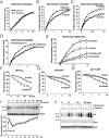
Fig. 1.
Fig. 1.
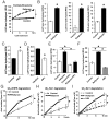
Raising cAMP with the PDE4 inhibitor rolipram or the activator of adenylate cyclase, forskolin, enhances the degradation of short- but not long-lived cell proteins as well as model substrates of the UPS and ubiquitin conjugates generally. (A) To label long-lived proteins selectively, differentiated C2C12 myotubes were incubated with [3H]tyrosine for 24 h and then washed with chase medium containing nonradioactive tyrosine (2 mM) for 2 h to allow breakdown of short-lived proteins (27). New chase media containing nonradioactive tyrosine (2 mM) and either DMSO (control) or rolipram (50 μM) was added, and media samples were collected at the times indicated. The radioactivity released from cell proteins was measured and plotted as a percentage of the total radioactivity in proteins at 0-time. Here and below error bars represents the SEM and n = 4. (B) Differentiated myotubes were pretreated with or without rolipram for 3 h. To follow degradation of short-lived and long-lived proteins, cells were then incubated with [3H]tyrosine for 10 min to label both short-lived and long-lived components and then washed with chase medium three times. Myotubes were incubated with media containing nonradioactive tyrosine (2 mM), CHX (100 μg/mL), and either rolipram or DMSO (control). The released radioactivity was measured as in A. (C) To follow degradation of short-lived and long-lived proteins in 293A cells, cells were incubated with [3H]tyrosine for 10 min to label both short-lived and long-lived components and then washed with chase medium three times. Cells were incubated as in B and either forskolin or DMSO (control). The released radioactivity was measured as in A. For B and C, because degradation of long-lived components was not affected by cAMP (A), the increase must be a result of breakdown of short-lived proteins. (D) The lysosomal V-ATPases inhibitor, concanamycin A (0.2 μM) does not block rolipram-stimulated degradation of short-lived proteins. Proteolysis was measured in myotubes as in A in the presence of concanamycin A. (E) The proteasome inhibitor, BTZ inhibits forskolin-stimulated degradation of short-lived proteins in 293A cells. Proteolysis was measured as in A in the presence or absence of forskolin, BTZ (0.1 μM) or combinations of these agents. (F–H) Forskolin treatment enhances the degradation of model substrates of the UPS. (F) 293T cells stably overexpressing GFP-CL1 or 293A cells were transfected with (G) Ub-R-GFP or (H) UbG76V-GFP were treated with or without forskolin in the presence of CHX to prevent protein synthesis. GFP fluorescence was measured in live cells at 395/509 nm. n = 16. (I and J) Forskolin treatment reduces rapidly the total level of ubiquitinated proteins in 293A cells, which slowly returns to control levels. Immunoblot analysis of total ubiquitin conjugates levels in 293A cells after treatment with (I) forskolin or (J) the proteasome inhibitor BTZ. Levels of Rpt5 and the α-subunits, α1, -2, -3, -5, -6, -7 (which react with anti-MCP antibody), were used as loading control. Line graph in I represents the levels of ubiquitinated proteins determined by densitometry. Error represents the mean of two observations.
Fig. 2.
Fig. 2.
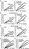
Rolipram-mediated activation of PKA in cells and PKA treatment of purified 26S promote the degradation of short peptides and ubiquitinated proteins. (A) Rolipram treatment of myotubes stimulates the peptidase activity of 26S proteasomes. Myotubes were incubated with or without rolipram, and at the indicated times, 26S proteasomes purified by the UBL method. The chymotrypsin-like peptidase activity was measured here and below (B–F) using suc-LLVY-amc and represented by relative fluorescence units (RFU). Here and below (A–I) error bars represent SEM and n = 3, *P < 0.01. (B) Myotubes were treated for 6 h with or without rolipram, 26S proteasomes purified, and their peptidase activities measured, the caspase-like activity with suc-LLE-amc, and trypsin-like with suc-VLR-amc. (C and D) Incubation of 26S proteasomes purified from control myotubes with (C) PKA increases and incubation with (D) PP1 reduces peptidase activity. Treatments were at 30 °C for 90 min. (E) The increased peptidase activity of 26S proteasomes purified from myotubes treated with rolipram was reversed by incubating them with PP1. Myotubes were treated with or without rolipram for 6 h, and 26S proteasomes were incubated with or without PP1 for 90 min. (F) Treatment of myotubes with the PKA inhibitor, H89, reduces the peptidase activity. Myotubes were also treated with or without rolipram, and H89 (10 μM) for 6 h. (G–I) 26S Proteasomes purified from myotubes treated with rolipram have a greater capacity to hydrolyze ubiquitinated proteins and treatment of cells with H89 reduces this activity. (G) Degradation of 32P-labeled Ub5-DHFR. (H and I) 35S-labeled ubiquitinated-Sic1 (Ubn-Sic1) by 26S proteasomes purified from myotubes treated as in A and F. Rates of degradation were measured by following the conversion of radiolabeled protein to TCA-soluble labeled material.
Fig. 3.
Fig. 3.
26S proteasomes from myotubes treated with rolipram have increased ATPase activity. (A) 26S proteasomes purified from myotubes treated with rolipram have increased ATPase activity. Myotubes were treated with or without rolipram for 6 h. Basal ATPase activity (Left) and hexa-Ub+casein-stimulated (Right) activity were measured by following the production of free phosphate using the malachite green method (35). Here and below (AE) error bars represent SEM and n = 3. *P < 0.01. (B and C) Treatment of purified 26S proteasomes with PKA increases and incubation with PP1 reduces ATPase activity. 26S from control myotubes were treated with (B) PKA or (C) PP1 for 90 min at 30 °C. Basal (Left) and hexa-Ub+casein-stimulated (Right) activities were measured as in A after 10 min incubation in the presence of PKA inhibitor, H89, or protein phosphatase inhibitor 2 (I-2) to avoid reincorporation or removal of phosphate group by the kinase or phosphatase. (D) The increased ATPase activity of 26S purified from myotubes treated with rolipram was reversed by incubating them with PP1. Myotubes were treated with or without rolipram for 6 h, 26S proteasomes treated with PKA as in B. ATPase activity of was measured as in A. (E) Treatment of myotubes with PKA inhibitor, H89, reduces the ATPase activity. Myotubes were treated with rolipram or H89 for 6 h, 26S proteasomes were purified, and their basal ATPase activity measured.
Fig. 4.
Fig. 4.
Forskolin promotes the degradation by the UPS of aggregation-prone proteins associated with neurodegenerative diseases. (A–D) Forskolin treatment decreased the levels of aggregation-prone proteins in both soluble and insoluble fraction. 293A cells were transfected with (A) Flag-FUS (WT or R495X), (B) TDP43 (WT or M337V), or (C) Tau (WT or P301L). After 48 h, cells were treated with or without forskolin (A–C) or H89 (D) for 5 h in the presence of CHX. Immunoblot analysis was performed on both 1% Triton-X 100-soluble and -insoluble fractions using antibodies against Flag-FUS (A and D), TDP43 (B), and total Tau and pTau(pS396/404) (C and D). Levels of Rpt5 and MCP in the soluble and actin in the insoluble fraction were used as loading controls. (E) Degradation of Tau(P301L) requires ubiquitin activation and proteasomes but not autophagy. 293A cells were transfected with the Tau mutant. After 48 h, cells were treated with forskolin, E1 (Ube1) inhibitor (ML00603997) (1 μM), MG132 (10 μM), concanamycin A (0.2 μM), or indicated combinations of these agents for 5 h in the presence of CHX. Immunoblot analysis was performed on total lysate (in RIPA buffer containing 0.1% SDS) with antibodies against Ub, Tau, pTau(pS396/404), or LC3. Rpt5 and MCP were used as loading controls.
Fig. 5.
Fig. 5.
After rolipram treatment more PKA catalytic subunit is bound to 26S proteasomes, and Rpn6 subunit (but not Rpt6) is phosphorylated. (A) More PKA is bound to proteasomes after raising the cAMP levels in myotubes. Equal amounts of 26S purified from myotubes treated with or without rolipram for 6 h were analyzed by immunoblot for Rpt6, pRpt6, Rpn6, PA200, PKAα (catalytic), and PKAIIα (regulatory) subunits. (B) Native gel electrophoresis of purified 26S proteasomes from myotubes treated with control or rolipram. In-gel proteasome activity was assayed using suc-LLVY-amc overlaid, followed by immunoblot against Rpn1, MCP, and PKAα (catalytic subunit). DC, doubly capped 26S proteasome; SC, singly capped. (C) Rolipram treatment of cells and PKA treatment of 26S proteasomes from untreated controls cause phosphorylation of Rpn6. 26S were purified from myotubes treated with or without rolipram or H89 for 6 h, or 26S proteasomes were purified from control myotubes and treated with PKA or PP1 for 90 min at 30 °C. Samples were subjected to Zn2+-Phos-tag SDS/PAGE (38) and followed by immunoblot analysis for Rpt6 and Rpn6. Rpt5 was used as loading control. (D) Overexpression of the catalytic subunit of PKA caused phosphorylation of Rpn6. 293A cells were transfected with a control vector, myc-AMPKα, or GFP-PKAα. Control transfected cells were treated with or without forskolin for 5 h, and 26S proteasomes were purified and analyzed as in C, followed by immunoblot analysis for Rpt6 and Rpn6. Rpn2 and Rpt5 were used as loading controls. (E) Pure Rpn6 expressed in E. coli was phosphorylated by PKA. Recombinant His-Rpn6 was incubated with or without PKA at 30 °C and was subjected to Zn2+-Phos-tag SDS/PAGE followed by immunoblot analysis of Rpn6 and His-tag. (F) PKA phosphorylates Rpn6 only at serine 14. 26S proteasomes were purified from 293A cells transfected with an empty control vector, Flag-Rpn6-WT, or -S14D or -S14A mutants and then treated with PKA as in C and was subjected to Zn2+-Phos-tag SDS/PAGE and followed by immunoblot analysis for Rpn6 and Flag. Rpn2 and Rpt5 were used as loading controls.
Fig. 6.
Fig. 6.
Overexpression of the phosphomimetic Rpn6-S14D mutant enhances the degradation of short-lived proteasome substrates and aggregation-prone proteins. (A) Overexpression of Rpn6-WT and -S14D reduces the levels of the UFD pathway substrate and ubiquitin conjugates compared with Rpn6-S14A. Cotransfection of UbG76V-GFP with a Flag-Rpn6-WT, -S14D, or -S14A into 293A cells. After 48 h, cells were subjected to immunoblot analysis for Ub, GFP, and Flag. Rpn2 and Rpt5 were used as the loading control. (B) Overexpression of Rpn6-S14D stimulates and S14A mutant reduces peptidase activity. 293A cells were transfected with Flag-Rpn6- WT, -S14D, or -S14A. Forty-eight hours posttransfection, the chymotrypsin-like peptidase activity was measured on proteasomes purified either by the UBL- (Left) or flag-method (Right). Here and below (B and C) error bars represent SEM. *P < 0.01. n = 3. (C) Overexpression of Rpn6-S14D stimulates and -S14A mutant reduces ATP hydrolysis. ATPase activity of purified proteasomes from transfected 293A cells as in B was measured as in Fig. 3. (D) Overexpression of Rpn6-S14D mutant decreased the levels of aggregation-prone proteins in both soluble and insoluble fractions. Cotransfection of GFP-SOD1(G93A) or Tau(P301L) with Flag-Rpn6- WT, -S14D, or -S14A into 293A cells. Immunoblot analysis was performed both in 1% Triton-X 100 soluble and insoluble fractions against GFP (GFP-SOD1-G93A) and Tau. Levels of Flag (Flag-Rpn6) in the soluble and actin in the insoluble fractions were used as loading control.

Similar articles

Cited by

References

    1. Goldberg AL. Protein degradation and protection against misfolded or damaged proteins. Nature. 2003;426(6968):895–899. - PubMed
    1. Hershko A, Ciechanover A. The ubiquitin system. Annu Rev Biochem. 1998;67:425–479. - PubMed
    1. Finley D. Recognition and processing of ubiquitin-protein conjugates by the proteasome. Annu Rev Biochem. 2009;78:477–513. - PMC - PubMed
    1. Peth A, Uchiki T, Goldberg AL. ATP-dependent steps in the binding of ubiquitin conjugates to the 26S proteasome that commit to degradation. Mol Cell. 2010;40(4):671–681. - PMC - PubMed
    1. Peth A, Kukushkin N, Bossé M, Goldberg AL. Ubiquitinated proteins activate the proteasomal ATPases by binding to Usp14 or Uch37 homologs. J Biol Chem. 2013;288(11):7781–7790. - PMC - PubMed

Publication types

LinkOut - more resources