. 2015 Dec 4;1(11):e1500737.
doi: 10.1126/sciadv.1500737. eCollection 2015 Dec.

Regulation of gene transcription by Polycomb proteins

Affiliations

Regulation of gene transcription by Polycomb proteins

Sergi Aranda et al. Sci Adv. .

Abstract

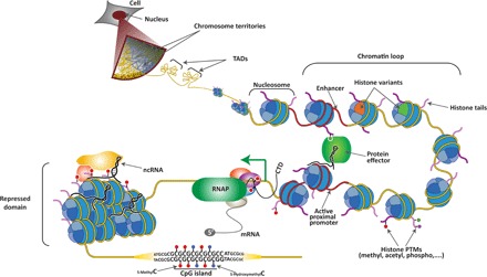
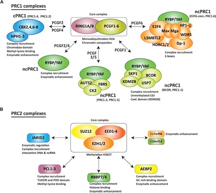
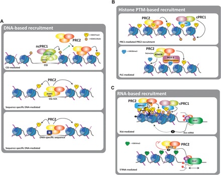
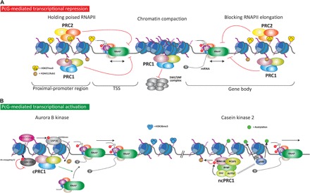
The Polycomb group (PcG) of proteins defines a subset of factors that physically associate and function to maintain the positional identity of cells from the embryo to adult stages. PcG has long been considered a paradigmatic model for epigenetic maintenance of gene transcription programs. Despite intensive research efforts to unveil the molecular mechanisms of action of PcG proteins, several fundamental questions remain unresolved: How many different PcG complexes exist in mammalian cells? How are PcG complexes targeted to specific loci? How does PcG regulate transcription? In this review, we discuss the diversity of PcG complexes in mammalian cells, examine newly identified modes of recruitment to chromatin, and highlight the latest insights into the molecular mechanisms underlying the function of PcGs in transcription regulation and three-dimensional chromatin conformation.

Keywords: Chromatin; Epigenetics; Gene Regulation.


Figures

Fig. 1
Fig. 1. Hierarchical layers of chromatin organization in mammalian cells.
Individual chromosomes cover a distinct region within the nucleus known as chromosome territory. At increasing resolution, chromosomes are composed of topologically associating domains (TADs), which are structural units defined by the high frequency of chromatin interactions between their loci that are partitioned by sharp boundaries. Within TADs, enhancer elements and active proximal promoters (both depicted in red) form chromatin loops, which are mediated and/or stabilized by protein effectors, noncoding RNAs (ncRNAs), and histone posttranslational modifications (PTMs). Enhancers and promoters are characterized by the presence of specific histone variants and PTMs on the histone tails. Upon transcription activation, elongating RNA polymerase II (RNAP, in green) is phosphorylated at Ser5 and Ser2 on its C-terminal domain (CTD) and begins to produce mRNA. Genomic regions that are transcriptionally silenced form repressed chromatin domains that are also stabilized by ncRNA and other repressive protein complexes. Finally, tracks of repetitive sequence are found in specific functional regions of the genome, including CpG islands (CGIs), in which cytosines can be modified (5-methylC and 5-hydroxymethylC).
Fig. 2
Fig. 2. PcG complexes in mammals.
(A and B) PcG complexes are classified into two major families: (A) PRC1 and (B) PRC2. Both families contain core subunits present in all the subcomplexes of the family. The interaction of the core complex with other accessory proteins defines the complete composition of each subcomplex. These accessory proteins have been found to regulate recruitment to specific chromatin domains and/or to modulate the catalytic activity of the core complex. PRC1 complexes are divided into cPRC1 and ncPRC1 (A). The core complex can associate with distinct Pcgf proteins, which allows for an alternative nomenclature. Therefore, Pcgf2 and Pcgf4 are present in the cPRC1 complexes (PRC1.2 and PRC1.4, respectively), Pcgf2 and Pcgf4 are also associated with ncPRC1-containing Rybp or YAF proteins, Pcgf3 and Pcgf5 are present in the ncPRC1 complexes (PRC1.3 and PRC1.5), Pcgf1 is present in the ncPRC1 complex PRC1.1 (also known as BCOR), and Pcgf6 is present in the ncPRC1 complex PRC1.6 (also known as E2F6.com). (B) The trimeric PRC2 core complex can associate with different proteins present in the PRC2 complex at the same time.
Fig. 3
Fig. 3. Mechanisms of PcG recruitment to chromatin.
(A to C) Three major mechanisms of recruitment of PcG complexes have been proposed: (A) a DNA-based mechanism in which PcG complexes are targeted to defined DNA sequences. DNA binding domains (DBD) present in different PcG complexes, such as Kdm2b or Aebp2, can mediate the recruitment to CGIs of CG-rich regions. Transient interaction with transcription factors (TF), such as Snail, can also mediate the recruitment of PcG to DNA-specific sequences; (B) histone modifications can also mediate the recruitment of PcG by its interaction with chromatin “readers” present in the PcG complexes, such as Cbx and PCL proteins; (C) ncRNAs also interact with PcG complexes and are required for their recruitment to chromatin. Two examples of this are PRC2 recruitment mediated by Xist during XCI and by short nascent transcripts from active promoters. In this latter case, interaction with 5′-nascent RNAs negatively regulates PRC2 methyltransferase activity.
Fig. 4
Fig. 4. Mechanisms of PcG-mediated transcription regulation.
PcG proteins mediate repression and activation of transcription. (A) Three major mechanisms of repression have been proposed. First, in bivalent promoters marked with both repressive (H3K27me3) and active (H3K4me3) histone PTMs, PcG complexes hold the poised RNAPII at transcriptional start site (TSS), thereby inhibiting its release. Second, PcG complex can compact chromatin. For PRC1, its ability to compact chromatin appears to be independent of its catalytic activity. Chromatin compaction is proposed to block the accessibility of chromatin remodeling complexes, such as the SWI/SNF complex, which is required during transcription activation. Third, deubiquitination and demethylation of histone H3 at gene bodies are required for efficient transcription elongation by RNAPII. Thus, the histone modifications imposed by PcG at gene bodies might prevent RNAPII processivity during transcription elongation. (B) PRC1 complexes can also regulate gene activation. Two different mechanisms have been proposed, both of which require the action of a protein kinase. The first mechanism involves the phosphorylation by Aurora B kinase of the deubiquitinase Usp16 and the E2-conjugating enzyme Ube2d3, which act in a coordinated manner to block PRC1 activity. Usp16 phosphorylation activates its deubiquitinase activity and hence leads to removal of the ubiquitin from H2AK119. In contrast, Ube2d3 phosphorylation inhibits its activity, thus impairing the E3 ligase activity of PRC1. Therefore, a catalytically impaired PRC1 complex would favor the recruitment of RNAPII to activate gene transcription. In the second mechanism, the CK2 within the PRC1.5 complex phosphorylates Ring1B on serine 168 and inhibits its catalytic activity. Additionally, the subunit Auts2 of the complex triggers the recruitment of the acetyltransferase P300, which acetylates histone tails and enhances transcription.
Fig. 5
Fig. 5. PcG shapes intra-TAD interactions.
In ESC nuclei, the genome is compartmentalized on the basis of the preferential interactions between genomic elements, forming multilooped structures called TADs (see text) of active chromatin (depicted in green) and transcriptionally repressed chromatin (in orange). Chromatin loops are flanked by insulator proteins such as the CTCF transcription factor (in gray). Top panel: Hypothetical chromosome conformation capture (3C) data showing pairwise interaction frequencies (in red) occurring between two active TADs (green) segregated from an inactive TAD (orange). The active and inactive TADs are densely marked by H3K4me3 and H3K27me3, respectively. Bottom panel: Upon ESCs differentiation, the overall TAD structure and location of TAD boundaries are not altered, but small rearrangements occur, which correlate with a redistribution of histone PTMs.

Similar articles

Cited by

References

    1. W. Flemming, Zellsubstanz, Kern und Zelltheilung (F. C. W. Vogel, Leipzig, 1882).
    1. Kouzarides T., Chromatin modifications and their function. Cell 128, 693–705 (2007). - PubMed
    1. Gorkin D. U., Leung D., Ren B., The 3D genome in transcriptional regulation and pluripotency. Cell Stem Cell 14, 762–775 (2014). - PMC - PubMed
    1. Dixon J. R., Selvaraj S., Yue F., Kim A., Li Y., Shen Y., Hu M., Liu J. S., Ren B., Topological domains in mammalian genomes identified by analysis of chromatin interactions. Nature 485, 376–380 (2012). - PMC - PubMed
    1. Gibcus J. H., Dekker J., The hierarchy of the 3D genome. Mol. Cell 49, 773–782 (2013). - PMC - PubMed

LinkOut - more resources