AICAR Protects against High Palmitate/High Insulin-Induced Intramyocellular Lipid Accumulation and Insulin Resistance in HL-1 Cardiac Cells by Inducing PPAR-Target Gene Expression
- PMID: 26649034
- PMCID: PMC4663352
- DOI: 10.1155/2015/785783
AICAR Protects against High Palmitate/High Insulin-Induced Intramyocellular Lipid Accumulation and Insulin Resistance in HL-1 Cardiac Cells by Inducing PPAR-Target Gene Expression
Abstract
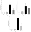
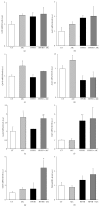
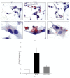
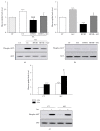
Here we studied the impact of 5-aminoimidazole-4-carboxamide riboside (AICAR), a well-known AMPK activator, on cardiac metabolic adaptation. AMPK activation by AICAR was confirmed by increased phospho-Thr(172)-AMPK and phospho-Ser(79)-ACC protein levels in HL-1 cardiomyocytes. Then, cells were exposed to AICAR stimulation for 24 h in the presence or absence of the AMPK inhibitor Compound C, and the mRNA levels of the three PPARs were analyzed by real-time RT-PCR. Treatment with AICAR induced gene expression of all three PPARs, but only the Ppara and Pparg regulation were dependent on AMPK. Next, we exposed HL-1 cells to high palmitate/high insulin (HP/HI) conditions either in presence or in absence of AICAR, and we evaluated the expression of selected PPAR-targets genes. HP/HI induced insulin resistance and lipid storage was accompanied by increased Cd36, Acot1, and Ucp3 mRNA levels. AICAR treatment induced the expression of Acadvl and Glut4, which correlated to prevention of the HP/HI-induced intramyocellular lipid build-up, and attenuation of the HP/HI-induced impairment of glucose uptake. These data support the hypothesis that AICAR contributes to cardiac metabolic adaptation via regulation of transcriptional mechanisms.
Figures


Similar articles
-
Prolonged AMPK activation increases the expression of fatty acid transporters in cardiac myocytes and perfused hearts.Mol Cell Biochem. 2006 Aug;288(1-2):201-12. doi: 10.1007/s11010-006-9140-8. Epub 2006 May 19. Mol Cell Biochem. 2006. PMID: 16710744
-
Small heterodimer partner (SHP) contributes to insulin resistance in cardiomyocytes.Biochim Biophys Acta Mol Cell Biol Lipids. 2017 May;1862(5):541-551. doi: 10.1016/j.bbalip.2017.02.006. Epub 2017 Feb 16. Biochim Biophys Acta Mol Cell Biol Lipids. 2017. PMID: 28214558
-
Restoring AS160 phosphorylation rescues skeletal muscle insulin resistance and fatty acid oxidation while not reducing intramuscular lipids.Am J Physiol Endocrinol Metab. 2009 Nov;297(5):E1056-66. doi: 10.1152/ajpendo.90908.2008. Epub 2009 Sep 1. Am J Physiol Endocrinol Metab. 2009. PMID: 19724017
-
Short-term AMP-regulated protein kinase activation enhances insulin-sensitive fatty acid uptake and increases the effects of insulin on fatty acid oxidation in L6 muscle cells.Exp Biol Med (Maywood). 2010 Apr;235(4):514-21. doi: 10.1258/ebm.2009.009228. Exp Biol Med (Maywood). 2010. PMID: 20407084
-
Overexpression of AMP-activated protein kinase or protein kinase D prevents lipid-induced insulin resistance in cardiomyocytes.J Mol Cell Cardiol. 2013 Feb;55:165-73. doi: 10.1016/j.yjmcc.2012.11.005. Epub 2012 Nov 15. J Mol Cell Cardiol. 2013. PMID: 23159540
Cited by
-
Mitochondrial Uncoupling Proteins: Subtle Regulators of Cellular Redox Signaling.Antioxid Redox Signal. 2018 Sep 1;29(7):667-714. doi: 10.1089/ars.2017.7225. Epub 2018 Mar 14. Antioxid Redox Signal. 2018. PMID: 29351723 Free PMC article. Review.
-
Proteomic investigation of human skeletal muscle before and after 70 days of head down bed rest with or without exercise and testosterone countermeasures.PLoS One. 2019 Jun 13;14(6):e0217690. doi: 10.1371/journal.pone.0217690. eCollection 2019. PLoS One. 2019. PMID: 31194764 Free PMC article.
-
Differential effects of AMP-activated protein kinase in isolated rat atria subjected to simulated ischemia-reperfusion depending on the energetic substrates available.Pflugers Arch. 2018 Feb;470(2):367-383. doi: 10.1007/s00424-017-2075-y. Epub 2017 Oct 14. Pflugers Arch. 2018. PMID: 29032506
-
Erythropoietin ameliorates hyperglycemia in type 1-like diabetic rats.Drug Des Devel Ther. 2016 Jun 3;10:1877-84. doi: 10.2147/DDDT.S105867. eCollection 2016. Drug Des Devel Ther. 2016. PMID: 27350742 Free PMC article.
-
Naoxintong Retards Atherosclerosis by Inhibiting Foam Cell Formation Through Activating Pparα Pathway.Curr Mol Med. 2018;18(10):698-710. doi: 10.2174/1566524019666190207143207. Curr Mol Med. 2018. PMID: 30734676 Free PMC article.
References
LinkOut - more resources
Full Text Sources
Other Literature Sources
Research Materials
Miscellaneous

