An unexpected journey: conceptual evolution of mechanoregulated potassium transport in the distal nephron
- PMID: 26632600
- PMCID: PMC4838068
- DOI: 10.1152/ajpcell.00328.2015
An unexpected journey: conceptual evolution of mechanoregulated potassium transport in the distal nephron
Abstract
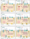
Flow-induced K secretion (FIKS) in the aldosterone-sensitive distal nephron (ASDN) is mediated by large-conductance, Ca(2+)/stretch-activated BK channels composed of pore-forming α-subunits (BKα) and accessory β-subunits. This channel also plays a critical role in the renal adaptation to dietary K loading. Within the ASDN, the cortical collecting duct (CCD) is a major site for the final renal regulation of K homeostasis. Principal cells in the ASDN possess a single apical cilium whereas the surfaces of adjacent intercalated cells, devoid of cilia, are decorated with abundant microvilli and microplicae. Increases in tubular (urinary) flow rate, induced by volume expansion, diuretics, or a high K diet, subject CCD cells to hydrodynamic forces (fluid shear stress, circumferential stretch, and drag/torque on apical cilia and presumably microvilli/microplicae) that are transduced into increases in principal (PC) and intercalated (IC) cell cytoplasmic Ca(2+) concentration that activate apical voltage-, stretch- and Ca(2+)-activated BK channels, which mediate FIKS. This review summarizes studies by ourselves and others that have led to the evolving picture that the BK channel is localized in a macromolecular complex at the apical membrane, composed of mechanosensitive apical Ca(2+) channels and a variety of kinases/phosphatases as well as other signaling molecules anchored to the cytoskeleton, and that an increase in tubular fluid flow rate leads to IC- and PC-specific responses determined, in large part, by the cell-specific composition of the BK channels.
Keywords: WNK kinases; cilia; kidney; mechanoregulation; potassium transport.
Copyright © 2016 the American Physiological Society.
Figures


Similar articles
-
The mechanosensitive BKα/β1 channel localizes to cilia of principal cells in rabbit cortical collecting duct (CCD).Am J Physiol Renal Physiol. 2017 Jan 1;312(1):F143-F156. doi: 10.1152/ajprenal.00256.2016. Epub 2016 Nov 2. Am J Physiol Renal Physiol. 2017. PMID: 27806944 Free PMC article.
-
L-WNK1 is required for BK channel activation in intercalated cells.Am J Physiol Renal Physiol. 2021 Aug 1;321(2):F245-F254. doi: 10.1152/ajprenal.00472.2020. Epub 2021 Jul 6. Am J Physiol Renal Physiol. 2021. PMID: 34229479 Free PMC article.
-
Going with the flow: New insights regarding flow induced K+ secretion in the distal nephron.Physiol Rep. 2024 Oct;12(20):e70087. doi: 10.14814/phy2.70087. Physiol Rep. 2024. PMID: 39428258 Free PMC article. Review.
-
Role of NKCC in BK channel-mediated net K⁺ secretion in the CCD.Am J Physiol Renal Physiol. 2011 Nov;301(5):F1088-97. doi: 10.1152/ajprenal.00347.2011. Epub 2011 Aug 3. Am J Physiol Renal Physiol. 2011. PMID: 21816753 Free PMC article.
-
Interacting influence of diuretics and diet on BK channel-regulated K homeostasis.Curr Opin Pharmacol. 2014 Apr;15:28-32. doi: 10.1016/j.coph.2013.11.001. Epub 2013 Dec 11. Curr Opin Pharmacol. 2014. PMID: 24721651 Free PMC article. Review.
Cited by
-
Inhibition of AT2R and Bradykinin Type II Receptor (BK2R) Compromises High K+ Intake-Induced Renal K+ Excretion.Hypertension. 2020 Feb;75(2):439-448. doi: 10.1161/HYPERTENSIONAHA.119.13852. Epub 2019 Dec 23. Hypertension. 2020. PMID: 31865783 Free PMC article.
-
Expression and localization of the mechanosensitive/osmosensitive ion channel TMEM63B in the mouse urinary tract.Physiol Rep. 2024 May;12(9):e16043. doi: 10.14814/phy2.16043. Physiol Rep. 2024. PMID: 38724885 Free PMC article.
-
WNK Kinases in Development and Disease.Curr Top Dev Biol. 2017;123:1-47. doi: 10.1016/bs.ctdb.2016.08.004. Epub 2016 Sep 28. Curr Top Dev Biol. 2017. PMID: 28236964 Free PMC article. Review.
-
Tissue Chips and Microphysiological Systems for Disease Modeling and Drug Testing.Micromachines (Basel). 2021 Jan 28;12(2):139. doi: 10.3390/mi12020139. Micromachines (Basel). 2021. PMID: 33525451 Free PMC article. Review.
-
Caveolae facilitate TRPV4-mediated Ca2+ signaling and the hierarchical activation of Ca2+-activated K+ channels in K+-secreting renal collecting duct cells.Am J Physiol Renal Physiol. 2018 Dec 1;315(6):F1626-F1636. doi: 10.1152/ajprenal.00076.2018. Epub 2018 Sep 12. Am J Physiol Renal Physiol. 2018. PMID: 30207167 Free PMC article.
References
-
- Adelman JP, Shen KZ, Kavanaugh MP, Warren RA, Wu YN, Lagrutta A, Bond CT, North RA. Calcium-activated potassium channels expressed from cloned complementary DNAs. Neuron 9: 209–216, 1992. - PubMed
-
- Alioua A, Tanaka Y, Wallner M, Hofmann F, Ruth P, Meera P, Toro L. The large conductance, voltage-dependent, and calcium-sensitive K+ channel, Hslo, is a target of cGMP-dependent protein kinase phosphorylation in vivo. J Biol Chem 273: 32950–32956, 1998. - PubMed
-
- Althaus M, Bogdan R, Clauss WG, Fronius M. Mechano-sensitivity of epithelial sodium channels (ENaCs): laminar shear stress increases ion channel open probability. FASEB J 21: 2389–2399, 2007. - PubMed
-
- Archer SL, Gragasin FS, Wu X, Wang S, McMurtry S, Kim DH, Platonov M, Koshal A, Hashimoto K, Campbell WB, Falck JR, Michelakis ED. Endothelium-derived hyperpolarizing factor in human internal mammary artery is 11,12-epoxyeicosatrienoic acid and causes relaxation by activating smooth muscle BK(Ca) channels. Circulation 107: 769–776, 2003. - PubMed
Publication types
MeSH terms
Substances
Grants and funding
LinkOut - more resources
Full Text Sources
Other Literature Sources
Medical
Miscellaneous

