Interferon-λ1 suppresses invasion and enhances autophagy in human osteosarcoma cell
- PMID: 26628983
- PMCID: PMC4658872
Interferon-λ1 suppresses invasion and enhances autophagy in human osteosarcoma cell
Abstract
Objective: The purpose of the present study was to determine whether type III IFN can modulate the autophagic response in human osteosarcoma cell.
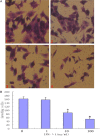
Methods: Human osteosarcoma cell were treated with Interferon-λ1. We investigated that Interferon-λ1 could inhibit the invasive ability of osteosarcoma cells by Matrigel invasion assay. Autophagy were assessed by acridine orange staining, MDC staining and Transmission electron microscopy.
Results: In this study, we found that Interferon-λ1 could inhibit the invasive ability of osteosarcoma cells. Acridine orange staining and MDC staining showed that Interferon-λ1 triggered the accumulation of acidic vesicular and autolysosomes in osteosarcoma cell. The acridine orange osteosarcoma cell ratios were 3.6 ± 0.5%, 4.5 ± 0.8%, and 12.4 ± 1.7% after treatment with 1, 10, and 100 ng/mL Interferon-λ1 for 48 h. Osteosarcoma cell cells treated with 100 ng/mL Interferon-λ1 for 48 h developed autophagy some-like characteristics, including single or double-membrane vacuoles containing intact and degraded cellular debris.
Conclusions: Interferon-λ1 could inhibit the invasive ability of osteosarcoma cells. Autophagy can be induced in a dose-dependent manner by treatment with Interferon-λ1 in osteosarcoma cell.
Keywords: Interferon-λ1; acridine orange; autophagy; transmission electron microscopy.
Figures


Similar articles
-
Interferon-alpha-2b induces autophagy in hepatocellular carcinoma cells through Beclin1 pathway.Cancer Biol Med. 2014 Mar;11(1):64-8. doi: 10.7497/j.issn.2095-3941.2014.01.006. Cancer Biol Med. 2014. PMID: 24738040 Free PMC article.
-
Interferon-α suppresses invasion and enhances cisplatin-mediated apoptosis and autophagy in human osteosarcoma cells.Oncol Lett. 2014 Mar;7(3):827-833. doi: 10.3892/ol.2013.1762. Epub 2013 Dec 16. Oncol Lett. 2014. PMID: 24527090 Free PMC article.
-
Downregulation of autophagy-related gene ATG5 and GABARAP expression by IFN-λ1 contributes to its anti-HCV activity in human hepatoma cells.Antiviral Res. 2017 Apr;140:83-94. doi: 10.1016/j.antiviral.2017.01.016. Epub 2017 Jan 26. Antiviral Res. 2017. PMID: 28131804
-
Ratiometric analysis of Acridine Orange staining in the study of acidic organelles and autophagy.J Cell Sci. 2016 Dec 15;129(24):4622-4632. doi: 10.1242/jcs.195057. Epub 2016 Nov 14. J Cell Sci. 2016. PMID: 27875278
-
Photodynamic therapy with the novel photosensitizer chlorophyllin f induces apoptosis and autophagy in human bladder cancer cells.Lasers Surg Med. 2014 Apr;46(4):319-34. doi: 10.1002/lsm.22225. Epub 2014 Jan 24. Lasers Surg Med. 2014. PMID: 24464873
Cited by
-
Osteosarcoma Overview.Rheumatol Ther. 2017 Jun;4(1):25-43. doi: 10.1007/s40744-016-0050-2. Epub 2016 Dec 8. Rheumatol Ther. 2017. PMID: 27933467 Free PMC article. Review.
-
New treatment alternatives for primary and metastatic colorectal cancer by an integrated transcriptome and network analyses.Sci Rep. 2024 Apr 16;14(1):8762. doi: 10.1038/s41598-024-59101-8. Sci Rep. 2024. PMID: 38627442 Free PMC article.
-
IFN-λ Modulates the Migratory Capacity of Canine Mammary Tumor Cells via Regulation of the Expression of Matrix Metalloproteinases and Their Inhibitors.Cells. 2021 Apr 23;10(5):999. doi: 10.3390/cells10050999. Cells. 2021. PMID: 33922837 Free PMC article.
References
-
- Ichihashi T, Asano A, Usui T, Takeuchi T, Watanabe Y, Yamano Y. Antiviral and antiproliferative effects of canine interferon-lambda1. Vet Immunol Immunopathol. 2013;156:141–146. - PubMed
-
- Li Q, Kawamura K, Tada Y, Shimada H, Hiroshima K, Tagawa M. Novel type III interferons produce anti-tumor effects through multiple functions. Front Biosci (Landmark Ed) 2013;18:909–918. - PubMed
LinkOut - more resources
Full Text Sources
