Review
. 2016 Feb;17(2):69-82.
doi: 10.1038/nrm.2015.8. Epub 2015 Dec 2.

Structure and function of ER membrane contact sites with other organelles

Affiliations
Review

Structure and function of ER membrane contact sites with other organelles

Melissa J Phillips et al. Nat Rev Mol Cell Biol. 2016 Feb.

Abstract

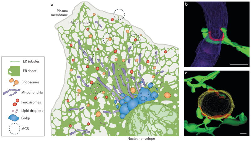
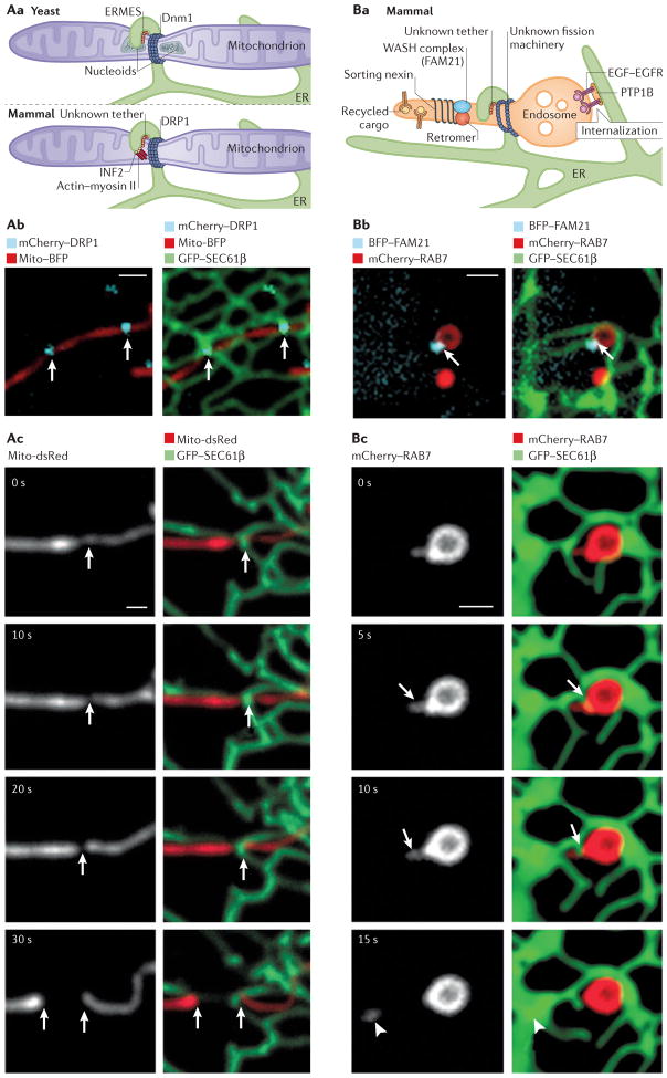
The endoplasmic reticulum (ER) is the largest organelle in the cell, and its functions have been studied for decades. The past several years have provided novel insights into the existence of distinct domains between the ER and other organelles, known as membrane contact sites (MCSs). At these contact sites, organelle membranes are closely apposed and tethered, but do not fuse. Here, various protein complexes can work in concert to perform specialized functions such as binding, sensing and transferring molecules, as well as engaging in organelle biogenesis and dynamics. This Review describes the structure and functions of MCSs, primarily focusing on contacts of the ER with mitochondria and endosomes.


Conflict of interest statement

statement The authors declare no competing interests.

Figures

Figure 1
Figure 1. Structure of endoplasmic reticulum (ER) membrane-contact sites (MCSs)
a | The ER consists of the nuclear envelope (outlined with a dashed line) and the peripheral ER, which spreads into the cytosol as a network of sheets and tubules. The peripheral ER forms MCSs with the plasma membrane, mitochondria, endosomes, peroxisomes, lipid droplets and the Golgi. b, c | Electron tomography reveals the three-dimensional structure of MCSs (coloured red) between ER tubules (green) and mitochondria (purple) in a yeast cell b) or an endosome (yellow) in an animal cell (c). Scale bars represent 200 nm in parts b–c. Image in parts b reproduced with permission from REF. , AAAS. Image in part c republished with permission of the American Society for Cell Biology, from Endoplasmic reticulum-endosome contact increases as endosomes traffic and mature. Friedman, J. R., Dibenedetto, J. R., West, M., Rowland, A. A. & Voeltz, G. K. Mol. Biol. Cell 24, 1030–1040 (2013); permission conveyed through Copyright Clearance Center, Inc.
Figure 2
Figure 2. Dynamics of endoplasmic reticulum (ER) membrane contact sites (MCSs)
a,b | Endosomes and mitochondria are tightly tethered to ER tubules even as they traffic. Time-lapse fluorescence images of ER and organelle dynamics in live Cos-7 cells expressing GFP–SEC61β (labelling ER in green) and (a) mito-BFP (labelling mitochondria in red) or (b) mCherry–RAB7 (labelling late endosomes (LEs) in red). Note how the contact sites are maintained as the apposing organelles move. Scale bars represent 1 μm. c | Endosomes mature as they traffic from the cell periphery along microtubules to the cell centre. ER–endosome contact increases as endosomes mature, with 53% of early endosomes (EEs; marked by RAB5) and 99% of LEs (marked by RAB7) remaining in contact with the ER during trafficking. d | Model of how cholesterol levels regulate the composition of ER–LEs MCSs and LE trafficking. When the LE contains high cholesterol levels (left panel), oxysterol-binding-related protein 1L (ORP1L) can bind to cholesterol on the LE membrane and does not associate with ER VAPs (VAMP-associated proteins). In addition, ORP1L interacts with RAB7 GTPase, which stimulates minus-end-directed LE trafficking through a complex that includes RILP (RAB-interacting lysosomal protein), the HOPS (homotypic fusion and vacuole protein sorting) complex, dynactin (p150Glued) and the motor protein dynein. At low cholesterol levels (right panel), ORP1L is not bound to cholesterol and instead interacts with ER VAPs. The ORP1L–VAP interaction displaces dynein from the endosome. e | Protrudin is an ER integral membrane protein that interacts with VAP and kinesin-1 (left panel). Protrudin binds to RAB7 and phosphatidylinositol-3-phosphate (PtdIns(3)P) on the LE membrane. Protrudin can bind to and transfer kinesin-1 to the LE protein FYCO1 (FYVE and coiled-coil domain-containing protein 1), and this promotes plus-end-directed microtubule trafficking of LEs (right panel).
Figure 3
Figure 3. Endoplasmic reticulum (ER) membrane contact sites (MCSs) function in lipid biosynthesis and exchange
a | ER– mitochondria MCSs are rich in lipids and lipid-synthesis enzymes. Lipids are transferred between organelles at MCSs. In one pathway, phosphatidic acid (PA) is converted to phosphatidylserine (PS) at the ER. PS is transferred to the inner mitochondria membrane (IMM) where it is converted to phosphatidylethanolamine (PE). PE is transferred back to the ER, where it is converted to phosphatidylcholine (PC). PC is likely to also be transported back to mitochondria. ER–mitochondria membrane-tethering proteins (such as the ER–mitochondrial encounter structure (ERMES) in yeast) may aid this process; however, the exact mechanism of their action is currently elusive, and a mammalian counterpart has not been identified. b | Various complexes sense, modify or potentially transfer lipids at ER–endosome MCSs. Oxysterol-binding-related protein 1L (ORP1L) and START domain-containing protein 3 (STARD3) on the endosome have both been shown to interact with ER-resident VAMP-associated proteins (VAPs), but how they aid the exchange of lipids at ER–endosome MCSs is unclear. Niemann–Pick type C2 protein (NPC2) resides in the endosome lumen and interacts with endosome membrane protein NPC1. NPC1 interacts with the ER protein ORP5. The interactions between NPC2, NPC1 and ORP5 provide a potential mechanism for cholesterol transfer between the endosome lumen and the ER (blow-up). In this model, NPC2 transfers cholesterol from the endosome lumen to NPC1 on the endosome membrane. ER-resident ORP5 then accepts cholesterol from NPC1 and may transfer the cholesterol to the ER for redistribution. c | Multiple potential lipid-transfer proteins localize to the Golgi membrane and interact with ER VAPs. These include the phosphatidylinositol-transfer protein NIR2 (PYK2 N-terminal domain-interacting receptor 2), the ceramide-transfer protein (CERT), the glycosylceramide-transfer protein Golgi-associated four-phosphate adaptor protein (FAPP2) and the cholesterol and phosphatidylinositol-4-phosphate (PtdIns(4)P)-transfer protein oxysterol-binding protein (OSBP). Studies specifically on OSBP (right panels) show that it associates with the Golgi membrane through PtdIns(4)P binding. The OSBP oxysterol-binding-related domain (ORD domain) can bind and transfer sterol from the ER to the Golgi and PtdIns(4)P from the Golgi to the ER. When PtdIns(4)P levels are depleted at the Golgi, OSBP dissociates from the Golgi membranes. PtdIns(4)P at the ER is converted back to PtdInsP by ER-associated PtdIns(4)P phosphatase. OMM, outer mitochondria membrane.
Figure 4
Figure 4. Calcium (Ca2+) exchange at endoplasmic reticulum (ER) membrane contact sites (MCSs)
a | The ER lumen is the major Ca2+ store in the cell, with a Ca2+ concentration ([Ca2+]) of ~60–500 μM). In the extracellular space, [Ca2+] is high (~1mM) compared to the intracellular cytosol (~100nM). Newly formed endosomes have taken up Ca2+ from the extracellular space, so the luminal [Ca2+] is close to the same as that of the extracellular space (~1 mM). Luminal Ca2+ is then released so that early endosomes have [Ca2+] ~0.5 μM and late endosomes have [Ca2+] ~2.5 μM. The ER–endosome MCS is a site of dynamic Ca2+ crosstalk. Endosomes may be able to sequester Ca2+ released from the ER. The ER transfers Ca2+ to mitochondria, with peak mitochondrial Ca2+ concentrations reaching 100 μM. b | ER Ca2+ released from the ER through inositol-1,4,5- trisphosphate receptors (Ins(1,4,5)P3Rs) provides a concentrated Ca2+ spike that can be taken up through the outer mitochondrial membrane (OMM) by VDACs (voltage dependent anion channels) and then through the inner mitochondrial membrane (IMM) by the mitochondrial Ca2+ uniporter (MCU) ion transporter into the mitochondrial matrix. The 75 kDa glucose-regulated protein (GRP75) functions as a chaperone, coupling Ins(1,4,5)P3R to the VDACs. c | Endosomes are capable of releasing Ca2+ though transient receptor potential channels (TRPs) or two-pore channels (TPCs). ER Ca2+ released from ER via Ins(1,4,5)P3Rs could be taken up into endosomes through unknown endosome Ca2+-uptake channels. Ca2+ release from endosomes can also stimulate Ca2+ release from the ER through Ins(1,4,5)P3Rs and vice versa.
Figure 5
Figure 5. Endoplasmic reticulum (ER) membrane contact sites (MCSs) define the timing and position of both mitochondrial and endosome fission
Aa. In yeast (top panel), mitochondrial ER-marked constriction and fission sites contain the ER–mitochondrial tethering complex (ERMES), mitochondrial nucleoid DNA and the fission-machinery protein dynamin-related protein 1 (Dnm1). In mammalian cells (bottom panel), an ER-localized inverted formin (INF2), actin and myosin II are candidates for driving ER-associated constriction of mitochondria. Then, the fission-machinery protein dynamin-related protein 1 (DRP1) is recruited by adaptor proteins to ER-marked constrictions, where it drives fission. Ab | Live confocal fluorescence microscopy images of a Cos7 cell expressing mito-BFP (mitochondria in red) and mCherry–DRP1 (in cyan), merged with GFP–SEC61 β (ER in green) in the right panel. ER tubules contact two mitochondrial constrictions labelled with DRP1, as marked by the white arrows. Ac. Live fluorescence microscopy, as in Ab, of a cell expressing mito-dsRed (mitochondria in grey in left panels, red in right panels) and GFP-–SEC61β (ER in green). Note that the ER tubule circumscribes the position of constriction and fission (white arrows) (t=30s). Ba. In ER-associated endosome fission in animal cells, cargo is sorted into tubules marked by the retromer, sorting nexins and WASH complex protein FAM21. ER tubules are recruited to these sorting domains by an unidentified tether, and fission is rapid following ER recruitment. Note that another ER–endosome MCS regulates dephosphorylation and internalization of epidermal growth factor receptor (EGFR) by ER-localized protein-Tyr phosphatase 1B (PTP1B). Bb. Live confocal fluorescence microscopy images of a Cos7 cell expressing mCherry–RAB7 (late endosome in red) and BFP–FAM21 (late endosome cargo-sorting domain in cyan), merged in the right panel with GFP–SEC61β (ER in green). The arrow marks a MCS between the tip of an ER tubule and the FAM21-labelled sorting domain on the late endosome. Bc. Time-lapsed images of a cell expressing mCherry– RAB7 (late endosome shown in grey in the left panels and red in the right panels) and GFP–SEC61β (ER in green) show ER tubule recruitment to the neck of the late endosome bud (t=5 s, arrow at the constriction), followed by fission (arrow, between t=10 s and t=15 s; bud marked by arrowhead, t=15 s). Scale bars in Ab, Ac and Bb, Bc represent 1 μm. Images in Ab courtesy of Jason Lee, University of Colorado Boulder, USA. Images in part Ac were adapted with permission from REF. , AAAS. Images in parts Bb and Bc were adapted with permission from REF. , Elsevier.

Similar articles

Cited by

References

    1. Fawcett DW. The Cell. W. B. Saunders; 1981.
    1. Ogata T, Yamasaki Y. Ultra-high-resolution scanning electron microscopy of mitochondria and sarcoplasmic reticulum arrangement in human red, white, and intermediate muscle fibers. Anat Rec. 1997;248:214–223. - PubMed
    1. Rolls MM, Hall DH, Victor M, Stelzer EHK, Rapoport TA. Targeting of rough endoplasmic reticulum membrane proteins and ribosomes in invertebrate neurons. Mol Biol Cell. 2002;13:1778–1791. - PMC - PubMed
    1. Shibata Y, Voeltz GK, Rapoport TA. Rough sheets and smooth tubules. Cell. 2006;126:435–439. - PubMed
    1. Shibata Y, et al. Mechanisms determining the morphology of the peripheral ER. Cell. 2010;143:774–788. - PMC - PubMed

Publication types

MeSH terms

LinkOut - more resources