. 2015 Nov 25;90(3):1657-67.
doi: 10.1128/JVI.02545-15. Print 2016 Feb 1.

An Adenovirus DNA Replication Factor, but Not Incoming Genome Complexes, Targets PML Nuclear Bodies

Affiliations

An Adenovirus DNA Replication Factor, but Not Incoming Genome Complexes, Targets PML Nuclear Bodies

Tetsuro Komatsu et al. J Virol. .

Abstract

Promyelocytic leukemia protein nuclear bodies (PML-NBs) are subnuclear domains implicated in cellular antiviral responses. Despite the antiviral activity, several nuclear replicating DNA viruses use the domains as deposition sites for the incoming viral genomes and/or as sites for viral DNA replication, suggesting that PML-NBs are functionally relevant during early viral infection to establish productive replication. Although PML-NBs and their components have also been implicated in the adenoviral life cycle, it remains unclear whether incoming adenoviral genome complexes target PML-NBs. Here we show using immunofluorescence and live-cell imaging analyses that incoming adenovirus genome complexes neither localize at nor recruit components of PML-NBs during early phases of infection. We further show that the viral DNA binding protein (DBP), an early expressed viral gene and essential DNA replication factor, independently targets PML-NBs. We show that DBP oligomerization is required to selectively recruit the PML-NB components Sp100 and USP7. Depletion experiments suggest that the absence of one PML-NB component might not affect the recruitment of other components toward DBP oligomers. Thus, our findings suggest a model in which an adenoviral DNA replication factor, but not incoming viral genome complexes, targets and modulates PML-NBs to support a conducive state for viral DNA replication and argue against a generalized concept that PML-NBs target incoming viral genomes.

Importance: The immediate fate upon nuclear delivery of genomes of incoming DNA viruses is largely unclear. Early reports suggested that incoming genomes of herpesviruses are targeted and repressed by PML-NBs immediately upon nuclear import. Genome localization and/or viral DNA replication has also been observed at PML-NBs for other DNA viruses. Thus, it was suggested that PML-NBs may immediately sense and target nuclear viral genomes and hence serve as sites for deposition of incoming viral genomes and/or subsequent viral DNA replication. Here we performed a detailed analyses of the spatiotemporal distribution of incoming adenoviral genome complexes and found, in contrast to the expectation, that an adenoviral DNA replication factor, but not incoming genomes, targets PML-NBs. Thus, our findings may explain why adenoviral genomes could be observed at PML-NBs in earlier reports but argue against a generalized role for PML-NBs in targeting invading viral genomes.


Figures

FIG 1
FIG 1
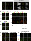
Incoming Ad genomes are not colocalized with PML-NBs. (A) IF analyses with Ad5-GFP. H1299 cells were either mock infected (first row) or infected with replication-deficient Ad5-GFP (second row) and at 1 hpi subjected to IF analyses using antibodies against PML-NB components (green) and protein VII (red, left panels). Dashed lines indicate the shapes of the nuclei indicated by DAPI (4′,6-diamidino-2-phenylindole) staining (not shown). For U2OS cells, cells were first transfected with the expression vectors for either EGFP alone or EGFP-ATRX (gray), and at 24 h posttransfection (hpt), IF analyses were performed as described above (right panels). (B) IF analyses with later time points. H1299 (left) or U2OS (right panels) cells were infected with Ad5-GFP and at 2 hpi (first row) and 4 hpi (second row) subjected to IF analyses. (C) IF analyses with Ad5. H1299 (left) or U2OS (right panels) cells were either mock infected (first row) or infected with replication-competent Ad5 and at 2 hpi (second row) and 4 hpi (third row) subjected to IF analyses. (D) IF analyses with human foreskin fibroblasts (HFFs). HFFs were either mock infected (first row) or infected with Ad5 (second row) and at 8 hpi subjected to IF analyses using antibodies against PML-NB components (green and blue) and protein VII (red). (E) IF analyses with Ad5-GFP-M1. H1299 (left panels) or U2OS (right panels) cells were infected with Ad5-GFP-M1, in which the PPxY motif of protein VI is mutated, and at 2 (first row) and 4 (second row) hpi subjected to IF analyses with the indicated antibodies. (F) IF analyses using BrdU-labeled viruses. To produce BrdU-labeled viruses, HEK293 cells were infected with Ad5, and 10 μM BrdU was added to the culture medium at 16 hpi. At 24 hpi, progeny viruses were released from infected cells. U2OS cells were infected with progeny viruses produced under the indicated conditions and at 2 hpi subjected to IF analyses using either anti-Ad5 (upper panels, cyan) or anti-PML (green) and anti-BrdU (red) antibodies (lower panels).
FIG 2
FIG 2
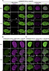
Incoming Ad genomes neither recruit components of nor are colocalized with PML-NBs in living cells. (A) Live-cell imaging using EGFP-TAF-Iβ. U2OS cells stably expressing EGFP-tagged TAF-Iβ (U2OS/EGFP-TAF-Iβ cells [green]) were first transiently transfected with the expression vectors for either mCherry-tagged PML (left panels) or Daxx (right panels, magenta) and then either mock infected (first row) or infected with Ad5-GFP (second through fourth rows) for live-cell imaging. Frames were taken every 3 s for 2 min (for mock-infected cells) or 3 min (for infected cells); snapshots from the movies are shown. Arrowheads indicate infection-specific TAF-I foci at the nuclear periphery. Full movies are provided as Movies S1 and S2 in the supplemental material. (B) Live-cell imaging using EGFP-ATRX. U2OS/mCherry-TAF-Iβ (magenta) cells were transiently transfected with the expression vector for EGFP-tagged ATRX (green) and then either mock infected or infected with Ad5-GFP for live-cell imaging. Full movies are provided as Movie S3 in the supplemental material. (C) Live-cell imaging using Ad5-GFP-M1. U2OS/EGFP-TAF-Iβ cells (green) were transiently transfected with the expression vector for mCherry-Daxx (magenta) and then either mock infected or infected with Ad5-GFP-M1 for live-cell imaging. Full movies are provided as Movie S4 in the supplemental material.
FIG 3
FIG 3
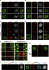
DBP targets PML-NBs in the absence of additional viral factors. U2OS cells were transfected with either an empty vector (first rows) or the vector for HA-DBP (second and third rows) and at 24 hpt subjected to IF analyses. HA-DBP was detected using either rabbit anti-HA (A and B, first columns, green), anti-DBP (C), or rat anti-HA (D) antibodies, while PML-NB components were stained with specific antibodies (second columns, red, PML, USP7, Sp100, and Daxx for panels A, B, C, and D, respectively). DAPI staining (gray) and merged images are shown in the third and fourth columns, respectively. Cells with small DBP puncta and large DBP structures are shown in the second and third rows, respectively. (E) Details for DBP structures. Higher-magnification images marked by squares are shown. (F) IF analyses using antibodies against HA and DBP. U2OS cells were transfected with either an empty vector (left) or the vector for HA-DBP (right panels) and at 24 hpt subjected to IF analyses using rabbit anti-HA (green, first column), rat anti-HA (red, second column), and mouse anti-DBP (cyan, third column) antibodies. DAPI staining is shown in the fourth columns (gray). (G) IF analyses using EGFP-tagged DBP. U2OS cells were transfected with the expression vectors for EGFP alone (first row, green) or EGFP-DBP (second row) and at 24 hpt subjected to IF analyses using anti-PML antibody (red).
FIG 4
FIG 4
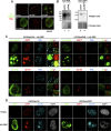
Depletion of one PML-NB component does not impair the recruitment of other factors into DBP structures. (A) IF analyses using HA-DBPΔC. U2OS cells were transfected with an empty vector (i), the vectors for HA-DBP (ii and iii), or HA-DBPΔC (iv to vi) and at 24 hpt subjected to IF analyses using anti-HA (green) and anti-PML (red) antibodies. (B) Western blotting with shRNA-treated cells. Cell lysates were prepared from U2OS cells transduced with either control shRNA-expressing (shCtrl, lanes 1 and 3), shPML-expressing (lane 2), or shUSP7-expressing (lane 4) lentiviral vectors and subjected to Western blot analyses using either anti-PML (left) or anti-USP7 (right panels) antibodies. Ponceau red staining is shown below as a loading control. (C) IF analyses with shPML-treated cells. U2OS/shCtrl (left) and U2OS/shPML (right panels) cells were transfected with an expression vector for HA-DBP, and IF analyses were carried out at 24 hpt using the indicated antibodies. Higher-magnification images marked by squares and merged images are also shown. (D) IF analyses with shUSP7-treated cells. U2OS/shCtrl (left panels) and U2OS/shUSP7 (right panels) cells were transfected with either an empty vector (first row) or a vector for HA-DBP (second row). At 24 hpt, IF analyses were carried out using the indicated antibodies.
FIG 5
FIG 5
Exogenously expressed DBP is not sufficient to recruit incoming viral genome complexes into PML-NBs. U2OS cells were transfected with either an empty vector (first and second rows) or the vector for HA-DBP (second through sixth rows) and at 24 hpt either mock infected (first, third, and fourth rows) or infected with Ad5 (second, fifth, and sixth rows). At 2 hpi, cells were subjected to IF analyses using anti-protein VII (green, first column), anti-HA (cyan, second column), and anti-PML (magenta, third column) antibodies. DAPI staining (gray) and merged images are shown in the fourth to sixth columns.

Similar articles

Cited by

References

    1. Lallemand-Breitenbach V, de Thé H. 2010. PML nuclear bodies. Cold Spring Harb Perspect Biol 2:a000661. doi:10.1101/cshperspect.a000661. - DOI - PMC - PubMed
    1. Ching RW, Dellaire G, Eskiw CH, Bazett-Jones DP. 2005. PML bodies: a meeting place for genomic loci? J Cell Sci 118:847–854. doi:10.1242/jcs.01700. - DOI - PubMed
    1. Schreiner S, Wodrich H. 2013. Virion factors that target Daxx to overcome intrinsic immunity. J Virol 87:10412–10422. doi:10.1128/JVI.00425-13. - DOI - PMC - PubMed
    1. Weidtkamp-Peters S, Lenser T, Negorev D, Gerstner N, Hofmann TG, Schwanitz G, Hoischen C, Maul G, Dittrich P, Hemmerich P. 2008. Dynamics of component exchange at PML nuclear bodies. J Cell Sci 121:2731–2743. doi:10.1242/jcs.031922. - DOI - PubMed
    1. Ishov AM, Maul GG. 1996. The periphery of nuclear domain 10 (ND10) as site of DNA virus deposition. J Cell Biol 134:815–826. doi:10.1083/jcb.134.4.815. - DOI - PMC - PubMed

Publication types

MeSH terms

Grants and funding

The funders had no role in study design, data collection and interpretation, or the decision to submit the work for publication.

LinkOut - more resources