

Therapeutic targeting of autophagy in cardiovascular disease
- PMID: 26602750
- PMCID: PMC4871782
- DOI: 10.1016/j.yjmcc.2015.11.019
Therapeutic targeting of autophagy in cardiovascular disease
Abstract
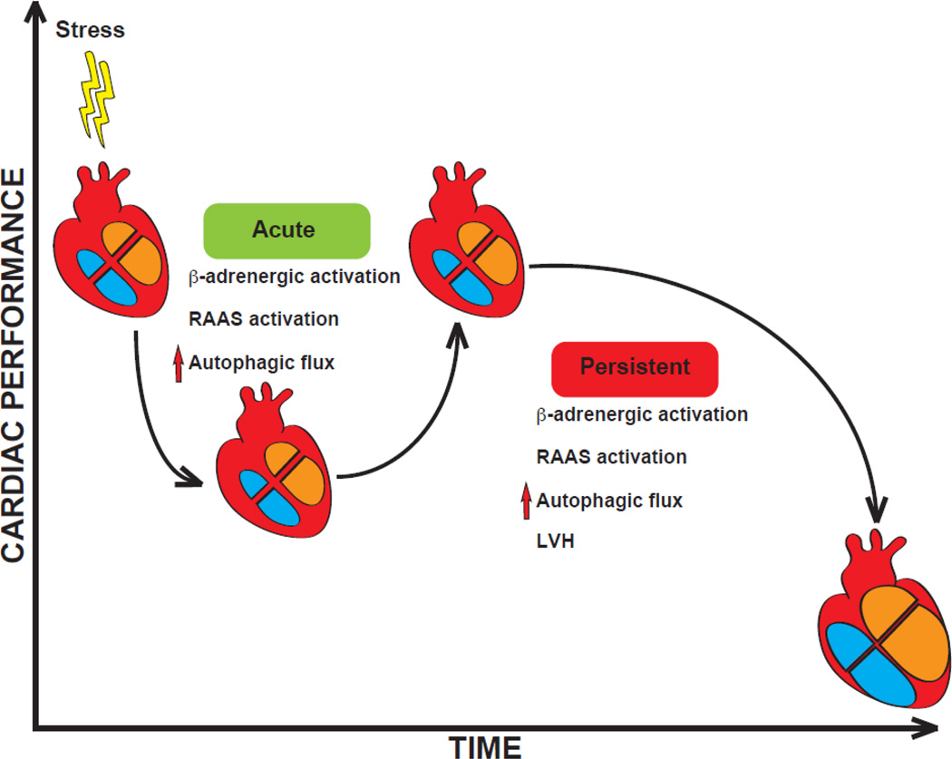
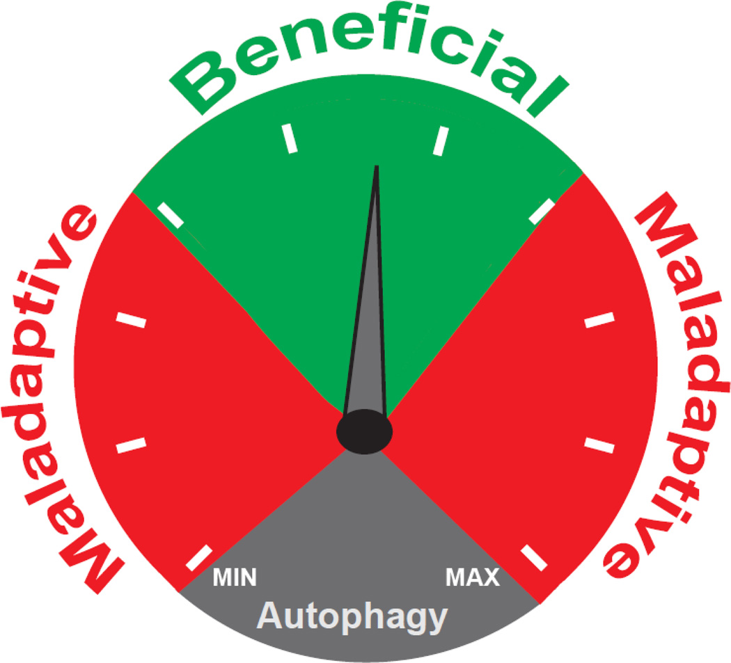
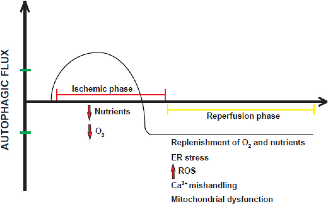
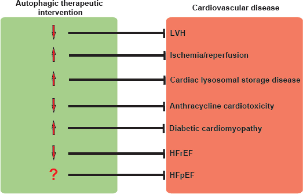
Autophagy is an evolutionarily ancient process of intracellular catabolism necessary to preserve cellular homeostasis in response to a wide variety of stresses. In the case of post-mitotic cells, where cell replacement is not an option, finely tuned quality control of cytoplasmic constituents and organelles is especially critical. And due to the ubiquitous and critical role of autophagic flux in the maintenance of cell health, it comes as little surprise that perturbation of the autophagic process is observed in multiple disease processes. A large body of preclinical evidence suggests that autophagy is a double-edged sword in cardiovascular disease, acting in either beneficial or maladaptive ways, depending on the context. In light of this, the autophagic machinery in cardiomyocytes and other cardiovascular cell types has been proposed as a potential therapeutic target. Here, we summarize current knowledge regarding the dual functions of autophagy in cardiovascular disease. We go on to analyze recent evidence suggesting that titration of autophagic flux holds potential as a novel treatment strategy.
Keywords: Cardiac hypertrophy; Heart failure; Remodeling.
Copyright © 2015 Elsevier Ltd. All rights reserved.
Conflict of interest statement
We declare no conflicts of interest.
Figures
Similar articles
-
Molecular mechanisms of autophagy in the cardiovascular system.Circ Res. 2015 Jan 30;116(3):456-67. doi: 10.1161/CIRCRESAHA.114.303788. Circ Res. 2015. PMID: 25634969 Free PMC article. Review.
-
Therapeutic targeting of autophagy: potential and concerns in treating cardiovascular disease.Circ Res. 2015 Jan 30;116(3):489-503. doi: 10.1161/CIRCRESAHA.116.303791. Circ Res. 2015. PMID: 25634972 Free PMC article. Review.
-
Endolysosomal two-pore channels regulate autophagy in cardiomyocytes.J Physiol. 2016 Jun 1;594(11):3061-77. doi: 10.1113/JP271332. Epub 2016 Feb 4. J Physiol. 2016. PMID: 26757341 Free PMC article.
-
Autophagic flux control in neurodegeneration: Progress and precision targeting-Where do we stand?Prog Neurobiol. 2017 Jun;153:64-85. doi: 10.1016/j.pneurobio.2017.03.006. Epub 2017 Apr 3. Prog Neurobiol. 2017. PMID: 28385648 Review.
-
Crosstalk between Autophagy and Apoptosis: Potential and Emerging Therapeutic Targets for Cardiac Diseases.Int J Mol Sci. 2016 Mar 3;17(3):332. doi: 10.3390/ijms17030332. Int J Mol Sci. 2016. PMID: 26950124 Free PMC article. Review.
Cited by
-
The role of captopril in leukotriene deficient type 1 diabetic mice.Sci Rep. 2023 Dec 13;13(1):22105. doi: 10.1038/s41598-023-49449-8. Sci Rep. 2023. PMID: 38092813 Free PMC article.
-
Dichloroacetate ameliorates myocardial ischemia-reperfusion injury via regulating autophagy and glucose homeostasis.Arch Med Sci. 2019 Aug 27;19(2):420-429. doi: 10.5114/aoms.2019.87503. eCollection 2023. Arch Med Sci. 2019. PMID: 37034518 Free PMC article.
-
Cardiac Shockwave Therapy - A Novel Therapy for Ischemic Cardiomyopathy?Front Cardiovasc Med. 2022 May 12;9:875965. doi: 10.3389/fcvm.2022.875965. eCollection 2022. Front Cardiovasc Med. 2022. PMID: 35647069 Free PMC article. Review.
-
Sodium (±)-5-bromo-2-(α-hydroxypentyl) benzoate ameliorates pressure overload-induced cardiac hypertrophy and dysfunction through inhibiting autophagy.J Cell Mol Med. 2019 Sep;23(9):6048-6059. doi: 10.1111/jcmm.14468. Epub 2019 Jun 20. J Cell Mol Med. 2019. PMID: 31222939 Free PMC article.
-
Cardiomyocyte ZKSCAN3 regulates remodeling following pressure-overload.Physiol Rep. 2023 May;11(9):e15686. doi: 10.14814/phy2.15686. Physiol Rep. 2023. PMID: 37144628 Free PMC article.
References
-
- Nabel EG, Braunwald E. A tale of coronary artery disease and myocardial infarction. The New England journal of medicine. 2012;366:54–63. - PubMed
-
- Hill JA, Olson EN. Cardiac plasticity. The New England journal of medicine. 2008;358:1370–1380. - PubMed
-
- Galluzzi L, Bravo-San Pedro JM, Kroemer G. Organelle-specific initiation of cell death. Nature cell biology. 2014;16:728–736. - PubMed
Publication types
MeSH terms
Substances
Grants and funding
LinkOut - more resources
Full Text Sources
Other Literature Sources
