. 2016 Jan 15;291(3):1336-47.
doi: 10.1074/jbc.M115.682997. Epub 2015 Nov 23.

Oxysterol-binding Protein Activation at Endoplasmic Reticulum-Golgi Contact Sites Reorganizes Phosphatidylinositol 4-Phosphate Pools

Affiliations

Oxysterol-binding Protein Activation at Endoplasmic Reticulum-Golgi Contact Sites Reorganizes Phosphatidylinositol 4-Phosphate Pools

Asako Goto et al. J Biol Chem. .

Abstract

Oxysterol-binding protein (OSBP) exchanges cholesterol and phosphatidylinositol 4-phosphate (PI-4P) at contact sites between the endoplasmic reticulum (ER) and the trans-Golgi/trans-Golgi network. 25-Hydroxycholesterol (25OH) competitively inhibits this exchange reaction in vitro and causes the constitutive localization of OSBP at the ER/Golgi interface and PI-4P-dependent recruitment of ceramide transfer protein (CERT) for sphingomyelin synthesis. We used PI-4P probes and mass analysis to determine how OSBP controls the availability of PI-4P for this metabolic pathway. Treatment of fibroblasts or Chinese hamster ovary (CHO) cells with 25OH caused a 50-70% reduction in Golgi-associated immunoreactive PI-4P that correlated with Golgi localization of OSBP. In contrast, 25OH caused an OSBP-dependent enrichment in Golgi PI-4P that was detected with a pleckstrin homology domain probe. The cellular mass of phosphatidylinositol monophosphates and Golgi PI-4P measured with an unbiased PI-4P probe (P4M) was unaffected by 25OH and OSBP silencing, indicating that OSBP shifts the distribution of PI-4P upon localization to ER-Golgi contact sites. The PI-4P and sterol binding activities of OSBP were both required for 25OH activation of sphingomyelin synthesis, suggesting that 25OH must be exchanged for PI-4P to be concentrated at contact sites. We propose a model wherein 25OH activation of OSBP promotes the binding and retention of PI-4P at ER-Golgi contact sites. This pool of PI-4P specifically recruits pleckstrin homology domain-containing proteins involved in lipid transfer and metabolism, such as CERT.

Keywords: 25-hydroxycholesterol; Golgi; cholesterol; endoplasmic reticulum (ER); fluorescence recovery after photobleaching (FRAP); lipid transport; oxysterol binding protein; sphingomyelin.


Figures

FIGURE 1.
FIGURE 1.
OSBP partially co-localizes with PI-4P in 25OH-treated cells. Primary fibroblasts were treated with 25OH (6 μm) for 2 h followed by immunostaining for PI-4P and either giantin (A), TGN46 (B), or OSBP (C). A and B, RGB line plots through regions of the Golgi apparatus are shown for selected merged images, and Pearson correlation coefficients are indicated on the merged image. D, Pearson correlation coefficients for OSBP co-localization with PI-4P are shown for fibroblasts treated with 25OH for up to 6 h. Results are the mean and S.D. for 10–16 fields of cells from three independent experiments.
FIGURE 2.
FIGURE 2.
25OH reduces immunodetection of PI-4P in primary fibroblasts. A, human primary fibroblasts were treated with 25OH (6 μm) for 2, 4, and 6 h. No addition (NA) controls received ethanol solvent for 6 h (solvent treatment for 0–6 h did not significantly effect PI-4P detection). Fixed cells were immunostained with primary antibodies against PI-4P and giantin (Golgi), and AlexaFluor-488 and -594 secondary antibodies, respectively. Similarly treated fibroblasts were immunostained for OSBP using AlexaFluor-584 (grayscale images). B, fluorescence intensity of PI-4P immunostaining in the cell (total), cytoplasm, and Golgi apparatus was quantified and expressed relative to the no addition controls. Results are the mean and S.D. of 3–4 experiments. C, immunostaining of PI-4P in the plasma membrane of fibroblasts treated with 25OH.
FIGURE 3.
FIGURE 3.
PI-4P immunostaining is dependent on OSBP expression and cholesterol content. A, fluorescence intensity of PI-4P immunostaining was quantified in CHO-shNT and CHO-shOSBP cells that were treated with 25OH for up to 6 h. Results are the mean and S.D. of four experiments using 15–20 fields of cells (*, p < 0.05; **, p < 0.01; ***, p < 0.005; #, p < 0.05 compared with no addition (6-h solvent-treated) CHO-shNT cells). The inset shows an immunoblot of OSBP in control and knockdown CHO cells relative to vimentin (Vim) expression. NA, no addition. B, CHO-K1 cells were incubated with methyl-β-cyclodextrin (5 mm), and the fluorescence intensity of PI-4P immunostaining was quantified as described in A (*, p < 0.01 compared with untreated cells at 0 h).
FIGURE 4.
FIGURE 4.
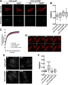
Differential detection of PI-4P in the Golgi apparatus by P4M and PH domain probes. A, confocal images of live CHO-shNT and CHO-shOSBP cells transiently co-expressing mCherry-P4M (mCh-P4M) and YFP-Golgi and treated with 25OH (6 μm) or no addition (solvent) for 4 h. B, quantification of the fluorescence intensity of mCherry-P4M in the Golgi apparatus of control and 25OH-treated cells. The box and whisker plots are for 15–25 cells from three experiments (box denotes the interquartile range with a line at the median value, and whiskers indicate the 10th and 90th percentile). C and D, FRAP analysis of Golgi-associated mCherry-P4M was conducted as described under “Experimental Procedures.” A typical photobleach-recovery sequence is shown in C for 25OH-treated CHO-shOSBP cells. The results in D are the means for 16–20 cells from two separate experiments. There was no significant difference between the four experimental groups, and thus error bars were excluded to improve clarity. E, confocal images of fixed CHO-shNT and CHO-shOSBP cells transiently expressing GFP-ORP4-PH and treated as described in A. F, fluorescence intensity analysis of GFP-ORP4-PH in the Golgi apparatus. Box and whisker plots are for 20–40 cells (p values were determined using one-way analysis of variance test). NA, no addition.
FIGURE 5.
FIGURE 5.
Mass analysis of PIP and PIP2 species in fibroblasts. A and B, mass of individual PIP (A) and PIP2 (B) species was quantified in fibroblasts treated with 25OH for 2, 4, and 6 h or solvent for 6 h (NA, no addition). C, total PIP and PIP mass in 25OH-treated fibroblasts derived from the data in A and B. Results are the mean and S.D. of four experiments (*, p < 0.05 compared with no addition).
FIGURE 6.
FIGURE 6.
Analysis of PIP and PIP2 mass in CHO cells. A and B, mass of PIP (A) and PIP2 (B) molecular species in CHO-shNT and CHO-shOSBP cells treated with 25OH (6 μm) or solvent control (NA, no addition) for 4 h. Results are the mean and S.D. of 4–5 experiments (*, p < 0.05). C, total PIP and PIP2 mass derived from the data in A and B. D, incorporation of [32P]phosphate into PIP and PIP2 of CHO-shNT or CHO-shOSBP cells treated with 25OH (6 μm) or solvent (NA, no addition) for 6 h. Results are the mean and S.D. of four experiments.
FIGURE 7.
FIGURE 7.
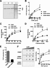
In vitro analysis of ligand-restricted mutants of OSBP. A, purified OSBP (lane 1), OSBP-HH/AA (lane 2), OSBP-RR/EE (lane 3), and OSBP-SBD (lane 4) (0.8 μg each) were separated by SDS-8% PAGE and stained with Coomassie Blue. B, indicated amounts of OSBP, OSBP-HH/AA, or OSBP-RR/EE were incubated with liposomes containing 0.5% [32P]PI-4P, and the extraction of radioactivity into the supernatant was measured. Results are the mean and S.D. of three experiments. C and D, 10 pmol of OSBP, OSBP-HH/AA, or OSBP-SBD (see legend in C) were assayed for specific binding of [3H]cholesterol and 25-[3H]hydroxycholesterol. Results are the mean and S.D. of three experiments. E, extraction of [32P]PI-4P from liposomes by OSBP-SBD (100 pmol). F, binding of 100 pmol of OSBP, OSBP HH/AA, or OSBP-RR/EE (see the legend in B) to PI-4P immobilized on nitrocellulose was quantified after immunoblotting with an anti-OSBP monoclonal antibody. Results are expressed relative to binding of OSBP to 500 pmol of PI-4P and are the mean and S.D. of three experiments.
FIGURE 8.
FIGURE 8.
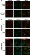
Immunofluorescence localization of PI-4P- and sterol-binding mutants of OSBP. CHO-shOSBP cells were transiently transfected with shRNA-resistant plasmids encoding mCherry-tagged OSBP, OSBP-SBD, and OSBP-HH/AA. A, localization of OSBP and OSBP-SBD with giantin in cells treated with 25OH or solvent (no addition, NA) for 1 h. B, cells transiently expressing OSBP-HH/AA and treated with or without 25OH were immunostained for giantin or VAPA using polyclonal antibodies and AlexaFluor-488-conjugated secondary antibodies. Confocal images are 0.5-μm sections captured using a ×63 objective.
FIGURE 9.
FIGURE 9.
Immunostaining of PI-4P in the Golgi apparatus is dependent on the PI-4P binding activity of OSBP. CHO-shOSBP cells were transfected with vectors expressing shRNA-resistant mCherry-tagged OSBP (A), OSBP-HH/AA (B), or OSBP-SBD (C) for 48 h. Cells were subsequently treated with 25OH (6 μm for 2 h) or solvent (NA, no addition) and immunostained for PI-4P. Asterisks in the PI-4P panels indicate cells that are expressing mCherry-OSBPs. Confocal images are 0.5 μm sections. D, quantification of PI-4P fluorescence intensity in non-transfected (NT) and OSBP-expressing cells. Results are the mean and S.D. for 15–20 fields of cells from four independent experiments.
FIGURE 10.
FIGURE 10.
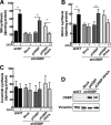
Activation of SM and glucosylceramide synthesis by 25OH requires the PI-4P binding activity of OSBP. shOSBP cells were transiently transfected with empty vector (mock) or shRNA-resistant pOSBP or pOSBP-HH/AA for 48 h. Incorporation of [3H]serine into SM (A), GlcCer (B), and ceramide (C) was measured in solvent control and 25OH-treated cells as described under “Experimental Procedures.” Results are the mean and S.D. of three experiments. *, p < 0.05. D, immunoblot of OSBP expression in lysates of CHO-shNT and CHO-shOSBP cell expressing shRNA-resistant cDNAs.
FIGURE 11.
FIGURE 11.
Redistribution of PI-4P at ER-Golgi contacts by oxysterol activation of OSBP. A, under basal conditions, PI-4P is in a free and unassociated state that is detected by a headgroup-specific antibody. OSBP is cytoplasmic or associated with VAPA (red bar) on the ER. 25OH binding to the OSBP OHD results in localization to ER-Golgi contact sites through PH domain binding to PI-4P. B, OSBP is fully engaged at ER-Golgi contact sites where 25OH is exchanged for PI-4P at the OHD, effectively blocking immunodetection. The PI-4P sequestered at contact sites acts as a recruiting platform for PH domain-containing proteins. OSBP is a dimer but monomers are shown for clarity.

Similar articles

Cited by

References

    1. Lagace T. A., and Ridgway N. D. (2013) The role of phospholipids in the biological activity and structure of the endoplasmic reticulum. Biochim. Biophys. Acta 1833, 2499–2510 - PubMed
    1. van Meer G., Voelker D. R., and Feigenson G. W. (2008) Membrane lipids: where they are and how they behave. Nat. Rev. Mol. Cell Biol. 9, 112–124 - PMC - PubMed
    1. Holthuis J. C., and Menon A. K. (2014) Lipid landscapes and pipelines in membrane homeostasis. Nature 510, 48–57 - PubMed
    1. Olkkonen V. M., and Li S. (2013) Oxysterol-binding proteins: sterol and phosphoinositide sensors coordinating transport, signaling and metabolism. Prog. Lipid Res. 52, 529–538 - PubMed
    1. Alpy F., and Tomasetto C. (2014) START ships lipids across interorganelle space. Biochimie 96, 85–95 - PubMed

Publication types

MeSH terms