Microglial P2Y12 receptors regulate microglial activation and surveillance during neuropathic pain
- PMID: 26576724
- PMCID: PMC4864135
- DOI: 10.1016/j.bbi.2015.11.007
Microglial P2Y12 receptors regulate microglial activation and surveillance during neuropathic pain
Abstract
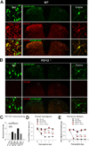
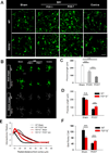
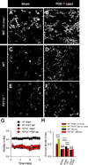
Microglial cells are critical in the pathogenesis of neuropathic pain and several microglial receptors have been proposed to mediate this process. Of these receptors, the P2Y12 receptor is a unique purinergic receptor that is exclusively expressed by microglia in the central nervous system (CNS). In this study, we set forth to investigate the role of P2Y12 receptors in microglial electrophysiological and morphological (static and dynamic) activation during spinal nerve transection (SNT)-induced neuropathic pain in mice. First, we found that a genetic deficiency of the P2Y12 receptor (P2Y12(-/-) mice) ameliorated pain hypersensitivities during the initiation phase of neuropathic pain. Next, we characterised both the electrophysiological and morphological properties of microglia in the superficial spinal cord dorsal horn following SNT injury. We show dramatic alterations including a peak at 3days post injury in microglial electrophysiology while high resolution two-photon imaging revealed significant changes of both static and dynamic microglial morphological properties by 7days post injury. Finally, in P2Y12(-/-) mice, these electrophysiological and morphological changes were ameliorated suggesting roles for P2Y12 receptors in SNT-induced microglial activation. Our results therefore indicate that P2Y12 receptors regulate microglial electrophysiological as well as static and dynamic microglial properties after peripheral nerve injury, suggesting that the microglial P2Y12 receptor could be a potential therapeutic target for the treatment of neuropathic pain.
Keywords: 2-Photon imaging; Electrophysiology; Microglia; Neuropathic pain; P2Y12 receptor; Surveillance.
Copyright © 2015 Elsevier Inc. All rights reserved.
Conflict of interest statement
Figures


Similar articles
-
Spinal Microgliosis Due to Resident Microglial Proliferation Is Required for Pain Hypersensitivity after Peripheral Nerve Injury.Cell Rep. 2016 Jul 19;16(3):605-14. doi: 10.1016/j.celrep.2016.06.018. Epub 2016 Jun 30. Cell Rep. 2016. PMID: 27373153 Free PMC article.
-
P2Y12 regulates microglia activation and excitatory synaptic transmission in spinal lamina II neurons during neuropathic pain in rodents.Cell Death Dis. 2019 Feb 18;10(3):165. doi: 10.1038/s41419-019-1425-4. Cell Death Dis. 2019. PMID: 30778044 Free PMC article.
-
Purinergic systems, neuropathic pain and the role of microglia.Exp Neurol. 2012 Apr;234(2):293-301. doi: 10.1016/j.expneurol.2011.09.016. Epub 2011 Sep 17. Exp Neurol. 2012. PMID: 21946271 Review.
-
Nerve injury-activated microglia engulf myelinated axons in a P2Y12 signaling-dependent manner in the dorsal horn.Glia. 2010 Nov 15;58(15):1838-46. doi: 10.1002/glia.21053. Glia. 2010. PMID: 20665560
-
The known knowns of microglia-neuronal signalling in neuropathic pain.Neurosci Lett. 2013 Dec 17;557 Pt A:37-42. doi: 10.1016/j.neulet.2013.08.037. Epub 2013 Aug 27. Neurosci Lett. 2013. PMID: 23994389 Review.
Cited by
-
Antinociceptive Effects of Aaptamine, a Sponge Component, on Peripheral Neuropathy in Rats.Mar Drugs. 2023 Feb 4;21(2):113. doi: 10.3390/md21020113. Mar Drugs. 2023. PMID: 36827154 Free PMC article.
-
Microglial P2Y12 receptor regulates ventral hippocampal CA1 neuronal excitability and innate fear in mice.Mol Brain. 2019 Aug 19;12(1):71. doi: 10.1186/s13041-019-0492-x. Mol Brain. 2019. PMID: 31426845 Free PMC article.
-
Regulation of cortical hyperexcitability in amyotrophic lateral sclerosis: focusing on glial mechanisms.Mol Neurodegener. 2023 Oct 19;18(1):75. doi: 10.1186/s13024-023-00665-w. Mol Neurodegener. 2023. PMID: 37858176 Free PMC article. Review.
-
Treatment of chronic neuropathic pain: purine receptor modulation.Pain. 2020 Jul;161(7):1425-1441. doi: 10.1097/j.pain.0000000000001857. Pain. 2020. PMID: 32187120 Free PMC article.
-
A review of non-prostanoid, eicosanoid receptors: expression, characterization, regulation, and mechanism of action.J Cell Commun Signal. 2022 Mar;16(1):5-46. doi: 10.1007/s12079-021-00630-6. Epub 2021 Jun 26. J Cell Commun Signal. 2022. PMID: 34173964 Free PMC article. Review.
References
-
- Akagi T, Matsumura Y, Yasui M, Minami E, Inoue H, Masuda T, Tozaki-Saitoh H, Tamura T, Mizumura K, Tsuda M, Kiyama H, Inoue K. Interferon regulatory factor 8 expressed in microglia contributes to tactile allodynia induced by repeated cold stress in rodents. J Pharmacol Sci. 2014;126:172–176. - PubMed
-
- Baron R. Mechanisms of disease: neuropathic pain--a clinical perspective. Nat Clin Pract Neurol. 2006;2:95–106. - PubMed
Publication types
MeSH terms
Substances
Grants and funding
LinkOut - more resources
Full Text Sources
Other Literature Sources
Medical

