

Relationship of Estimated SHIV Acquisition Time Points During the Menstrual Cycle and Thinning of Vaginal Epithelial Layers in Pigtail Macaques
- PMID: 26562699
- PMCID: PMC4646715
- DOI: 10.1097/OLQ.0000000000000367
Relationship of Estimated SHIV Acquisition Time Points During the Menstrual Cycle and Thinning of Vaginal Epithelial Layers in Pigtail Macaques
Abstract
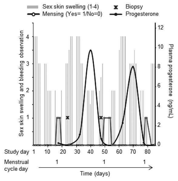
Background: HIV acquisition in the female genital tract remains incompletely understood. Quantitative data on biological HIV risk factors, the influence of reproductive hormones, and infection risk are lacking. We evaluated vaginal epithelial thickness during the menstrual cycle in pigtail macaques (Macaca nemestrina). This model previously revealed increased susceptibility to vaginal infection during and after progesterone-dominated periods in the menstrual cycle.
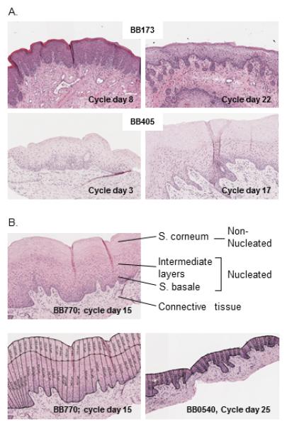
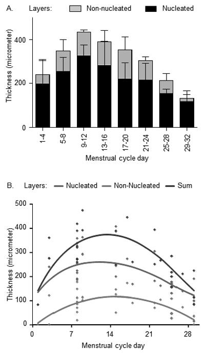
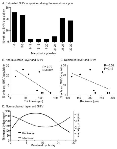
Methods: Nucleated and nonnucleated (superficial) epithelial layers were quantitated throughout the menstrual cycle of 16 macaques. We examined the relationship with previously estimated vaginal SHIVSF162P3 acquisition time points in the cycle of 43 different animals repeatedly exposed to low virus doses.
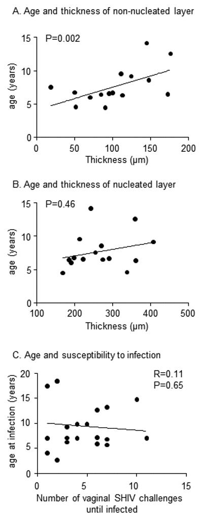
Results: In the luteal phase (days 17 to cycle end), the mean vaginal epithelium thinned to 66% of mean follicular thickness (days 1-16; P = 0.007, Mann-Whitney test). Analyzing 4-day segments, the epithelium was thickest on days 9 to 12 and thinned to 31% thereof on days 29 to 32, with reductions of nucleated and nonnucleated layers to 36% and 15% of their previous thickness, respectively. The proportion of animals with estimated SHIV acquisition in each cycle segment correlated with nonnucleated layer thinning (Pearson r = 0.7, P < 0.05, linear regression analysis), but not nucleated layer thinning (Pearson r = 0.6, P = 0.15).
Conclusions: These data provide a detailed picture of dynamic cycle-related changes in the vaginal epithelium of pigtail macaques. Substantial thinning occurred in the superficial, nonnucleated layer, which maintains the vaginal microbiome. The findings support vaginal tissue architecture as susceptibility factor for infection and contribute to our understanding of innate resistance to SHIV infection.
Figures
Similar articles
-
SHIV susceptibility changes during the menstrual cycle of pigtail macaques.J Med Primatol. 2014 Oct;43(5):310-6. doi: 10.1111/jmp.12124. Epub 2014 Apr 29. J Med Primatol. 2014. PMID: 24779484 Free PMC article.
-
High susceptibility to repeated, low-dose, vaginal SHIV exposure late in the luteal phase of the menstrual cycle of pigtail macaques.J Acquir Immune Defic Syndr. 2011 Aug 1;57(4):261-4. doi: 10.1097/QAI.0b013e318220ebd3. J Acquir Immune Defic Syndr. 2011. PMID: 21546848
-
Analysis of putative mucosal SHIV susceptibility factors during repeated DMPA treatments in pigtail macaques.J Med Primatol. 2015 Oct;44(5):286-95. doi: 10.1111/jmp.12188. Epub 2015 Aug 4. J Med Primatol. 2015. PMID: 26238265
-
Comparison of the vaginal environment of Macaca mulatta and Macaca nemestrina throughout the menstrual cycle.Am J Reprod Immunol. 2014 Apr;71(4):322-9. doi: 10.1111/aji.12201. Epub 2014 Feb 13. Am J Reprod Immunol. 2014. PMID: 24521395 Free PMC article. Review.
-
Target cells in vaginal HIV transmission.Microbes Infect. 2003 Jan;5(1):59-67. doi: 10.1016/s1286-4579(02)00056-4. Microbes Infect. 2003. PMID: 12593974 Review.
Cited by
-
Nonhuman primate models for the evaluation of HIV-1 preventive vaccine strategies: model parameter considerations and consequences.Curr Opin HIV AIDS. 2016 Nov;11(6):546-554. doi: 10.1097/COH.0000000000000311. Curr Opin HIV AIDS. 2016. PMID: 27559710 Free PMC article. Review.
-
Depot medroxyprogesterone acetate (DMPA) enhances susceptibility and increases the window of vulnerability to HIV-1 in humanized mice.Sci Rep. 2021 Feb 16;11(1):3894. doi: 10.1038/s41598-021-83242-9. Sci Rep. 2021. PMID: 33594113 Free PMC article.
-
High-Resolution Quantitative Mapping of Macaque Cervicovaginal Epithelial Thickness: Implications for Mucosal Vaccine Delivery.Front Immunol. 2021 Jun 28;12:660524. doi: 10.3389/fimmu.2021.660524. eCollection 2021. Front Immunol. 2021. PMID: 34262561 Free PMC article.
-
The impact of pregnancy on anti-HIV activity of cervicovaginal secretions.Am J Obstet Gynecol. 2016 Dec;215(6):748.e1-748.e12. doi: 10.1016/j.ajog.2016.06.057. Epub 2016 Jul 5. Am J Obstet Gynecol. 2016. PMID: 27393267 Free PMC article.
-
Characterization of the Genital Mucosa Immune Profile to Distinguish Phases of the Menstrual Cycle: Implications for HIV Susceptibility.J Infect Dis. 2019 Feb 23;219(6):856-866. doi: 10.1093/infdis/jiy585. J Infect Dis. 2019. PMID: 30383238 Free PMC article.
References
-
- Shattock RJ, Moore JP. Inhibiting sexual transmission of HIV-1 infection. Nature reviews. Microbiology. 2003;2003(1):25–34. - PubMed
-
- Marx PA, Spira AI, Gettie A, et al. Progesterone implants enhance SIV vaginal transmission and early virus load. Nature medicine. 1996;1996(10):1084–1089. - PubMed
-
- Poonia B, Walter L, Dufour J, Harrison R, Marx PA, Veazey RS. Cyclic changes in the vaginal epithelium of normal rhesus macaques. J Endocrinol. 2006;2006(3):829–835. - PubMed
Publication types
MeSH terms
Grants and funding
LinkOut - more resources
Full Text Sources
