Metastatic spread in patients with non-small cell lung cancer is associated with a reduced density of tumor-infiltrating T cells
- PMID: 26541588
- PMCID: PMC11028782
- DOI: 10.1007/s00262-015-1768-3
Metastatic spread in patients with non-small cell lung cancer is associated with a reduced density of tumor-infiltrating T cells
Abstract
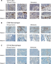
Tumor-infiltrating lymphocytes play an important role in cell-mediated immune destruction of cancer cells and tumor growth control. We investigated the heterogeneity of immune cell infiltrates between primary non-small cell lung carcinomas (NSCLC) and corresponding metastases. Formalin-fixed, paraffin-embedded primary tumors and corresponding metastases from 34 NSCLC patients were analyzed by immunohistochemistry for CD4, CD8, CD11c, CD68, CD163 and PD-L1. The percentage of positively stained cells within the stroma and tumor cell clusters was recorded and compared between primary tumors and metastases. We found significantly fewer CD4(+) and CD8(+) T cells within tumor cell clusters as compared with the stromal compartment, both in primary tumors and corresponding metastases. CD8(+) T cell counts were significantly lower in metastatic lesions than in the corresponding primary tumors, both in the stroma and the tumor cell islets. Of note, the CD8/CD4 ratio was significantly reduced in metastatic lesions compared with the corresponding primary tumors in tumor cell islets, but not in the stroma. We noted significantly fewer CD11c(+) cells and CD68(+) as well as CD163(+) macrophages in tumor cell islets compared with the tumor stroma, but no difference between primary and metastatic lesions. Furthermore, the CD8/CD68 ratio was higher in primary tumors than in the corresponding metastases. We demonstrate a differential pattern of immune cell infiltration in matched primary and metastatic NSCLC lesions, with a significantly lower density of CD8(+) T cells in metastatic lesions compared with the primary tumors. The lower CD8/CD4 and CD8/CD68 ratios observed in metastases indicate a rather tolerogenic and tumor-promoting microenvironment at the metastatic site.
Keywords: Anti-tumor immunity; Immune cells; Metastasis; Non-small cell lung cancer; Primary tumor.
Conflict of interest statement
The authors declare that they have no conflict of interest.
Figures


Similar articles
-
The implications of clinical risk factors, CAR index, and compositional changes of immune cells on hyperprogressive disease in non-small cell lung cancer patients receiving immunotherapy.BMC Cancer. 2021 Jan 5;21(1):19. doi: 10.1186/s12885-020-07727-y. BMC Cancer. 2021. PMID: 33402127 Free PMC article.
-
Distribution of CD4(+) and CD8(+) T cells in tumor islets and stroma from patients with non-small cell lung cancer in association with COPD and smoking.Medicina (Kaunas). 2015 Nov;51(5):263-71. doi: 10.1016/j.medici.2015.08.002. Epub 2015 Sep 21. Medicina (Kaunas). 2015. PMID: 26674143
-
Composition of the immune microenvironment differs between carcinomas metastatic to the lungs and primary lung carcinomas.Ann Diagn Pathol. 2018 Apr;33:62-68. doi: 10.1016/j.anndiagpath.2017.12.004. Epub 2017 Dec 13. Ann Diagn Pathol. 2018. PMID: 29566951
-
Genomic profiles and their associations with TMB, PD-L1 expression, and immune cell infiltration landscapes in synchronous multiple primary lung cancers.J Immunother Cancer. 2021 Dec;9(12):e003773. doi: 10.1136/jitc-2021-003773. J Immunother Cancer. 2021. PMID: 34887263 Free PMC article.
-
Immunotherapy in NSCLC patients with brain metastases. Understanding brain tumor microenvironment and dissecting outcomes from immune checkpoint blockade in the clinic.Cancer Treat Rev. 2020 Sep;89:102067. doi: 10.1016/j.ctrv.2020.102067. Epub 2020 Jul 7. Cancer Treat Rev. 2020. PMID: 32682248 Review.
Cited by
-
Heterogeneous Tumor-Immune Microenvironments between Primary and Metastatic Tumors in a Patient with ALK Rearrangement-Positive Large Cell Neuroendocrine Carcinoma.Int J Mol Sci. 2020 Dec 19;21(24):9705. doi: 10.3390/ijms21249705. Int J Mol Sci. 2020. PMID: 33352665 Free PMC article.
-
Living prosthetic breast for promoting tissue regeneration and inhibiting tumor recurrence.Bioeng Transl Med. 2022 Sep 20;8(5):e10409. doi: 10.1002/btm2.10409. eCollection 2023 Sep. Bioeng Transl Med. 2022. PMID: 37693055 Free PMC article.
-
Immune Checkpoint Imaging in Oncology: A Game Changer Toward Personalized Immunotherapy?J Nucl Med. 2020 Aug;61(8):1137-1144. doi: 10.2967/jnumed.119.237891. Epub 2020 Jan 10. J Nucl Med. 2020. PMID: 31924724 Free PMC article. Review.
-
Sintilimab plus docetaxel as second-line therapy of advanced non-small cell lung cancer without targetable mutations: a phase II efficacy and biomarker study.BMC Cancer. 2022 Sep 5;22(1):952. doi: 10.1186/s12885-022-10045-0. BMC Cancer. 2022. PMID: 36064386 Free PMC article. Clinical Trial.
-
The lung microenvironment: an important regulator of tumour growth and metastasis.Nat Rev Cancer. 2019 Jan;19(1):9-31. doi: 10.1038/s41568-018-0081-9. Nat Rev Cancer. 2019. PMID: 30532012 Free PMC article. Review.
References
-
- Audia S, Nicolas A, Cathelin D, et al. Increase of CD4+ CD25+ regulatory T cells in the peripheral blood of patients with metastatic carcinoma: a Phase I clinical trial using cyclophosphamide and immunotherapy to eliminate CD4+ CD25+ T lymphocytes. Clin Exp Immunol. 2007;150:523–530. doi: 10.1111/j.1365-2249.2007.03521.x. - DOI - PMC - PubMed
Publication types
MeSH terms
LinkOut - more resources
Full Text Sources
Medical
Research Materials

