. 2015 Nov 4;35(44):14727-39.
doi: 10.1523/JNEUROSCI.1304-15.2015.

Cell-Permeable Peptide Targeting the Nrf2-Keap1 Interaction: A Potential Novel Therapy for Global Cerebral Ischemia

Affiliations

Cell-Permeable Peptide Targeting the Nrf2-Keap1 Interaction: A Potential Novel Therapy for Global Cerebral Ischemia

Jingyi Tu et al. J Neurosci. .

Abstract

The current study examined efficacy of a small Tat (trans-activator of transcription)-conjugated peptide activator of the Nrf2 (nuclear factor-E2-related factor-2) antioxidant/cell-defense pathway as a potential injury-specific, novel neuroprotectant against global cerebral ischemia (GCI). A competitive peptide, DEETGE-CAL-Tat, was designed to facilitate Nrf2 activation by disrupting interaction of Nrf2 with Keap1 (kelch-like ECH-associated protein 1), a protein that sequesters Nrf2 in the cytoplasm and thereby inactivates it. The DEETGE-CAL-Tat peptide contained the critical sequence DEETGE for the Nrf2-Keap1 interaction, the cell transduction domain of the HIV-Tat protein, and the cleavage sequence of calpain, which is sensitive to Ca(2+) increase and allows injury-specific activation of Nrf2. Using an animal model of GCI, we demonstrated that pretreatment with the DEETGE-CAL-Tat peptide markedly decreased Nrf2 interaction with Keap1 in the rat hippocampal CA1 region after GCI, and enhanced Nrf2 nuclear translocation and DNA binding. The DEETGE-CAL-Tat peptide also induced Nrf2 antioxidant/cytoprotective target genes, reduced oxidative stress, and induced strong neuroprotection and marked preservation of hippocampal-dependent cognitive function after GCI. These effects were specific as control peptides lacked neuroprotective ability. Intriguingly, the DEETGE-CAL-Tat peptide effects were also injury specific, as it had no effect upon neuronal survival or cognitive performance in sham nonischemic animals. Of significant interest, peripheral, postischemia administration of the DEETGE-CAL-Tat peptide from days 1-9 after GCI also induced robust neuroprotection and strongly preserved hippocampal-dependent cognitive function. Based on its robust neuroprotective and cognitive-preserving effects, and its unique injury-specific activation properties, the DEETGE-CAL-Tat peptide represents a novel, and potentially promising new therapeutic modality for the treatment of GCI.

Significance statement: The current study demonstrates that DEETGE-CAL-Tat, a novel peptide activator of a key antioxidant gene transcription pathway in the hippocampus after global cerebral ischemia, can exert robust neuroprotection and preservation of cognitive function. A unique feature of the peptide is that its beneficial effects are injury specific. This feature is attractive as it targets drug activation specifically in the site of injury, and likely would lead to a reduction of undesirable side effects if translatable to the clinic. Due to its injury-specific activation, robust neuroprotection, and cognitive-preserving effects, this novel peptide may represent a much-needed therapeutic advance that could have efficacy in the treatment of global cerebral ischemia.

Keywords: cardiac arrest; cognitive defect; hippocampus CA1 region; neuroprotection; peptide; reactive oxidative species.


Figures

Figure 1.
Figure 1.
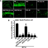
Examination of the neuroprotective ability of the DEETGE-CAL-Tat peptide and various control peptides following GCI. Adult male rats were injected intracebroventrically with either DEETGE-CAL-Tat peptide or control peptides (CAL-Scr-Tat, DEETGE-Tat, CAL-Tat, Tat) at 30 min before GCI and the neuroprotective effect was examined in the hippocampal CA1 region. A, Representative hippocampal CA1 sections from sham-operated (Sh), DEETGE-CAL-Tat-treated (50 μg), vehicle-treated (Veh; 0.9% NaCl), or control-treated (CAL-Scr-Tat, DEETGE-Tat, CAL-Tat, Tat; 50 μg) animals were subjected to immunofluorescent staining for NeuN (green, indicating surviving cells) at 7 d after GCI reperfusion. B, Quantification was performed by counting the number of NeuN-positive neurons per 250 μm length in the medial CA1 pyramidal cell layer (n = 5–6). *p < 0.05 compared with Sh group, #p < 0.05 compared with Veh group. Scale bar, 50 μm. Magnification, 40×.
Figure 2.
Figure 2.
Dose–response characteristics for DEETGE-CAL-Tat peptide neuroprotection of hippocampal CA1 neurons following GCI. Adult male rats were injected intracebroventrically with various doses of the DEETGE-CAL-Tat peptide at 30 min before GCI and the neuroprotective effect examined in the hippocampal CA1 region at 7 d after GCI. A, Representative hippocampal CA1 sections from sham (Sh), I/R, DEETGE-CAL-Tat (30, 50, 100 μg), or vehicle (Veh; 0.9% NaCl) were subjected to double staining for NeuN (green, indicating surviving cells) and TUNEL (red, indicating apoptotic cells). B, C, Quantification was performed by counting the number of NeuN-positive neurons (B) and TUNEL-positive neurons (C) per 250 μm length in the medial CA1 pyramidal cell layer (n = 5–6). *p < 0.05 compared with Sh group, #p < 0.05 compared with Veh groups. Scale bar, 50 μm. Magnification, 40×.
Figure 3.
Figure 3.
DEETGE-CAL-Tat peptide treatment preserves hippocampal-dependent cognitive function following GCI. A, B, Latency trial (A) and probe trial (B) results of sham (Sh), I/R, vehicle (Veh, 0.9% saline), and DEETGE-CAL-Tat (50 μg) pretreatment animals in the MWM. A, Time [in seconds (sec)] spent finding the submerged platform at day 7, 8, and 9 after ischemic injury. B, Exploration time spent in the quadrant that initially contained the platform at day 9 following reperfusion. C, Representative traces indicating the sample paths of the rats from the maze latency trials (a–d) and the swimming traces from probe trials (e–h). a, e: Sh; b, f: I/R; c, g: Veh; d, h: DEETGE-CAL-Tat). Data are expressed as mean ± SD from five different animals. *p < 0.05 vs Sh group; #p < 0.05 vs Veh group.
Figure 4.
Figure 4.
DEETGE-CAL-Tat peptide treatment does not affect neuronal survival or cognitive function in nonischemic animals. A, Representative hippocampal CA1 sections from sham and DEETGE-CAL-Tat (50 μg) treatment (nonischemic) groups were subjected to CV staining. B, C, Latency trial (B) and probe trial (C) results in the MWM for the two groups. B, Time [in seconds (sec)] spent finding the submerged platform at 7, 8, and 9 d after sham surgery. C, Exploration time spent in the quadrant that initially contained the platform at 9 d following reperfusion. D, Representative traces indicating the sample paths of the rats from the maze latency trials (a, b), and the swimming traces from probe trials (c, d). a, c: sham; b, d: DEETGE-CAL-Tat. Data are expressed as mean ± SD from five different animals. n.s., No significant change between the two groups. Magnification: 4× in left line and 20× in right line. Scale bar, 50 μm.
Figure 5.
Figure 5.
DEETGE-CAL-Tat peptide treatment enhances Nrf2 nuclear translocation and DNA binding in the hippocampal CA1 region following GCI. The cytoplasm or nuclear extracts were subjected to Western blot analysis for Nfr2 at indicated time points after reperfusion with or without DEETGE-CAL-Tat pretreatment. A, B, Data analyses of Nrf2 protein from the cytoplasmic extracts (A) or nuclear extracts (B) were expressed as ratios to β-actin or NeuN and the analysis was represented by means ± SD from four independent animals. C, Confocal analysis for NeuN (green), Nrf2 (red), and merged images in the hippocampal CA1 region following sham (Sh) treatment, 24 h after reperfusion with DEETGE-CAL-Tat treatment, or 24 h after reperfusion without DEETGE-CAL-Tat treatment. D, Western blot analysis of DEETGE-CAL-Tat effect upon total Nrf2 protein levels in the hippocampal CA1 region after GCI. E, Quantification of Western blot data shown in D. F, Effect of DEETGE-CAL-Tat upon DNA-binding activity of Nrf2, as determined using a TransAM Nrf2 assay kit. *p < 0.05 compared with the same time point for groups without DEETGE-CAL-Tat treatment, #p < 0.05 compared with Sh group. Magnification, 40×. Scale bar, 50 μm.
Figure 6.
Figure 6.
DEETGE-CAL-Tat peptide decreases interaction of Keap1 with Nrf2 in the cytoplasm at 24 h after GCI reperfusion. A, IP for Keap1 and immunoblotting for Nrf2 reveals that DEETGE-CAL-Tat peptide treatment markedly reduces the interaction of Keap1 with Nrf2 in the cytoplasm of hippocampal CA1 region samples. B, Immunoblotting of the samples for either β-actin, a cytoplasmic marker, or for NeuN, a nuclear marker, showing the purity of the cytoplasmic and nuclear fractions. C, Duo-Link II in situ proximity ligation assay using Keap1 and Nrf2 antibodies reveals that Duolink puncta in the hippocampal CA1 regions at 24 h after GCI is markedly decreased by DEETGE-CAL-Tat peptide treatment, compared with vehicle and I/R controls. For clarity, both color and black-and-white images of the Duolink puncta are provided. D, Quantification of the Duolink puncta in C confirms that DEETGE-CAL-Tat peptide treatment significantly reduced interaction of Keap1 and Nrf2 in the hippocampal CA1 region at 24 h after GCI, compared with vehicle and I/R controls. *p < 0.05 compared with all other groups. N = 5–6/group. Scale bar, 50 μm, Magnification, 40×.
Figure 7.
Figure 7.
DEETGE-CAL-Tat peptide treatment enhances expression of Nrf2-regulated antioxidant genes in the hippocampal CA1 region following GCI. A, Total protein from sham (Sh), I/R, and DEETGE-CAL-Tat treatment rats were subjected to Western blotting for the Nrf2-regulated genes: HO-1, NQO1, GPx1, SOD2. β-Actin expression was used for the loading control. B–E, Quantification of blots depicted in A. Data are expressed as fold changes versus Sh (means ± SD, n = 4 in each group). *p < 0.05 compared with the same time point of group without DEETGE-CAL-Tat treatment (I/R).
Figure 8.
Figure 8.
DEETGE-CAL-Tat peptide treatment attenuates oxidative stress damage in the hippocampal CA1 region following GCI. A, Coronal hippocampal CA1 sections obtained from sham-treated (Sh), I/R-treated (Veh), or DEETGE-CAL-Tat (50 μg)-treated animals at reperfusion 24 or 72 h after GCI were subjected to immunostaining for the oxidative stress markers 4HNE or 8OHdG (red). DAPI was converted to green for better viewing. B, Fluorescence intensity for 4HNE or 8OHdG staining, expressed as arbitrary units ratio to Sh, was measured in hippocampal CA1 region, and data shown are representative as mean ± SD from five independent animals (n = 5 in each group). *p < 0.05 versus Sh groups, #p < 0.05 versus I/R (Veh) without DEETGE-CAL-Tat treatment (−DEETGE-CAL-Tat). Magnification, 40×. Scale bar, 50 μm.
Figure 9.
Figure 9.
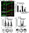
Post-treatment with the DEETGE-CAL-Tat peptide enhances neuronal survival and hippocampal-dependent cognitive function following GCI. DEETGE-CAL-Tat (120 or 240 μg/d) or vehicle (Veh; 0.9% NaCl) was administrated subcutaneously using a minipump (Alzet 2002, 0.5 μl/h for 14 d) beginning at 1 d after reperfusion. The MWM was performed during days 7–9, and double staining for NeuN (green) and TUNEL (red) at 9 d after ischemia. A, Representative hippocampal CA1 area stained with NeuN (green) and TUNEL (red). B, Quantification was performed by counting the number of NeuN-positive cells and TUNEL-positive cells per 250 μm length in the medial CA1 pyramidal cell layer (n = 5. Scale bar, 50 μm). *p < 0.05 compared with sham (Sh) group, #p < 0.05 compared with vehicle (Veh) groups. C, MWM results. Time [in seconds (sec)] spent finding the submerged platform at day 7, 8, and 9 after ischemic injury. D, Exploration time spent in the quadrant that initially contained the platform at day 9 following reperfusion. E–F, Representative traces indicating the sample paths of the rats from the maze latency trials (a–c) and the swimming traces from probe trials (d–f). a, d: Sh; b, e: Veh; c, f: 240 μg/d DEETGE-CAL-Tat treatment. Data are expressed as mean ± SD from five different animals. *p < 0.05 versus Sh group, and #p < 0.05 versus Veh group.

Similar articles

Cited by

References

    1. Beerens AM, Al Hadithy AF, Rots MG, Haisma HJ. Protein transduction domains and their utility in gene therapy. Curr Gene Ther. 2003;3:486–494. doi: 10.2174/1566523034578258. - DOI - PubMed
    1. Brillman J. Central nervous system complications in coronary artery bypass graft surgery. Neurol Clin. 1993;11:475–495. - PubMed
    1. Calabrese B, Saffin JM, Halpain S. Activity-dependent dendritic spine shrinkage and growth involve downregulation of cofilin via distinct mechanisms. PloS One. 2014;9:e94787. doi: 10.1371/journal.pone.0094787. - DOI - PMC - PubMed
    1. Cao G, Pei W, Ge H, Liang Q, Luo Y, Sharp FR, Lu A, Ran R, Graham SH, Chen J. In vivo delivery of a Bcl-xL fusion protein containing the TAT protein transduction domain protects against ischemic brain injury and neuronal apoptosis. J Neurosci. 2002;22:5423–5431. - PMC - PubMed
    1. Chen HH, Chen YT, Huang YW, Tsai HJ, Kuo CC. 4-Ketopinoresinol, a novel naturally occurring ARE activator, induces the Nrf2/HO-1 axis and protects against oxidative stress-induced cell injury via activation of PI3K/AKT signaling. Free Radic Biol Med. 2012;52:1054–1066. doi: 10.1016/j.freeradbiomed.2011.12.012. - DOI - PubMed

Publication types

MeSH terms

Substances