. 2015 Dec;138(Pt 12):3699-715.
doi: 10.1093/brain/awv293. Epub 2015 Oct 28.

Opposing effects of Apoe/Apoa1 double deletion on amyloid-β pathology and cognitive performance in APP mice

Affiliations

Opposing effects of Apoe/Apoa1 double deletion on amyloid-β pathology and cognitive performance in APP mice

Nicholas F Fitz et al. Brain. 2015 Dec.

Abstract

ATP binding cassette transporter A1 (encoded by ABCA1) regulates cholesterol efflux from cells to apolipoproteins A-I and E (ApoA-I and APOE; encoded by APOA1 and APOE, respectively) and the generation of high density lipoproteins. In Abca1 knockout mice (Abca1(ko)), high density lipoproteins and ApoA-I are virtually lacking, and total APOE and APOE-containing lipoproteins in brain substantially decreased. As the ε4 allele of APOE is the major genetic risk factor for late-onset Alzheimer's disease, ABCA1 role as a modifier of APOE lipidation is of significance for this disease. Reportedly, Abca1 deficiency in mice expressing human APP accelerates amyloid deposition and behaviour deficits. We used APP/PS1dE9 mice crossed to Apoe and Apoa1 knockout mice to generate Apoe/Apoa1 double-knockout mice. We hypothesized that Apoe/Apoa1 double-knockout mice would mimic the phenotype of APP/Abca1(ko) mice in regards to amyloid plaques and cognitive deficits. Amyloid pathology, peripheral lipoprotein metabolism, cognitive deficits and dendritic morphology of Apoe/Apoa1 double-knockout mice were compared to APP/Abca1(ko), APP/PS1dE9, and single Apoa1 and Apoe knockouts. Contrary to our prediction, the results demonstrate that double deletion of Apoe and Apoa1 ameliorated the amyloid pathology, including amyloid plaques and soluble amyloid. In double knockout mice we show that (125)I-amyloid-β microinjected into the central nervous system cleared at a rate twice faster compared to Abca1 knockout mice. We tested the effect of Apoe, Apoa1 or Abca1 deficiency on spreading of exogenous amyloid-β seeds injected into the brain of young pre-depositing APP mice. The results show that lack of Abca1 augments dissemination of exogenous amyloid significantly more than the lack of Apoe. In the periphery, Apoe/Apoa1 double-knockout mice exhibited substantial atherosclerosis and very high levels of low density lipoproteins compared to APP/PS1dE9 and APP/Abca1(ko). Plasma level of amyloid-β42 measured at several time points for each mouse was significantly higher in Apoe/Apoa1 double-knockout then in APP/Abca1(ko) mice. This result demonstrates that mice with the lowest level of plasma lipoproteins, APP/Abca1(ko), have the lowest level of peripheral amyloid-β. Unexpectedly, and independent of amyloid pathology, the deletion of both apolipoproteins worsened behaviour deficits of double knockout mice and their performance was undistinguishable from those of Abca1 knockout mice. Finally we observed that the dendritic complexity in the CA1 region of hippocampus but not in CA2 is significantly impaired by Apoe/Apoa1 double deletion as well as by lack of ABCA1.

In conclusion: (i) plasma lipoproteins may affect amyloid-β clearance from the brain by the 'peripheral sink' mechanism; and (ii) deficiency of brain APOE-containing lipoproteins is of significance for dendritic complexity and cognition.

Keywords: ABCA1; APOA1; APOE; Alzheimer’s disease; behaviour.


Figures

None
See Corona and Landreth (doi:10.1093/awv300) for a scientific commentary on this article. The ABCA1 transporter regulates cholesterol efflux to the lipid carriers, ApoE and ApoA1. Fitz et al. compare the phenotype of Apoe/Apoa1 double knockout mice to that of Abca1-/- animals. Surprisingly, double deletion of Apoe/Apoa1 decreases amyloid pathology and increases amyloid-β clearance, but worsens cognitive deficits and reduces dendritic complexity.
Figure 1
Figure 1
Lack of both APOE and ApoA-I in double knockout mice significantly decreases the level of amyloid-β plaques. Compact amyloid was visualized using X-34 (A–C) and diffuse amyloid plaques were stained with 6E10 anti-amyloid-β antibody (D–F). Analysis is by one-way ANOVA with Tukey’s post-test for multiple comparisons (shown on each graph). For both staining we used 6-7-month-old male and female mice: APP/WT (n = 10), APP/Abca1ko (n = 10), APP/DKO (n = 9), APP/Eko (n = 10), APP/Ako (n = 9). (A) Representative X-34 pictures for all genotypes. Quantification of X-34 staining in cortex (B) and in hippocampus (C). ANOVA for cortex and hippocampus is P < 0.0001. Post-test for hippocampus, APP/DKO versus APP/WT P < 0.05; APP/Eko versus APP/WT, P < 0.05. (D) Representative 6E10 staining for all genotypes. Quantification of 6E10 staining in cortex (E) and in hippocampus (F). ANOVA for cortex and hippocampus is P < 0.01.
Figure 2
Figure 2
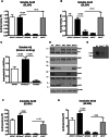
Insoluble amyloid-β is increased in APP/Abca1ko and decreased in APP/DKO mice. Soluble and insoluble proteins were extracted from cortex and hippocampus of 6–7-month-old male and female mice as described in the methods and normalized to total protein (TP). (A) Soluble amyloid-β40 (Aβ40) and (B) amyloid-β42 (Aβ42) levels were determined by ELISA. For A and B, n = 8–13 mice per group. (C) Soluble amyloid-β in APP/WT, APP/Abca1ko and APP/DKO was also measured by western blotting. n = 10–12 per group. (D) The level of ABCA1, full length APP (APPfl), APOE, ApoA-I, CTF-α and soluble amyloid-β proteins was measured by western blotting. Pictures are representative of at least six mice per group. (E) Native gel electrophoresis of CSF from APP/WT, APP/Abca1ko and APP/DKO followed by western blotting for APOE. Loaded were: for APP/WT, 1 µl; for APP/Abca1ko and APP/DKO 10 µl per lane. Native markers are shown on the left. (F) Insoluble amyloid-β40 and (G) insoluble amyloid-β42 was measured by ELISA. For F and G, n = 8–13 per group. Analysis is by one-way ANOVA (For A, B, C, F and G, P < 0.0001) followed by Tukey’s post-test (shown on the graphs).
Figure 3
Figure 3
Soluble amyloid-β oligomers are increased in APP/Abca1ko and unchanged in APP/DKO mice. Amyloid-β oligomers were measured in the soluble fraction (cortex and hippocampus) by dot blot using conformation specific A-11 antibody. The intensity was normalized on total protein (TP) as measured by Coomassie blue staining. For comparison, dot blotting of the same samples probed with 6E10 antibody is shown. Analysis is by one-way ANOVA, P < 0.05, followed by Tukey’s post-test. n = 10 male and female mice per group. N.S. = not significant.
Figure 4
Figure 4
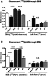
Abca1 deficiency but not the lack of Apoe and Apoa1 delays amyloid-β40 and amyloid-β42 clearance from the brain. (A and B) 5-6-month-old wild type (WT), Abca1ko, DKO, Eko and Ako mice were injected with 12 nmol 125I-amyloid-β40 (Aβ40, A) or 125I-amyloid-β42 (Aβ42, B) into the caudate-putamen. Clearance was assessed 30 min following this injection. 14C-inulin was co-injected in each experiment to assess bulk flow elimination from the interstitial fluid. n = 3–5 mice per group. One-way ANOVA followed by Tukey’s post-test was used to analyse the difference. N.S. = not significant; BBB = blood–brain barrier.
Figure 5
Figure 5
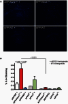
Effects of Abca1, Apoa1 and Apoe genotypes on the dissemination of exogenous amyloid-β seeds: Three-month-old pre-depositing APP mice of different genotypes were injected with amyloid-β seeds from 23-month-old APP23 mouse (labelled as ‘+’) and amyloid-β plaques evaluated 3 months later using X-34 staining. Control mice were injected with brain homogenate from 24-month-old wild-type mouse (labelled as ‘−’). Analysis is by one-way ANOVA (P < 0.001) with Tukey’s post-test (shown on the graph). n per group: APP/WT ( + ) = 8; APP/WT (−) = 6; APP/Abca1ko = 6 per each group; APP/DKO and APP/Eko = 4 mice/group and APP/Ako = 3 mice/group.
Figure 6
Figure 6
Effects of Abca1, Apoa1 and Apoe deletion on peripheral lipoproteins and amyloid-β level in plasma. (A) Shown is fast protein liquid chromatography lipid profiles for APP/WT, APP/Abca1ko, APP/DKO, APP/Eko and APP/Ako mice. Filtered mouse serum was fractionated by size exclusion chromatography on Superose 6 columns in PBS, running at a 0.2 ml/min flow rate. Fractions were collected and assayed for cholesterol. Used were pooled samples of five mice per group. (B) VLDL/LDL and high density lipoprotein fractions were separated from serum of the same genotypes shown on A using kit from Abcam. Quantification of cholesterol level for VLDL/LDL fraction is shown. (C) Kinetic of amyloid-β42 level in plasma: blood was withdrawn from the lateral saphenous vein of APP/WT, APP/Abca1ko, APP/DKO mice at age of 4, 6 and 7 months and amyloid-β level determined by ELISA. n = 6–9 mice per group. Analysis for each time point is by one-way ANOVA (P < 0.05 for 4 and 7 months). *P < 0.05 by Tukey’s post-test when APP/Abca1ko was compared to APP/DKO is shown on the graph. (D) Ratio of amyloid-β42 in plasma and total level of VLDL/LDL fractions as a fold of APP/WT level. For amyloid-β42 was used the mean value from all time points for the respective genotype.
Figure 7
Figure 7
Effects of Abca1, Apoa1 and Apoe deletion on contextual and cued fear conditioning memory. Seven-month-old mice of various genotypes were examined for changes in memory performance using a contextual cued fear conditioning paradigm. Twenty-four hours after the initial acquisition of the contextual fear conditioning (A) the contextual test demonstrates that deletion of Abca1 or ApoA1 and Apoe in APP expressing mice significantly worsens memory performance. Forty-eight hours after the initial acquisition the cued phase of test showed no statistical differences between genotype (B). Similar change in memory performance of non-APP expressing mice that had an Abca1 or ApoA1 and Apoe deletion was observed in the contextual (C) but not in cued (D) phase of training. Analysis by one-way ANOVA with Tukey’s post-test. n = 13–25 male and female mice per genotype. N.S. = not significant.
Figure 8
Figure 8
Deletion of Apoa1 and Apoe significantly affect dendritic morphology in CA1 but not in CA2 region of hippocampus. MAP2 and H3342 staining were used for dendritic tree reconstruction in the hippocampal CA, and CA2 regions of APP/WT, APP/Abca1ko and APP/DKO mice. Data were analysed from a total of four images (×60 confocal imaging) from each of the three sections for each mouse (n = 5 per group). The total dendritic length, branch points and segments were quantified using Imaris filament tracing macros and were normalized to the total H3342 positive nuclei of the CA1 (A and B) and CA2 (C and D) regions. Analysis by one-way ANOVA with Tukey’s post-test. Panel A shows representative images for CA1 region and B shows the quantification of neurites length, branches and segments. In C, representative images for CA2 region and D the quantification of neurites length, branches and segments in this region. Note that deletion of Abca1 as well as the deletion of Apoa1 and Apoe significantly impacts neurite length, segments and branch points only in the CA1 region of the hippocampus. N.S. = not significant.
Figure 8
Figure 8
Deletion of Apoa1 and Apoe significantly affect dendritic morphology in CA1 but not in CA2 region of hippocampus. MAP2 and H3342 staining were used for dendritic tree reconstruction in the hippocampal CA, and CA2 regions of APP/WT, APP/Abca1ko and APP/DKO mice. Data were analysed from a total of four images (×60 confocal imaging) from each of the three sections for each mouse (n = 5 per group). The total dendritic length, branch points and segments were quantified using Imaris filament tracing macros and were normalized to the total H3342 positive nuclei of the CA1 (A and B) and CA2 (C and D) regions. Analysis by one-way ANOVA with Tukey’s post-test. Panel A shows representative images for CA1 region and B shows the quantification of neurites length, branches and segments. In C, representative images for CA2 region and D the quantification of neurites length, branches and segments in this region. Note that deletion of Abca1 as well as the deletion of Apoa1 and Apoe significantly impacts neurite length, segments and branch points only in the CA1 region of the hippocampus. N.S. = not significant.

Comment in

Similar articles

Cited by

References

    1. Abad-Rodriguez J, Ledesma MD, Craessaerts K, Perga S, Medina M, Delacourte A, et al. Neuronal membrane cholesterol loss enhances amyloid peptide generation. J Cell Biol 2004; 167: 953–60. - PMC - PubMed
    1. Bales KR, Verina T, Dodel RC, Du Y, Altstiel L, Bender M, et al. Lack of apolipoprotein E dramatically reduces amyloid beta-peptide deposition. Nat Genet 1997; 17: 263–4. - PubMed
    1. Bell RD, Sagare AP, Friedman AE, Bedi GS, Holtzman DM, Deane R, et al. Transport pathways for clearance of human Alzheimer's amyloid beta-peptide and apolipoproteins E and J in the mouse central nervous system. J Cereb Blood Flow Metab 2007; 27: 909–18. - PMC - PubMed
    1. Bien-Ly N, Gillespie AK, Walker D, Yoon SY, Huang Y. Reducing human apolipoprotein E levels attenuates age-dependent Ab accumulation in mutant human amyloid precursor protein transgenic mice. J Neurosci 2012; 32: 4803–11. - PMC - PubMed
    1. Breslow JL. Mouse models of atherosclerosis. Science 1996; 272: 685–8. - PubMed

Publication types

MeSH terms