. 2015 Oct 26:12:87.
doi: 10.1186/s12977-015-0216-y.

Latent HIV-1 is activated by exosomes from cells infected with either replication-competent or defective HIV-1

Affiliations

Latent HIV-1 is activated by exosomes from cells infected with either replication-competent or defective HIV-1

Claudia Arenaccio et al. Retrovirology. .

Abstract

Background: Completion of HIV life cycle in CD4(+) T lymphocytes needs cell activation. We recently reported that treatment of resting CD4(+) T lymphocytes with exosomes produced by HIV-1 infected cells induces cell activation and susceptibility to HIV replication. Here, we present data regarding the effects of these exosomes on cells latently infected with HIV-1.

Results: HIV-1 latently infecting U937-derived U1 cells was activated upon challenge with exosomes purified from the supernatant of U937 cells chronically infected with HIV-1. This effect was no more detectable when exosomes from cells infected with HIV-1 strains either nef-deleted or expressing a functionally defective Nef were used, indicating that Nef is the viral determinant of exosome-induced HIV-1 activation. Treatment with either TAPI-2, i.e., a specific inhibitor of the pro-TNFα-processing ADAM17 enzyme, or anti-TNFα Abs abolished HIV-1 activation. Hence, similar to what previously demonstrated for the exosome-mediated activation of uninfected CD4(+) T lymphocytes, the Nef-ADAM17-TNFα axis is part of the mechanism of latent HIV-1 activation. It is noteworthy that these observations have been reproduced using: (1) primary CD4(+) T lymphocytes latently infected with HIV-1; (2) exosomes from both primary CD4(+) T lymphocytes and macrophages acutely infected with HIV-1; (3) co-cultures of HIV-1 acutely infected CD4(+) T lymphocytes and autologous lymphocytes latently infected with HIV-1, and (4) exosomes from cells expressing a defective HIV-1.

Conclusions: Our results strongly suggest that latent HIV-1 can be activated by TNFα released by cells upon ingestion of exosomes released by infected cells, and that this effect depends on the activity of exosome-associated ADAM17. These pieces of evidence shed new light on the mechanism of HIV reactivation in latent reservoirs, and might also be relevant to design new therapeutic interventions focused on HIV eradication.


Figures
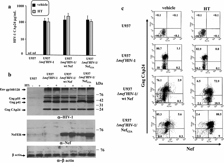
Fig. 1
Fig. 1
Characterization of HIV-1 chronically infected U937-based cell lines expressing Nef in a regulatable way. a Determination of viral release from HIV-1 chronically infected U937-based cell lines by HIV-1 CAp24 ELISA. Cultures of 106 cells/mL of U937 cells either uninfected or chronically infected with Δnef HIV-1, the latter stably transfected with vectors expressing either ER alone (U937 Δnef HIV-1), ER fused with wt Nef (U937 Δnef HIV-1/wt Nef), or ER fused with the Nef G2A mutant (U937 Δnef HIV-1/NefG2A), were carried out for 48 h with either HT or equal volume of vehicle. Afterwards, supernatants were harvested, clarified, and viral contents measured in terms of concentration of CAp24. Shown are the mean values +SD as calculated from duplicate conditions run in seven independent experiments. Nd: not detectable. b Western blot analysis for expression of HIV-1 related products in the HIV-1 chronically infected U937 cell lines either untreated (−) or treated (+) with HT for 48 h. Cell lysates from the U937-based cell lines were resolved in 12 % SDS-PAGE and probed for Gag, Env (upper panel), and Nef expression (middle panel). Signals were normalized by β-actin detection (lower panel). On the right of each panel, molecular weight markers are given in kilodaltons (kDa). On the left, migrations of relevant products are indicated. The results are representative of five independent experiments. c FACS analysis for the expression of both Gag and Nef products. The different U937-based cell lines were incubated for 48 h with HT or equal volume of vehicle, then permeabilized and labeled with both anti-Gag and—Nef mAbs. Percentages of events are reported in the respective quadrants. The results are representative of two independent experiments
Fig. 2
Fig. 2
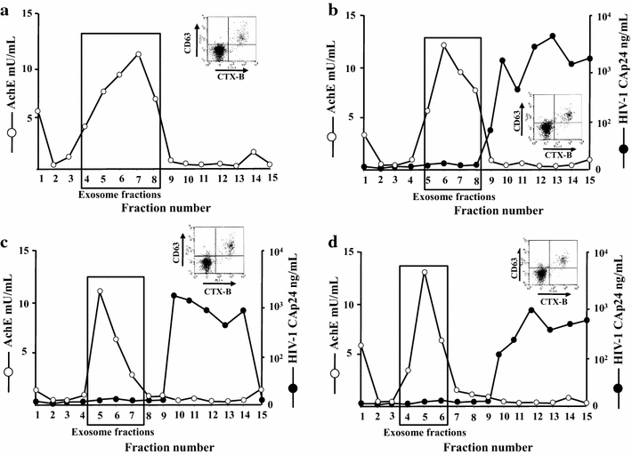
Purification and characterization of exosomes from parental and HIV-1 chronically infected U937 cells expressing Nef in a regulatable way. AchE activity and, for HIV-1 infected cells only, HIV-1 Gag CAp24 contents were measured in fractions from 6 to 18 % iodixanol gradients loaded with vesicles obtained by differential centrifugations of supernatants from a U937, b U937 ΔnefHIV-1, c U937 ΔnefHIV-1/wtNef, and d U937 ΔnefHIV-1/NefG2A cells after treatment with HT for 48 h. In the insets, shown are FACS analyses for the presence of both GM1 and CD63 in nanovesicles recovered from pools of AchE positive fractions of iodixanol gradients. Vesicles were bound to aldehyde/sulfate latex beads and then labeled with FITC-conjugated CTX-B and PE-conjugated anti-CD63 mAb. The results are representative of two independent experiments
Fig. 3
Fig. 3
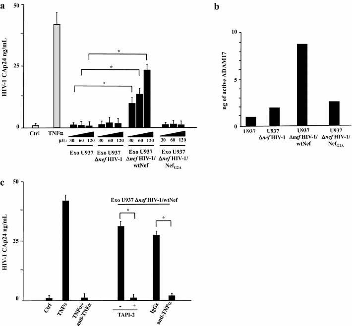
HIV-1 latently infecting U1 cells is activated upon challenge with exosomes from HIV-1 infected cells in a Nef-, TNFα-, and ADAM17-dependent manner. a Different amounts of exosomes (i.e., from 30 to 120 μU of AchE activity) purified from supernatants of the indicated cell lines were used to challenge 5 × 104 U1 cells in U-bottom 96 well plates. After 24 h, the cells were extensively washed, and the amount of released HIV-1 was measured in terms of CAp24 concentration in the supernatants, after additional 24 h. As a control, cells were left untreated (Ctrl) or treated with 100 ng/mL of recombinant TNFα. The results are the mean values + SD from five independent experiments carried out with duplicates. *p < 0.05. b ADAM17 activity detected in 1 mU of exosomes purified from the supernatants of the indicated U937-derived cell lines. Shown are mean amounts of active ADAM17 + SD detected in quadruplicate samples from a representative exosome preparation from each cell line. c Effects of TAPI-2 and anti-TNFα on the activation of latent HIV-1 induced by exosomes from HIV-1 expressing cells. U1 cells (5 × 104/condition) were challenged with 100 μU of exosomes from HIV-1 infected cells expressing wt Nef. Then, cells were left untreated or incubated in the presence of either 1 μM TAPI-2, 160 ng/mL of anti-TNFα neutralizing antibodies, or equivalent amounts of unrelated, isotype-specific IgGs. As a control, cells were treated with 100 ng/mL of recombinant TNFα in the presence or not of the anti-TNFα neutralizing antibodies. After 24 h, cells from all conditions were extensively washed, re-seeded in the appropriate conditions, and the amount of HIV-1 released in supernatants was measured as CAp24 concentration after additional 24 h. The results are the mean values + SD from three independent experiments carried out with duplicates. *p < 0.05
Fig. 4
Fig. 4
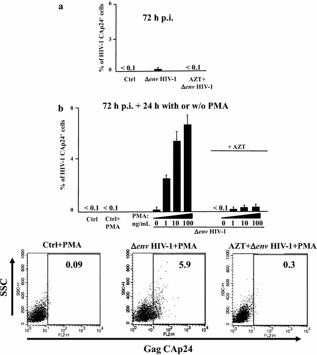
Set up of a system of HIV-1 latent infection. Untouched CD4+ T lymphocytes were purified from PBMCs of healthy donors and challenged by spinoculation with (VSV-G) Δenv HIV-1 in the presence or not of AZT. As a control, conditions with unchallenged CD4+ T lymphocytes (Ctrl) were also run. After 48 h, cells were extensively washed and then left in culture for additional 24 h. a Intracytoplasmic CAp24 FACS analysis of CD4+ T lymphocytes 72 h post-infection. The results are the mean values + SD from nine independent experiments carried out with duplicates. b Intracytoplasmic CAp24 FACS analysis of CD4+ T lymphocytes which, 72 h post-infection, were either activated for 24 h by the indicated doses of PMA+ 0.5 μg/μL of ionomycin (PMA) or left untreated. Ctrl: uninfected CD4+ T lymphocytes. The results are the mean values + SD from three independent experiments carried out with duplicates. In the bottom panels, shown are representative raw results obtained by the intracytoplasmic CAp24 FACS analysis of CD4+ T lymphocytes either uninfected (Ctrl), or latently infected with (VSV-G) Δenv HIV-1 in the presence or not of AZT, and treated for 24 h with PMA + ionomycin
Fig. 5
Fig. 5
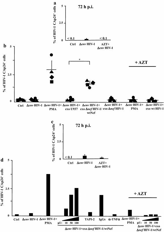
HIV-1 latently infecting primary CD4+ T lymphocytes is activated upon challenge with exosomes from HIV-1 infected cells. Untouched CD4+ T lymphocytes isolated from PBMCs of four healthy donors were challenged by spinoculation with (VSV-G) ΔenvHIV-1 in the presence or not of AZT. As control, conditions with unchallenged CD4+ T lymphocytes (Ctrl) were included. After 48 h, cells were extensively washed and then left in culture for additional 24 h. a Intracytoplasmic CAp24 FACS analysis of CD4+ T lymphocytes 72 h post-infection. The results are the mean values + SD calculated after challenge of CD4+ T lymphocytes from four healthy donors in duplicate conditions. b Intracytoplasmic CAp24 FACS analysis of HIV-1 latently infected CD4+ T lymphocytes 24 h after spinoculation with 100 μU of exosomes purified from either uninfected U937 cells or their counterpart chronically infected with ΔnefHIV-1 and expressing or not wt Nef. As control, cells were treated with 10 ng/mL of PMA+ 0.5 μg/μL of ionomycin (PMA) or left untreated (Ctrl). In addition, CD4+ T lymphocytes originally challenged with (VSV-G) ΔenvHIV-1 in the presence of AZT were treated with either PMA + ionomycin or exosomes from HIV-1 infected U937 cells expressing wt Nef. Shown are the results calculated as mean percentage values of triplicate cultures of CD4+ T lymphocytes from each donor. The inter-donor mean values + SD are also presented. *p < 0.05. The same experiments whose data are shown on panels a and b have been reproduced using PBMCs from two healthy donors and three doses (from 10 to 100 μU) of exosomes from cells infected with ΔnefHIV-1 and expressing wt Nef. In addition, CD4+ T quiescent lymphocytes challenged with 100 μU of exosomes were treated with either TAPI-2, anti-TNFα Abs, or isotype matched IgGs soon after the exosome challenge. c Intracytoplasmic CAp24 FACS analysis of CD4+ T lymphocytes 72 h post-infection. The results are the mean values + SD calculated after challenge of CD4+ T lymphocytes from two healthy donors in duplicate conditions. d Intracytoplasmic CAp24 FACS analysis of HIV-1 latently infected CD4+ T lymphocytes 24 h after spinoculation with the exosomes. As control, cells were treated with 10 ng/mL of PMA+ 0.5 μg/μL of ionomycin (PMA) or left untreated (Ctrl). In addition, CD4+ T lymphocytes originally challenged with (VSV-G) ΔenvHIV-1 in the presence of AZT were treated with either PMA+ ionomycin or exosomes. Shown are the results calculated as mean percentage values of duplicate cultures of CD4+ T lymphocytes from each donor
Fig. 6
Fig. 6
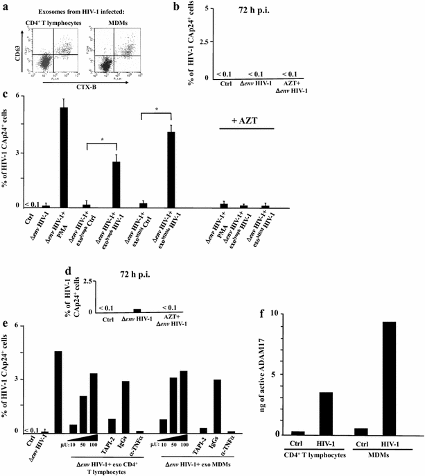
Exosomes from HIV-1 productively infected primary cells activates latent HIV-1 infecting primary CD4+ T lymphocytes. Untouched CD4+ T lymphocytes from PBMCs of healthy donors were challenged by spinoculation with (VSV-G) ΔenvHIV-1 in the presence or not of AZT. As a control, conditions with unchallenged CD4+ T lymphocytes (Ctrl) were included. After 48 h, cells were extensively washed, and then left in culture for additional 24 h. Afterwards, exosome challenge was performed as described below. a FACS analysis for the presence of both GM1 and CD63 in nanovesicles recovered from pools of AchE positive fractions of iodixanol gradients loaded with nanovesicle pellets from supernatants of either CD4+ T lymphocytes or MDMs infected with (VSV-G) wtHIV-1. Vesicles were bound to aldehyde/sulfate latex beads and then labeled with FITC-conjugated CTX-B and PE-conjugated anti-CD63 mAb. b Intracytoplasmic CAp24 FACS analysis of CD4+ T lymphocytes 72 h post-infection. The results are expressed as mean HIV-1 Gag+ percentage calculated after challenge of CD4+ T lymphocytes from three healthy donors analyzed in duplicate conditions. c Intracytoplasmic CAp24 FACS analysis of HIV-1 latently infected CD4+ T lymphocytes 24 h after spinoculation with 100 μU of exosomes purified from HIV-1 infected CD4+ T lymphocytes or MDMs. As a control, the cells were challenged with equivalent amounts of exosomes from uninfected CD4+ T lymphocytes or MDMs. CD4+ T lymphocytes were also treated with PMA + ionomycin (PMA) or left untreated (Ctrl). In addition, CD4+ T lymphocytes originally challenged with (VSV-G) ΔenvHIV-1 in the presence of AZT were treated with either PMA + ionomycin or exosomes from HIV-1 infected cells, i.e. CD4+ T lymphocytes and MDMs. The results are reported as mean percentage values + SD of duplicate cultures of CD4+ T lymphocytes from three healthy donors. *p < 0.05. The same experiment whose results are shown in the panel C has been conducted with CD4+ T lymphocytes from two healthy donors which were latently infected with ΔenvHIV-1 in the presence or not of AZT. Panel d reports the mean percentage values of HIV-1 expressing cells 72 h after challenge. Then, cells were challenged with three doses of each exosome preparation, i.e., from 10 to 100 μU, or with PMA as a control. In addition, cell challenged with 100 μU of exosomes were treated with either TAPI-2, anti-TNFα Abs, or isotype matched IgGs soon after exosome challenge. The results calculated as mean percentage values of duplicate cultures of CD4+ T lymphocytes from each donor are shown in panel e, f. ADAM17 activity detected in 1 mU of exosomes purified from the supernatants of both uninfected or HIV-1 infected CD4+ T lymphocytes and MDMs. Shown are mean values of ng of active ADAM17 detected in quadruplicate samples from a representative exosome preparation from each cell culture
Fig. 7
Fig. 7
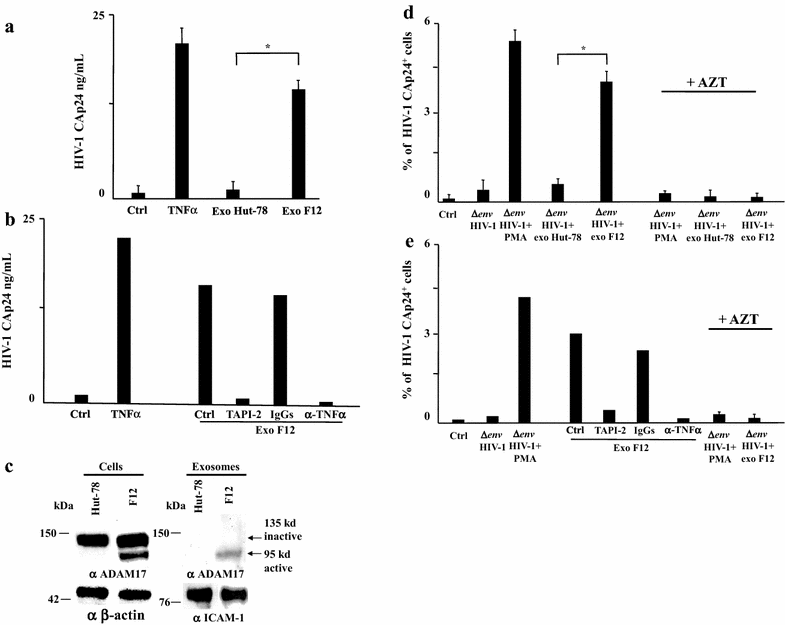
Latent HIV-1 is activated by exosomes from cells infected with defective HIV-1. a A total of 120 μU of exosomes purified from the supernatants of either parental Hut-78 or Hut-78/F12 cells were used to challenge 5 × 104 U1 cells in U-bottom 96 well plates. After 24 h, cells were extensively washed, and released HIV-1 was measured as CAp24 concentration in cell supernatants after additional 24 h. As a control, cells were either left untreated (Ctrl) or treated with 100 ng/mL of recombinant TNFα. The results are expressed as mean values + SD from six independent experiments carried out with duplicates. *p < 0.05. b The same experiment whose data are reported on panel a has been reproduced on U1 treated with either TAPI-2, anti-TNFα Abs, or isotype matched IgGs soon after the exosome challenge. Shown are the results calculated as mean values from two experiments with duplicates. c Western blot analysis for the expression of ADAM17 in either Hut-78 or Hut-78/F12 cells. Signals from cellular ADAM17 were normalized with β-actin signals, whereas exosome preparations were also probed for the presence of ICAM-1. On the left of each panel, molecular weight markers are given in kDa. On the right, arrows identify both inactive and active ADAM17 forms. The results are representative of two independent experiments. d Effects of exosomes from Hut-78/F12 cells on HIV-1 latently infecting primary CD4+ T lymphocytes. A total of 5 × 104 of untouched primary CD4+ T lymphocytes was challenged by spinoculation with (VSV-G) ΔenvHIV-1 in the presence or not of AZT. As a control, conditions with unchallenged CD4+ T lymphocytes (Ctrl) were included. After 48 h, HIV-1 latently infected cells were extensively washed and then left in culture for additional 24 h. Afterwards, cells were challenged with 120 μU of exosomes from either Hut-78 or Hut-78/F12 cells. Shown are the percentages of HIV-1 Gag expressing cells as detected by intracytoplasmic CAp24 FACS analysis 24 h post-exosome challenge. As a control, cells were either treated with PMA + ionomycin (PMA), or left untreated (Ctrl). In addition, CD4+ T lymphocytes challenged with (VSV-G) ΔenvHIV-1 in the presence of AZT were treated with either PMA + ionomycin or exosomes from either Hut-78 or Hut-78/F12 cells. Shown are the results calculated as mean percentage values + SD of triplicate cultures of CD4+ T lymphocytes from three donors. *p < 0.05. e The same experiment whose data are reported in panel d has been reproduced using CD4+ T lymphocytes treated with either TAPI-2, anti-TNFα Abs, or isotype matched IgGs soon after exosome challenge. The results are shown as mean percentage values from two experiments with duplicate cultures

Similar articles

Cited by

References

    1. Siliciano RF, Greene WC. HIV latency. Cold Spring Harb Perspect Med. 2011;1(1):a007096. doi: 10.1101/cshperspect.a007096. - DOI - PMC - PubMed
    1. Swiggard WJ, Baytop C, Yu JJ, Dai J, Li C, Schretzenmair R, et al. Human immunodeficiency virus type 1 can establish latent infection in resting CD4+ T cells in the absence of activating stimuli. J Virol. 2005;79(22):14179–14188. doi: 10.1128/JVI.79.22.14179-14188.2005. - DOI - PMC - PubMed
    1. Siliciano JD, Siliciano RF. AIDS/HIV. Rekindled HIV infection. Science. 2014;345(6200):1005–1006. doi: 10.1126/science.1259452. - DOI - PubMed
    1. Siliciano JD, Kajdas J, Finzi D, Quinn TC, Chadwick K, Margolick JB, et al. Long-term follow-up studies confirm the stability of the latent reservoir for HIV-1 in resting CD4+ T cells. Nat Med. 2003;9(6):727–728. doi: 10.1038/nm880. - DOI - PubMed
    1. Chomont N, El-Far M, Ancuta P, Trautmann L, Procopio FA, Yassine-Diab B, et al. HIV reservoir size and persistence are driven by T cell survival and homeostatic proliferation. Nat Med. 2009;15(8):893–900. doi: 10.1038/nm.1972. - DOI - PMC - PubMed

Publication types

MeSH terms