

Sporulation Temperature Reveals a Requirement for CotE in the Assembly of both the Coat and Exosporium Layers of Bacillus cereus Spores
- PMID: 26497467
- PMCID: PMC4702650
- DOI: 10.1128/AEM.02626-15
Sporulation Temperature Reveals a Requirement for CotE in the Assembly of both the Coat and Exosporium Layers of Bacillus cereus Spores
Abstract
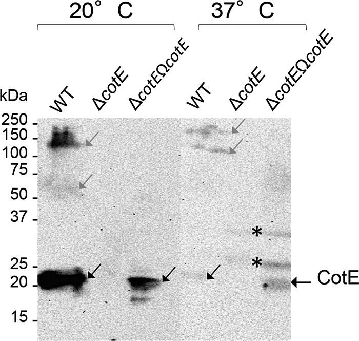
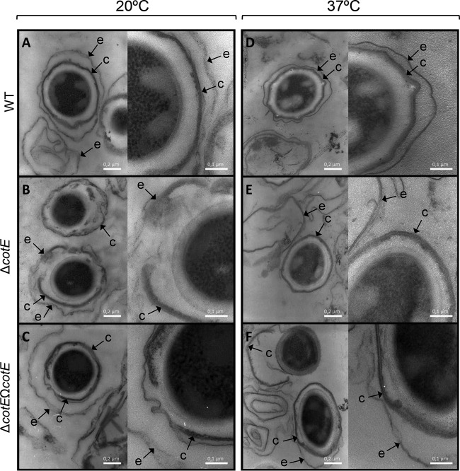
The Bacillus cereus spore surface layers consist of a coat surrounded by an exosporium. We investigated the interplay between the sporulation temperature and the CotE morphogenetic protein in the assembly of the surface layers of B. cereus ATCC 14579 spores and on the resulting spore properties. The cotE deletion affects the coat and exosporium composition of the spores formed both at the suboptimal temperature of 20°C and at the optimal growth temperature of 37°C. Transmission electron microscopy revealed that ΔcotE spores had a fragmented and detached exosporium when formed at 37°C. However, when produced at 20°C, ΔcotE spores showed defects in both coat and exosporium attachment and were susceptible to lysozyme and mutanolysin. Thus, CotE has a role in the assembly of both the coat and exosporium, which is more important during sporulation at 20°C. CotE was more represented in extracts from spores formed at 20°C than at 37°C, suggesting that increased synthesis of the protein is required to maintain proper assembly of spore surface layers at the former temperature. ΔcotE spores formed at either sporulation temperature were impaired in inosine-triggered germination and resistance to UV-C and H2O2 and were less hydrophobic than wild-type (WT) spores but had a higher resistance to wet heat. While underscoring the role of CotE in the assembly of B. cereus spore surface layers, our study also suggests a contribution of the protein to functional properties of additional spore structures. Moreover, it also suggests a complex relationship between the function of a spore morphogenetic protein and environmental factors such as the temperature during spore formation.
Copyright © 2015, American Society for Microbiology. All Rights Reserved.
Figures

Similar articles
-
The Morphogenetic Protein CotE Positions Exosporium Proteins CotY and ExsY during Sporulation of Bacillus cereus.mSphere. 2021 Apr 21;6(2):e00007-21. doi: 10.1128/mSphere.00007-21. mSphere. 2021. PMID: 33883264 Free PMC article.
-
Stoichiometry, Absolute Abundance, and Localization of Proteins in the Bacillus cereus Spore Coat Insoluble Fraction Determined Using a QconCAT Approach.J Proteome Res. 2018 Feb 2;17(2):903-917. doi: 10.1021/acs.jproteome.7b00732. Epub 2018 Jan 4. J Proteome Res. 2018. PMID: 29260567 Free PMC article.
-
The ExsA protein of Bacillus cereus is required for assembly of coat and exosporium onto the spore surface.J Bacteriol. 2005 Jun;187(11):3800-6. doi: 10.1128/JB.187.11.3800-3806.2005. J Bacteriol. 2005. PMID: 15901704 Free PMC article.
-
Structure, assembly, and function of the spore surface layers.Annu Rev Microbiol. 2007;61:555-88. doi: 10.1146/annurev.micro.61.080706.093224. Annu Rev Microbiol. 2007. PMID: 18035610 Review.
-
Clostridium difficile spore biology: sporulation, germination, and spore structural proteins.Trends Microbiol. 2014 Jul;22(7):406-16. doi: 10.1016/j.tim.2014.04.003. Epub 2014 May 7. Trends Microbiol. 2014. PMID: 24814671 Free PMC article. Review.
Cited by
-
Comparison of one-step with two-step production of Bacillus atrophaeus spores for use as bioindicators.Microbiologyopen. 2022 Dec;11(6):e1332. doi: 10.1002/mbo3.1332. Microbiologyopen. 2022. PMID: 36479624 Free PMC article.
-
Food Sensing: Detection of Bacillus cereus Spores in Dairy Products.Biosensors (Basel). 2020 Feb 25;10(3):15. doi: 10.3390/bios10030015. Biosensors (Basel). 2020. PMID: 32106440 Free PMC article. Review.
-
Targeting the Impossible: A Review of New Strategies against Endospores.Antibiotics (Basel). 2023 Jan 26;12(2):248. doi: 10.3390/antibiotics12020248. Antibiotics (Basel). 2023. PMID: 36830159 Free PMC article. Review.
-
Insights into the Structure and Protein Composition of Moorella thermoacetica Spores Formed at Different Temperatures.Int J Mol Sci. 2022 Jan 4;23(1):550. doi: 10.3390/ijms23010550. Int J Mol Sci. 2022. PMID: 35008975 Free PMC article.
-
Changes in the Spore Proteome of Bacillus cereus in Response to Introduction of Plasmids.Microorganisms. 2022 Aug 24;10(9):1695. doi: 10.3390/microorganisms10091695. Microorganisms. 2022. PMID: 36144297 Free PMC article.
References
Publication types
MeSH terms
Substances
Grants and funding
LinkOut - more resources
Full Text Sources
Other Literature Sources
