. 2016 Jan;173(2):305-18.
doi: 10.1111/bph.13372. Epub 2015 Dec 19.

CREB, AP-1, ternary complex factors and MAP kinases connect transient receptor potential melastatin-3 (TRPM3) channel stimulation with increased c-Fos expression

Affiliations

CREB, AP-1, ternary complex factors and MAP kinases connect transient receptor potential melastatin-3 (TRPM3) channel stimulation with increased c-Fos expression

Sandra Rubil et al. Br J Pharmacol. 2016 Jan.

Abstract

Background and purpose: The rise in intracellular Ca(2+) stimulates the expression of the transcription factor c-Fos. Depending on the mode of entry of Ca(2+) into the cytosol, distinct signal transducers and transcription factors are required. Here, we have analysed the signalling pathway connecting a Ca(2+) influx via activation of transient receptor potential melastatin-3 (TRPM3) channels with enhanced c-Fos expression.

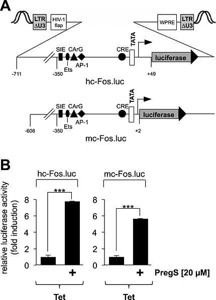
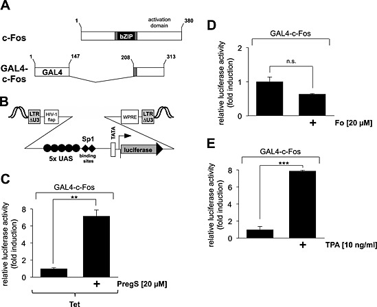
Experimental approach: Transcription of c-Fos promoter/reporter genes that were integrated into the chromatin via lentiviral gene transfer was analysed in HEK293 cells overexpressing TRPM3. The transcriptional activation potential of c-Fos was measured using a GAL4-c-Fos fusion protein.

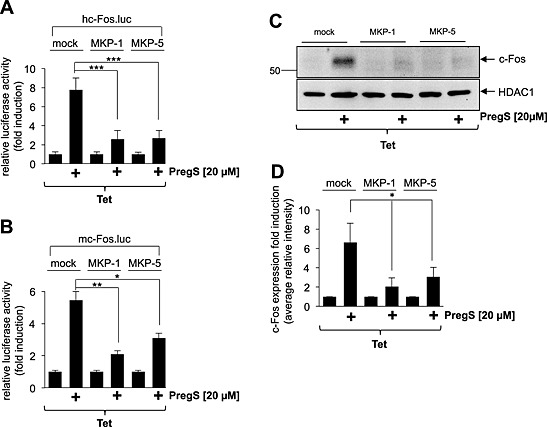
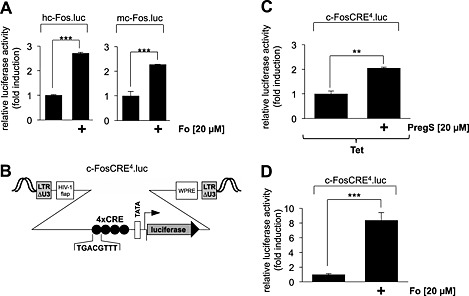
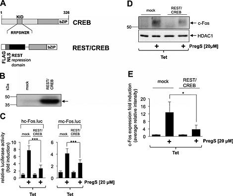
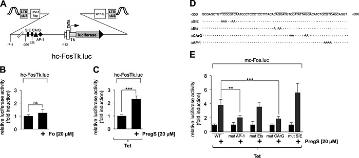
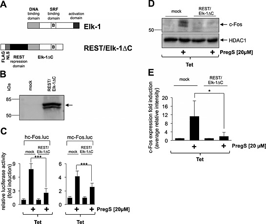
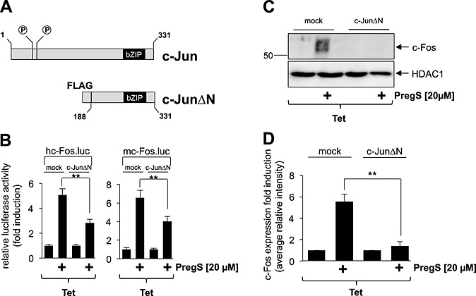
Key results: The signalling pathway connecting TRPM3 stimulation with enhanced c-Fos expression requires the activation of MAP kinases. On the transcriptional level, three Ca(2+) -responsive elements, the cAMP-response element and the binding sites for the serum response factor (SRF) and AP-1, are essential for the TRPM3-mediated stimulation of the c-Fos promoter. Ternary complex factors are additionally involved in connecting TRPM3 stimulation with the up-regulation of c-Fos expression. Stimulation of TRPM3 channels also increases the transcriptional activation potential of c-Fos.

Conclusions and implications: Signalling molecules involved in connecting TRPM3 with the c-Fos gene are MAP kinases and the transcription factors CREB, SRF, AP-1 and ternary complex factors. As c-Fos constitutes, together with other basic region leucine zipper transcription factors, the AP-1 transcription factor complex, the results of this study explain TRPM3-induced activation of AP-1 and connects TRPM3 with the biological functions regulated by AP-1.


Figures

Figure 1
Figure 1
Stimulation of TRPM3 activates transcription of c‐Fos promoter/luciferase reporter genes. (A) Schematic representation of integrated proviruses encoding a human c‐Fos promoter/luciferase reporter gene (hc‐Fos.luc) or a murine c‐Fos promoter/luciferase reporter gene (mc‐Fos.luc). The landmark transcription factor binding sites are depicted, including the SIS‐inducible element (SIE), Ets, the CArG box, the AP‐1 binding site, and the CRE. (B) HEK293 cells containing a tetracycline‐inducible TRPM3 transcription unit were infected with recombinant lentiviruses encoding either a human c‐Fos promoter/luciferase reporter gene (hc‐Fos.luc) or a murine c‐Fos promoter/luciferase reporter gene (mc‐Fos.luc) respectively. The cells were serum starved for 24 h in the presence of tetracycline (1 μg·mL−1) and then stimulated with pregnenolone sulfate (PregS, 20 μM) for 24 h. Cell extracts were prepared and analysed for luciferase activities. Luciferase activity was normalized to the protein concentration. Data shown are mean ± SD from three independent experiments performed in quadruplicate (***P < 0.001).
Figure 2
Figure 2
MAPKs connect TRPM3 activation with stimulation of c‐Fos promoter activity and c‐Fos expression. (A, B) HEK293 cells containing a tetracycline‐inducible TRPM3 transcription unit were infected with a recombinant lentivirus containing the luciferase reporter gene under the control of either the human (A) or murine (B) c‐Fos promoter. Cells were infected with a lentivirus encoding either MKP‐1 or MKP‐5. The transgenes were expressed under the control of the human ubiquitin‐C promoter. As a control, cells were infected with a lentivirus generated with the lentiviral transfer plasmid pFUW (mock). The cells were serum starved for 24 h in the presence of tetracycline (1 μg·mL−1) and then stimulated with pregnenolone sulfate (PregS, 20 μM) for 24 h. Cell extracts were prepared and analysed for luciferase activities. Luciferase activity was normalized to the protein concentration. Data shown are mean ± SD of three experiments performed in quadruplicate (*P < 0.05, **P < 0.01, ***P < 0.001). (C) Expression of either MKP‐1 or MKP‐5 attenuates c‐Fos expression in pregnenolone sulfate‐stimulated HEK293 cells expressing TRPM3. HEK293 cells containing a tetracycline‐inducible TRPM3 transcription unit were infected with a recombinant lentivirus encoding either MKP‐1 or MKP‐5. As a control, cells were infected with lentiviral stocks prepared with the lentiviral transfer vector pFUW (mock). Cells were serum starved for 24 h in the presence of tetracycline (1 μg·mL−1) and then stimulated with pregnenolone sulfate (PregS, 20 μM). Nuclear extracts were prepared and subjected to western blot analysis using an antibody directed against c‐Fos. The antibody directed against HDAC1 was used as a loading control. (D) Quantification of c‐Fos expression. The intensity of the c‐Fos signal was normalized to the intensity of the HDAC1 control. Data shown are mean ± SD of three independent experiments (*P < 0.05).
Figure 3
Figure 3
Stimulation of HEK293 cells expressing TRPM3 with pregnenolone sulfate induces transcription mediated by the CRE derived from the human c‐Fos gene. (A) Up‐regulation of c‐Fos promoter activity by elevated intracellular cAMP concentrations. HEK293 cells containing a tetracycline‐inducible TRPM3 transcription unit were infected with recombinant lentiviruses encoding a luciferase reporter gene under the control of the human (left panel) or murine (right panel) c‐Fos promoter respectively. The cells were serum starved for 24 h and then stimulated with forskolin (Fo, 20 μM) for 24 h. Cell extracts were prepared and analysed for luciferase activities. Luciferase activity was normalized to the protein concentration. Data shown are mean ± SD from three independent experiments performed in quadruplicate (***P < 0.001). (B) Schematic representation of the integrated provirus encoding a c‐Fos CRE/luciferase reporter gene. The regulatory region of the reporter gene contains a minimal promoter consisting of the human immunodeficiency virus TATA box, the adenovirus major late promoter initiator element and four copies of CRE derived from the human c‐Fos gene. (C) HEK293 cells containing a tetracycline‐inducible TRPM3 transcription unit were infected with recombinant lentiviruses containing the CRE/luciferase reporter gene c‐FosCRE4.luc. The cells were serum starved for 24 h in the presence of tetracycline (1 μg·mL−1) and then stimulated with pregnenolone sulfate (PregS, 20 μM) for 24 h. Cell extracts were prepared and analysed for luciferase activities. Luciferase activity was normalized to the protein concentration. Data shown are mean ± SD from three independent experiments performed in quadruplicate (**P < 0.01). (D) Cells were infected with recombinant lentiviruses encoding the reporter gene c‐FosCRE4.luc, serum starved for 24 h and stimulated with Fo (20 μM) for 24 h. Cell extracts were prepared and analysed for luciferase activities. Luciferase activity was normalized to the protein concentration. Data shown are mean ± SD from three independent experiments performed in quadruplicate (***P < 0.001).
Figure 4
Figure 4
A dominant‐negative CREB mutant attenuates the up‐regulation of c‐Fos promoter activity and c‐Fos expression in HEK293 cells expressing activated TRPM3 channels. (A) Schematic representation of CREB and the dominant‐negative mutant REST/CREB. The phosphorylation‐dependent transcriptional activation domain of CREB [kinase‐inducible domain (KID)] is depicted. The bZIP domain is located on the C‐terminus. The mutant lacks the KID domain but retains the DNA‐binding and dimerization domains. The mutant is expressed as a fusion protein together with a transcriptional repression domain derived from the transcriptional repressor REST. (B) Western blot analysis of HEK293 cells either mock infected or infected with recombinant lentiviruses encoding REST/CREB. Western blots were probed with an antibody against the FLAG tag. Molecular mass markers in kDa are shown on the left. (C) HEK293 cells containing a tetracycline‐inducible transcription unit were double infected with a lentivirus encoding either human c‐Fos promoter‐controlled (left panel) or murine c‐Fos promoter‐controlled luciferase reporter gene (right panel) and with a lentivirus encoding REST/CREB. As a control, cells were infected with lentiviral stocks prepared with the lentiviral transfer vector pFUW (mock). The cells were serum starved for 24 h in the presence of tetracycline (1 μg·mL−1) and then stimulated with pregnenolone sulfate (PregS, 20 μM) for 24 h. Cell extracts were prepared and analysed for luciferase activities. Luciferase activity was normalized to the protein concentration. Data shown are mean ± SD from three independent experiments performed in quadruplicate (***P < 0.001). (D) Expression of REST/CREB attenuates c‐Fos expression in pregnenolone sulfate‐stimulated HEK293 cells expressing TRPM3. HEK293 cells containing a tetracycline‐inducible TRPM3 transcription unit were infected with a recombinant lentivirus encoding REST/CREB. As a control, cells were infected with lentiviral stocks prepared with the lentiviral transfer vector pFUW (mock). Cells were serum starved for 24 h in the presence of tetracycline (1 μg·mL−1) and then stimulated with pregnenolone sulfate (PregS, 20 μM). Nuclear extracts were prepared and subjected to western blot analysis using an antibody directed against the c‐Fos protein. The antibody directed against HDAC1 was used as a loading control. (E) Quantification of c‐Fos expression. The intensity of the c‐Fos signal was normalized to the intensity of the HDAC1 control. Data shown are mean ± SD from three independent experiments (*P < 0.05).
Figure 5
Figure 5
Mutational analysis identifies the AP‐1 binding site and the CArG box as pregnenolone sulfate‐responsive elements within the c‐Fos promoter. (A) Schematic representation of the integrated provirus encoding a truncated c‐Fos promoter/luciferase reporter gene lacking the CRE. (B) HEK293 cells containing a tetracycline‐inducible TRPM3 transcription unit were infected with recombinant lentiviruses containing a luciferase reporter gene under the control of the truncated human c‐Fos promoter. The cells were serum starved for 24 h and then stimulated with forskolin (Fo, 20 μM) for 24 h. Cell extracts were prepared and analysed for luciferase activities. Luciferase activity was normalized to the protein concentration. Data shown are mean ± SD from three independent experiments performed in quadruplicate (n.s., not significant). (C) HEK293 cells containing a tetracycline‐inducible TRPM3 transcription unit were infected with a recombinant lentivirus containing the truncated human c‐Fos promoter/luciferase reporter gene hc‐FosTK.luc. The cells were serum starved for 24 h in the presence of tetracycline (1 μg·mL−1) and then stimulated with pregnenolone sulfate (PregS, 20 μM) for 24 h. Cell extracts were prepared and analysed for luciferase activities. Luciferase activity was normalized to the protein concentration. Data shown are mean ± SD from three independent experiments performed in quadruplicate (***P < 0.001). (D) Sequence of the murine c‐Fos promoter from −350 to −280, depicting mutated base pairs of the SIS‐inducible element (SIE), Ets and SRF and AP‐1 binding sites respectively. (E) HEK293 cells containing a tetracycline‐inducible TRPM3 transcription unit were infected with recombinant lentiviruses encoding either the wild‐type murine c‐Fos promoter/luciferase reporter gene or one of the mutated murine c‐Fos promoter/luciferase reporter genes. The cells were serum starved for 24 h in the presence of tetracycline (1 μg·mL−1) and then stimulated with pregnenolone sulfate (PregS, 20 μM) for 24 h. Cell extracts were prepared and analysed for luciferase activities. Luciferase activity was normalized to the protein concentration. Data shown are mean ± SD from three independent experiments performed in quadruplicate (***P 3< 0.001).
Figure 6
Figure 6
Expression of a dominant‐negative mutant of Elk‐1 attenuates the up‐regulation of c‐Fos promoter activity and c‐Fos expression in HEK293 cells expressing activated TRPM3 channels. (A) Schematic representation of Elk‐1 and the dominant‐negative mutant REST/Elk‐1ΔC. The DNA‐binding domain of Elk‐1 is located on the N‐terminus. The transcriptional activation domain is located on the C‐terminus. A regulatory domain lies within this transcriptional activation domain encompassing the key phosphoacceptor serine residues S383 and S389. Elk‐1 binds with its B‐domain to SRF, leading to the formation of the ternary Elk‐1–SRF complex. The B‐domain also couples the C‐terminal phosphorylation of Elk‐1 with enhanced DNA binding via the Ets domain. The dominant‐negative mutant REST/Elk‐1∆C lacks the phosphorylation‐regulated activation domain but retains the DNA‐binding and SRF‐binding domains. The mutant is expressed as a fusion protein together with a transcriptional repression domain of REST. (B) Western blot analysis of HEK293 cells that were either mock infected or infected with a recombinant lentivirus encoding REST/Elk‐1ΔC. The Western blot was probed with an antibody against the FLAG tag. Molecular mass markers in kDa are shown on the left. (C) HEK293 cells were double infected with a lentivirus containing the luciferase reporter gene under the control of either the human (left panel) or murine (right panel) c‐Fos promoter. In addition, cells were infected with a lentivirus encoding REST/Elk‐1ΔC. As a control, cells were infected with lentiviral stocks prepared with the lentiviral transfer vector pFUW (mock). The cells were serum starved for 24 h in the presence of tetracycline (1 μg·mL−1) and then stimulated with pregnenolone sulfate (PregS, 20 μM) for 24 h. Cell extracts were prepared and analysed for luciferase activities. Luciferase activity was normalized to the protein concentration. Data shown are mean ± SD from three independent experiments performed in quadruplicate (***P < 0.001). (D) Expression of REST/Elk‐1ΔC attenuates c‐Fos expression in pregnenolone sulfate‐stimulated HEK293 cells expressing TRPM3. HEK293 cells containing a tetracycline‐inducible TRPM3 transcription unit were infected with a recombinant lentivirus encoding REST/Elk‐1ΔC. As a control, cells were infected with lentiviral stocks prepared with the lentiviral transfer vector pFUW (mock). Cells were serum starved for 24 h in the presence of tetracycline (1 μg·mL−1) and then stimulated with pregnenolone sulfate (PregS, 20 μM). Nuclear extracts were prepared and subjected to Western blot analysis using an antibody directed against the c‐Fos protein. The antibody directed against HDAC1 was used as a loading control. (E) Quantification of c‐Fos expression. The intensity of the c‐Fos signal was normalized to the intensity of the HDAC1 control. Data shown are mean ± SD of three experiments (*P < 0.05).
Figure 7
Figure 7
The transcription factor c‐Jun connects TRPM3 activation with increased c‐Fos promoter activity and c‐Fos expression. (A) Modular structure of c‐Jun and the dominant‐negative form c‐JunΔN. The mutant encompasses amino acid residues 188 to 331 of c‐Jun, retaining the bZIP domain responsible for DNA binding and dimerization, but lacking the NH2‐terminal transcriptional activation domain. (B) HEK293 cells containing a tetracycline‐inducible TRPM3 transcription unit were infected with a lentivirus encoding the luciferase reporter gene under the control of the human (left panel) or murine (right panel) c‐Fos promoter. In addition, cells were infected with a lentivirus encoding the c‐Jun mutant c‐JunΔN. As a control, cells were infected with lentiviral stocks prepared with the lentiviral transfer vector pFUW (mock). The cells were serum starved for 24 h in the presence of tetracycline (1 μg·mL−1) and then stimulated with pregnenolone sulfate (PregS, 20 μM) for 24 h. Cell extracts were prepared and analysed for luciferase activities. Luciferase activity was normalized to the protein concentration. Data shown are mean ± SD of three experiments performed in quadruplicate (**P < 0.01). (C) Expression of c‐JunΔN attenuates c‐Fos expression in pregnenolone sulfate‐stimulated HEK293 cells expressing TRPM3. HEK293 cells containing a tetracycline‐inducible TRPM3 transcription unit were infected with a recombinant lentivirus encoding c‐JunΔN. As a control, cells were infected with lentiviral stocks prepared with the lentiviral transfer vector pFUW (mock). Cells were serum starved for 24 h in the presence of tetracycline (1 μg·mL−1) and then stimulated with pregnenolone sulfate (PregS, 20 μM). Nuclear extracts were prepared and subjected to Western blot analysis using an antibody directed against the c‐Fos protein. The antibody directed against HDAC1 was used as a loading control. (D) Quantification of c‐Fos expression. The intensity of the c‐Fos signal was normalized to the intensity of the HDAC1 control. Data shown are mean ± SD of three independent experiments (**P < 0.01).
Figure 8
Figure 8
Stimulation of TRPM3 channels up‐regulates the transcriptional activation potential of c‐Fos. (A) Schematic representation of the modular structure of c‐Fos and GAL4‐c‐Fos. The bZIP DNA‐binding and dimerization domains are depicted. The transcriptional activation domain is localized on the C‐terminus. The GAL4‐c‐Fos fusion protein lacks the bZIP domain but retains the C‐terminal activation domain of c‐Fos. The truncated c‐Fos protein is expressed as a fusion protein together with the N‐terminal DNA‐binding domain of GAL4. (B) Schematic representation of the integrated provirus encoding a luciferase reporter gene under the control of the minimal promoter, consisting of five GAL4 binding sites [upstream activating sequence (UAS)], two Sp1 binding sites, a TATA box and an initiator element. (C) HEK293 cells containing a tetracycline‐inducible TRPM3 expression unit were double infected with a lentivirus containing the GAL4‐responsive luciferase reporter gene and a lentivirus encoding GAL4‐c‐Fos. The cells were serum starved for 24 h in the presence of tetracycline (1 μg·mL−1) and then stimulated with pregnenolone sulfate (PregS, 20 μM) for 24 h. Cell extracts were prepared and analysed for luciferase activities. Luciferase activity was normalized to the protein concentration. Data shown are mean ± SD from three independent experiments performed in quadruplicate (**P < 0.01). (D, E) HEK293 cells containing a tetracycline‐inducible TRPM3 expression unit were double infected with a lentivirus containing the GAL4‐responsive luciferase reporter gene and a lentivirus encoding GAL4‐c‐Fos. The cells were serum starved for 24 h and then stimulated with either forskolin (Fo, 20 μM) (D) or TPA (10 ng·mL−1) (E) for 24 h. Cell extracts were prepared and analysed for luciferase activities. Luciferase activity was normalized to the protein concentration. Data shown are mean ± SD of three experiments performed in quadruplicate (n.s., not significant; **P < 0.01).

Similar articles

Cited by

References

    1. Alexander SPH, Benson HE, Faccenda E, Pawson AJ, Sharman JL, Spedding M, et al (2013a). The Concise Guide to PHARMACOLOGY 2013/14:ligand‐gated ion channels. Br J Pharmacol 170: 1582–1606. - PMC - PubMed
    1. Alexander SPH, Benson HE, Faccenda E, Pawson AJ, Sharman JL, Spedding M, et al (2013b). The Concise Guide to PHARMACOLOGY 2013/14:enzymes. Br J Pharmacol 170: 1797–1867. - PMC - PubMed
    1. Bennett TM, Mackay DS, Siegfried CJ, Shiels A (2014). Mutation of the melastatin‐related cation channel, TRPM3, underlies inherited cataract and glaucoma. PLoS One 9: e104000. - PMC - PubMed
    1. Brown RL, Xiong W‐H, Peters JH, Tekmen‐Clark M, Strycharska I, Reed BT, et al. (2015). TRPM3 expression in mouse retina. PLoS One. 10: e0117615 - PMC - PubMed
    1. Cahill MA, Janknecht R, Nordheim A (1995). Signal uptake by the c‐fos serum response element In: Baeuerle PA. Inducible Gene Expression, Vol. 2 Birkhäuser: Boston/Basel/Berlin, pp. 39–72.

Publication types

MeSH terms