. 2015 Nov 15;128(22):4220-34.
doi: 10.1242/jcs.175372. Epub 2015 Oct 12.

α-Arrestins participate in cargo selection for both clathrin-independent and clathrin-mediated endocytosis

Affiliations

α-Arrestins participate in cargo selection for both clathrin-independent and clathrin-mediated endocytosis

Derek C Prosser et al. J Cell Sci. .

Abstract

Clathrin-mediated endocytosis (CME) is a well-studied mechanism to internalize plasma membrane proteins; however, to endocytose such cargo, most eukaryotic cells also use alternative clathrin-independent endocytic (CIE) pathways, which are less well characterized. The budding yeast Saccharomyces cerevisiae, a widely used model for studying CME, was recently shown to have a CIE pathway that requires the GTPase Rho1, the formin Bni1, and their regulators. Nevertheless, in both yeast and mammalian cells, the mechanisms underlying cargo selection in CME and CIE are only beginning to be understood. For CME in yeast, particular α-arrestins contribute to recognition of specific cargos and promote their ubiquitylation by recruiting the E3 ubiquitin protein ligase Rsp5. Here, we show that the same α-arrestin-cargo pairs promote internalization through the CIE pathway by interacting with CIE components. Notably, neither expression of Rsp5 nor its binding to α-arrestins is required for CIE. Thus, α-arrestins are important for cargo selection in both the CME and CIE pathways, but function by distinct mechanisms in each pathway.

Keywords: Internalization; Plasma membrane; Protein trafficking; Ubiquitin ligase; Yeast.


Conflict of interest statement

Competing interests

The authors declare no competing or financial interests.

Figures
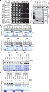
Fig. 1.
Fig. 1.
α-Arrestins interact with the Rho1 GEF Rom2 and the GTPase Rho1. (A) Yeast two-hybrid analyses of α-arrestin Aly2 fusions to the Gal4 TAD with Rom2 fused to Gal4 DBD. PJ69-4a cells containing the indicated plasmids grown on the indicated media for 4 days at 30°C. (B-D) Purified GST or GST-fused GTPases (Coomassie-Blue-stained gels) incubated with [35S]Met-labeled α-arrestins. Co-purifying α-arrestins (top panels) are detected and quantified relative to the amount of GST or GST–GTPase. A representative experiment from at least three replicates is shown. (C) Detection of GST–Rho1, nucleotide-free GST–Rho1G22A or constitutively active GST–Rho1Q68L. (D) GST–Rho1 incubated in nucleotide-free buffer or with GTPγS or GDPβS to assess nucleotide specificity of α-arrestins binding to Rho1. (E) Co-purification of HA-Rho1 with GST or GST-α-arrestins extracted from BJ5459 GEV cells assessed by immunoblotting. Red dots indicate full-length α-arrestins; yellow dots indicate full-length GTPase; white lines indicate gel cropping; molecular masses are indicated in kilodaltons.
Fig. 2.
Fig. 2.
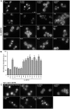
Overexpression of specific α-arrestins promotes internalization of Ste3–GFP in CME-deficient cells. (A) WT, 4Δ+Ent1 and 4Δ+ENTH1 cells expressing Ste3–GFP transformed with vector or high-copy plasmids expressing α-arrestins as indicated and imaged by fluorescence microscopy. (B) WT, 4Δ+Ent1 and 4Δ+ENTH1 cells expressing Ste3-pHluorin transformed as in A, and whole-cell fluorescence quantified (arbitrary units, a.u.; ***P<0.001 compared with WT and 4Δ+Ent1 with vector; †††P<0.001 compared with 4Δ+ENTH1 with vector). (C) aly1Δ aly2Δ ldb19Δ cells generated in WT, 4Δ+Ent1 and 4Δ+ENTH1 strains expressing Ste3–GFP and transformed with vector or high-copy ROM1, ALY1, ALY2, or LDB19 plasmids. Scale bars: 2 µm.
Fig. 3.
Fig. 3.
Latrunculin A treatment to assess requirement for F-actin in Ste3–GFP endocytosis. WT, 4Δ+Ent1 and 4Δ+ENTH1 cells expressing Ste3–GFP transformed with vector or high-copy plasmids expressing the indicated α-arrestins. Cells were imaged by fluorescence microscopy before (Untreated) or 2 h after (LatA) addition of 200 µM LatA. Following treatment, LatA was washed out, and endocytosis was allowed to resume for 2 h before imaging (Washout). Scale bar: 2 µm.
Fig. 4.
Fig. 4.
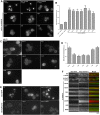
α-Arrestin-stimulated internalization of Ste3–GFP in CME-deficient cells requires the formin Bni1 but not clathrin and fails to correct cortical actin patch immobility. (A) bni1Δ and WT, 4Δ+Ent1 or 4Δ+ENTH1 strains expressing Ste3–GFP and transformed with vector or the indicated high-copy plasmids examined by fluorescence microscopy. (B) Quantification of fluorescence intensity in bni1Δ, bni1Δ 4Δ+Ent1 and bni1Δ 4Δ+ENTH1 strains expressing Ste3-pHluorin transformed as in A (***P<0.001 compared with bni1Δ and bni1Δ 4Δ+Ent1 with vector). (C) chc1Δ cells expressing Ste3–pHluorin and transformed with vector or the indicated high-copy plasmids examined by fluorescence microscopy. (D) Quantification of fluorescence intensity from cells as shown in C (***P<0.001 compared with chc1Δ with vector). (E) Localization of Ste3–GFP and Ste3–pHluorin in WT and chc1Δ cells, as well as in chc1Δ cells transformed with a centromeric CHC1 plasmid. (F) Kymographs derived from time-lapse TIR-FM imaging of WT, 4Δ+Ent1 and 4Δ+ENTH1 cells expressing Sla1–GFP (green) and Abp1–mCherry (red) transformed with the indicated plasmids. Images were captured every second for 120 s. Scale bars: 2 µm.
Fig. 5.
Fig. 5.
The Rsp5 ubiquitin ligase is dispensable for α-arrestin-stimulated internalization of Ste3–GFP. (A) Schematic of α-arrestin primary structure. The arrestin fold (red hexagon) is indicated according to Lin et al. (2008) for Ldb19 and as predicted for Aly1 and Aly2 by Phyre2 (Kelley and Sternberg, 2009; O'Donnell et al., 2010). Green ovals indicate L/PPxY Rsp5-binding motifs and blue rectangle denotes the calcineurin-binding site in Aly1. (B) WT, 4Δ+Ent1 and 4Δ+ENTH1 cells expressing Ste3–GFP and transformed with vector or the indicated high-copy plasmids imaged by fluorescence microscopy. (C) Quantification of fluorescence intensity in WT, 4Δ+Ent1 and 4Δ+ENTH1 cells expressing Ste3–pHluorin transformed as in B (***P<0.001 compared with WT or 4Δ+Ent1 cells with vector; †††P<0.001 compared with 4Δ+ENTH1 with vector). (D) Cell extracts from rsp5Δ cells expressing plasmid-borne Mga2ΔTMD or HA–Rsp5 resolved by SDS-PAGE and probed with anti-Rsp5 or anti-G6PDH antibodies. (E) rsp5Δ cells expressing Mga2ΔTMD and Ste3–GFP transformed with vector or the indicated high-copy plasmids and examined by fluorescence microscopy. Scale bars: 2 µm.
Fig. 6.
Fig. 6.
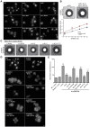
Aly1, Aly2 and Ldb19 facilitate internalization of Ste3 by CME and the endocytic function of Aly1 requires its calcineurin-mediated dephosphorylation. (A) Ste3–GFP expressing WT cells and mutant cells bearing the gene deletions indicated examined by fluorescence microscopy. (B) Mating factor-a pheromone sensitivity of cells with the indicated genotype was assessed using an agar diffusion assay for a-factor-induced growth arrest. One of four replicates is shown where 20 µl of a-factor was spotted on the filter disk. The diameter of the zone of growth inhibition was measured across a range of a-factor concentrations for these strains and the diameter of the halo versus a-factor concentration is plotted in the lower panel. Error bars represent the standard deviations (n=4). (C) Mating factor-a pheromone sensitivity assays for sst2Δ aly1Δ aly2Δ ldb19Δ yeast containing either vector or a centromeric plasmid expressing the indicated α-arrestin allele. (D) WT, 4Δ+Ent1 and 4Δ+ENTH1 cells expressing Ste3–GFP transformed with vector or high-copy ROM1 or the indicated ALY1 plasmids examined by fluorescence microscopy. (E) Quantification of fluorescence intensity in WT, 4Δ+Ent1 and 4Δ+ENTH1 cells expressing Ste3–pHluorin transformed as in D (***P<0.001 compared with WT and 4Δ+Ent1 with vector; †††P<0.001 compared with 4Δ+ENTH1 with vector). Scale bars: 2 µm.
Fig. 7.
Fig. 7.
α-Arrestins have the same cargo-selective roles during both CIE and CME. (A) WT, 4Δ+Ent1 and 4Δ+ENTH1 cells expressing Mup1-pHluorin transformed with vector or the indicated high-copy plasmids, grown in the absence of methionine imaged by fluorescence microscopy 0 or 30 min after addition of 20 µg/ml methionine. (B) Quantification of fluorescence intensity from experiments shown in A. Values are presented as % internalization after 30 min treatment with methionine (n=4; ***P<0.001 compared with WT; P<0.05 compared with 4Δ+ENTH1 with vector). (C) WT, 4Δ+Ent1 and 4Δ+ENTH1 cells expressing Ste2–GFP were transformed with vector or the indicated high-copy plasmids and localization was assessed by fluorescence microscopy. LDB19, ROD1, ROG3 and their respective PPxY-less mutants are included. Scale bars: 2 µm.
Fig. 8.
Fig. 8.
Schematic depiction of the roles played by α-arrestins as cargo-selective regulators of CME and CIE. (A) For CME, α-arrestins bind Rsp5 through their L/PPxY motifs and recruit the ligase to cargo proteins. Rsp5 ubiquitylates both the α-arrestin and the cargo to stimulate CME (Alvaro et al., 2014; Lin et al., 2008; Nikko and Pelham, 2009; Nikko et al., 2008; O'Donnell et al., 2013). (B) For CIE, Rsp5 and Rsp5-binding motifs in α-arrestins are dispensable. Instead, we propose that α-arrestins bind cargo proteins and help recruit the Rho1 GTPase and its GEFs, Rom1 or Rom2. Recruitment of Rho1 to the site of cargo internalization stimulates localized activation of the formin Bni1 and subsequent actin nucleation. This model incorporates cargo selection, GTPase activity and actin nucleation, all of which are key features needed to stimulate endocytosis.

Similar articles

Cited by

References

    1. Aghamohammadzadeh S., Smaczynska-de Rooij I. I. and Ayscough K. R. (2014). An Abp1-dependent route of endocytosis functions when the classical endocytic pathway in yeast is inhibited. PLoS ONE 9, e103311 10.1371/journal.pone.0103311 - DOI - PMC - PubMed
    1. Aguilar R. C., Longhi S. A., Shaw J. D., Yeh L.-Y., Kim S., Schön A., Freire E., Hsu A., McCormick W. K., Watson H. A. et al. (2006). Epsin N-terminal homology domains perform an essential function regulating Cdc42 through binding Cdc42 GTPase-activating proteins. Proc. Natl. Acad. Sci. USA 103, 4116-4121. 10.1073/pnas.0510513103 - DOI - PMC - PubMed
    1. Alvarez C. E. (2008). On the origins of arrestin and rhodopsin. BMC Evol. Biol. 8, 222 10.1186/1471-2148-8-222 - DOI - PMC - PubMed
    1. Alvaro C. G., O'Donnell A. F., Prosser D. C., Augustine A. A., Goldman A., Brodsky J. L., Cyert M. S., Wendland B. and Thorner J. (2014). Specific alpha-arrestins negatively regulate saccharomyces cerevisiae pheromone response by down-modulating the G-protein-coupled receptor Ste2. Mol. Cell. Biol. 34, 2660-2681. 10.1128/MCB.00230-14 - DOI - PMC - PubMed
    1. Aubry L. and Klein G. (2013). True arrestins and arrestin-fold proteins: a structure-based appraisal. Prog. Mol. Biol. Transl. Sci. 118, 21-56. 10.1016/B978-0-12-394440-5.00002-4 - DOI - PubMed

Publication types

MeSH terms