. 2015 Dec 1;408(1):41-55.
doi: 10.1016/j.ydbio.2015.10.005. Epub 2015 Oct 9.

The endocytic recycling regulatory protein EHD1 Is required for ocular lens development

Affiliations

The endocytic recycling regulatory protein EHD1 Is required for ocular lens development

Priyanka Arya et al. Dev Biol. .

Abstract

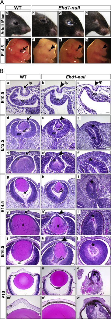
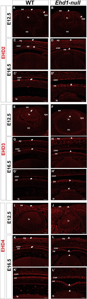
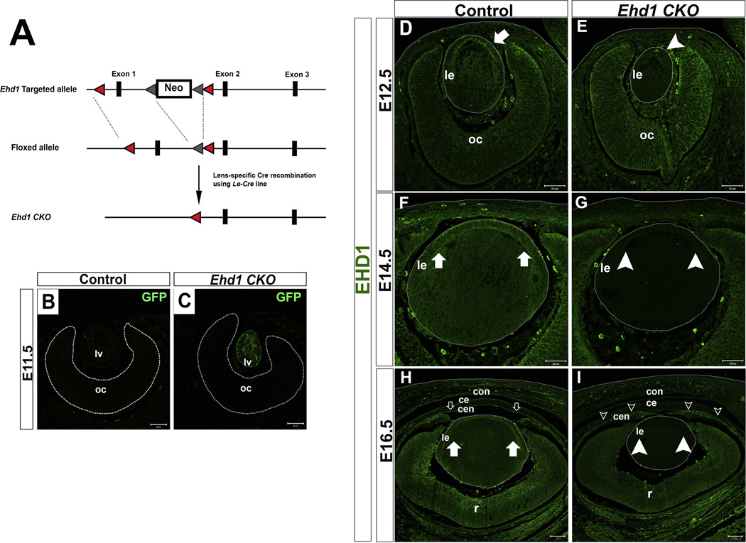
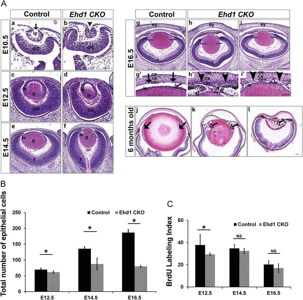
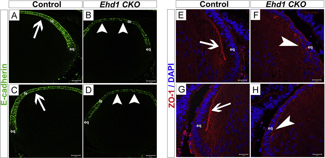
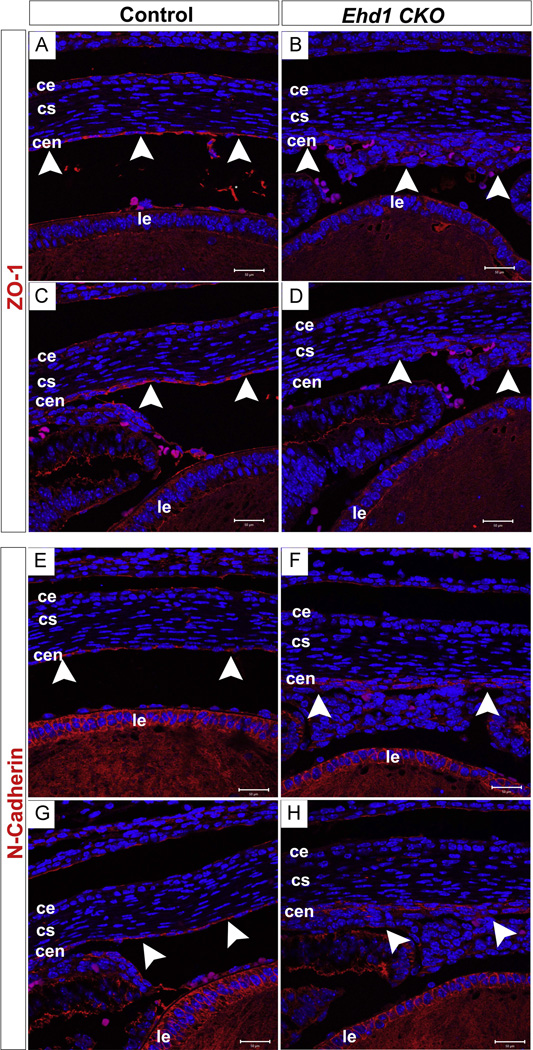
The C-terminal Eps15 homology domain-containing (EHD) proteins play a key role in endocytic recycling, a fundamental cellular process that ensures the return of endocytosed membrane components and receptors back to the cell surface. To define the in vivo biological functions of EHD1, we have generated Ehd1 knockout mice and previously reported a requirement of EHD1 for spermatogenesis. Here, we show that approximately 56% of the Ehd1-null mice displayed gross ocular abnormalities, including anophthalmia, aphakia, microphthalmia and congenital cataracts. Histological characterization of ocular abnormalities showed pleiotropic defects that include a smaller or absent lens, persistence of lens stalk and hyaloid vasculature, and deformed optic cups. To test whether these profound ocular defects resulted from the loss of EHD1 in the lens or in non-lenticular tissues, we deleted the Ehd1 gene selectively in the presumptive lens ectoderm using Le-Cre. Conditional Ehd1 deletion in the lens resulted in developmental defects that included thin epithelial layers, small lenses and absence of corneal endothelium. Ehd1 deletion in the lens also resulted in reduced lens epithelial proliferation, survival and expression of junctional proteins E-cadherin and ZO-1. Finally, Le-Cre-mediated deletion of Ehd1 in the lens led to defects in corneal endothelial differentiation. Taken together, these data reveal a unique role for EHD1 in early lens development and suggest a previously unknown link between the endocytic recycling pathway and regulation of key developmental processes including proliferation, differentiation and morphogenesis.

Keywords: Apoptosis; Corneal endothelium; EHD1; Endocytic recycling; Lens development; Lens epithelium; Polarity; Proliferation.


Figures

Fig. 1
Fig. 1. Defective ocular development in Ehd1-null mice
1A: Gross anatomical features of eye structures of Ehd1-null adult mice (b, c, d) and E14.5 embryos (f, g, h) were compared to control adult mice (a) and embryos (e). Shown are examples of microphthalmia (b), cataract (c) and anophthalmia (d) in Ehd1-null mice. At embryonic day E14.5, smaller eyes and irregular retinal-pigmented epithelium (RPE) are visible in Ehd1-null embryos (f, g, h) compared to littermate wild type control (e). 1B: Histological analyses of formalin-fixed, paraffin-embedded sections depicting examples of: smaller lens pits in Ehd1-null (b, c) compared to WT (a) at E10.5; and lens stalk persistence (e, e’ h, h’, k, arrowheads), hyaloid vasculature persistence (e, e’, h, h’, k, open arrowheads), aphakia (f, f’, i, i’, l, asterisk) in Ehd1-null compared to WT controls (d, d’ g, g’ j, arrows) at E12.5, E14.5, E16.5; normal architecture of the lens and the retina with a smaller lens (n, n’) and a severely malformed residual eye in Ehd1-null mice (o, o’, asterisk) at P10 vs. a well-formed lens, cornea and distinct lamination of neural retina in WT eyes (m, m’). Abbreviations: c, cornea; le, lens epithelium; lf, lens fiber; lp, lens pit; oc, optic cup; cells; r, retina. Scale bars are 50 µm in panels (a, b, c, d’, e’, f’, g’, h’, i’), 100 µm in panels (d, e, f, j, k, l, m’, n’, o’) and 200 µm in panels (g, h, i, m, n, o).
Fig. 2
Fig. 2. EHD1 expression during mouse eye development
Formalin-fixed, paraffin-embedded 4 µm thick eye tissue sections, at the indicated embryonic time points, were stained with anti-EHD1 (red) and anti-E-cadherin (green) antibodies and visualized by confocal fluorescence microscopy. In control embryos, EHD1 expression is observed in the lens pit and the underlying optic cup at E10.5 (A, C); in the surface ectoderm, the epithelial cells and the underlying optic cup (G, I) at E12.5; in the overlying ectoderm and the lens epithelial cells (M, O) at E14.5 and in the eyelids, the corneal epithelium, corneal stroma and the lens epithelium (U, W) at E16.5. Colocalization (yellow) is observed along the cells of the lens pit (C, arrows) and the lens epithelium (I, O, W, arrows) in control embryos. EHD1 staining is not observed in Ehd1-null at E10.5 (D, F), at E12.5 (J, L), E14.5 (P, R) and E16.5 (X, Z). E-cadherin colocalization with EHD1 is not observed in Ehd1-null embryos (F, L, Z, arrowheads). The dotted line demarcates the lens pit, the surface ectoderm and the lens epithelium. Abbreviations: ce, corneal epithelium; cen, corneal endothelium; con, conjunctival epithelium; le, lens epithelium; lf, lens fiber cells; lv, lens vesicle; lp, lens pit; oc, optic cup. Scale bar is 20 µm.
Fig. 3
Fig. 3. Expression of EHD family members is not altered in developing eyes of Ehd1-null mice
Formalin-fixed, paraffin-embedded tissue sections, at the indicated embryonic time points, were stained with anti-EHD2, anti-EHD3, anti-EHD4 antibodies and visualized by confocal fluorescence microscopy. In control embryos, EHD2 expression is observed in the surface ectoderm (A, arrows), blood vessels of the vitreous, the optic cup (A), the retinal pigmented epithelium (A, arrows) at E12.5 and in the eyelids (C, arrows), the corneal epithelium (C, arrows) at E16.5. EHD2 expression pattern in Ehd1-null embryos (B, D, arrowheads) is comparable to that in controls. EHD3 expression is observed in: the overlying surface ectoderm, the lens vesicle, the optic cup of WT (E) and Ehd1-null embryos (F) at E12.5, and in the eyelids, the corneal epithelium, the lens epithelium and surrounding mesenchymal tissues of WT (G, G’ arrows) and Ehd1-null eyes (H, H’ arrowheads). Similarly, EHD4 expression is seen in: the surface ectoderm and lens vesicle of WT (I, arrow) and Ehd1-null embryos (J, arrowhead) and in the eyelids, corneal epithelium and in the lens epithelium at E16.5 in WT (K, K’ arrow) and Ehd1-null (L, L’ arrowheads). Abbreviations: ce, corneal epithelium; cen, corneal endothelium; con, conjunctival epithelium; ey, eyelids; le, lens epithelium; lp, lens pit; lv, lens vesicle; oc, optic cup; rpe, retinal pigmented epithelium; se, surface ectoderm. Scale bar is 50 µm in panels C, D and 20 µm in the remaining panels.
Fig. 4
Fig. 4. Conditional deletion of Ehd1 in the mouse lens
A: Schematic of the floxed Ehd1 allele with a Neo cassette surrounded by FRT recombination sites (grey triangles) and loxP recombination sites surrounding exon 1 (red triangles) (top), floxed allele after genetic transgenic FLP recombinase-mediated removal of the Neo cassette (middle) and the mutant allele lacking exon 1 sequences (called Ehd1 CKO) expected to be generated upon Le-Cre driven Cre recombinase expression (bottom). B, C: Formalin-fixed paraffin-embedded sections of E11.5 embryonic eyes of control (B; floxed mice lacking Le-Cre) or Ehd1 CKO mice (C) were subjected to staining with anti-GFP antibody (green) followed by confocal imaging. Lens-specific expression of GFP in Ehd1 CKO mice confirms the specificity of Le-Cre transgene in our stocks. D-I: Control sections (D, F, H) or Ehd1 CKO (E, G, I) embryonic eyes at the indicated ages were stained with an anti-EHD1 antibody and analyzed by confocal microscopy. Loss of EHD1 staining is seen specifically in the developing lens in Ehd1 CKO embryos (E, G, I, arrowheads) while staining in retina is intact and comparable to that in control embryos (D, F, H). EHD1 expression is also retained in the neural crest derived corneal endothelial cells as seen in E16.5 Ehd1 CKO (I, open arrowheads) vs. control (H, open arrows) embryos. A dotted line demarcates the lens boundary in panels D, E, F, G, H, I. Abbreviations: ce, corneal epithelium; cen, corneal endothelium; con, conjunctival epithelium; le, lens epithelium; lv, lens vesicle; oc, optic cup; r, retina. Scale bar is 50 µm in panels B-G and 100 µm in panels H, I.
Fig. 5
Fig. 5. Lens development defects in Ehd1 CKO mice
A: H & E sections of embryonic (a–i) or 6-month old (j–l) eyes from control (a, c, e, g, g’, j) or Ehd1 CKO mice (b, d, f, h, i, h’, i’, k, l) at E10.5 (a, b), E12.5 (c, d), E14.5 (e, f), E16.5 (g–i) and 6-months of age (j–l). Smaller lens pit in E10.5 Ehd1 CKO (b, arrowhead) compared to control (a, arrow) embryo is indicated. Smaller lenses are seen in Ehd1 CKO embryos at E12.5 (d), E14.5 (f) and E16.5 (h, i, h’, i’). At E16.5, Ehd1 CKO embryonic lenses show lens epithelial thinning, aberrant epithelial cell shape (open arrowheads), and absence of corneal endothelium (downward arrowheads). Open arrows in (panels k, l) represent vacuolated lenses in adult Ehd1 CKO mice. The dotted lines on two sides of lens in panels g-i represents the equator region. g’, h’, i’ panels are higher magnification images of segments from g, h and i panels, respectively. Abbreviations: ce, corneal epithelium; cs, corneal stroma; cen, corneal endothelium; ey, eyelids; le, lens epithelium; lf, lens fiber cells; lp, lens pit; oc, optic cup; r, retina. Scale bar is 100 µm. B: Lens epithelial cell numbers in control (black bars) and Ehd1 CKO eyes (grey bars) at E12.5, E14.5, E16.5 were quantified and performed as described in Methods. Error bars indicate SEM. *p < 0.001 C: BrdU positive lens epithelial cell nuclei were counted in control and Ehd1 CKO embryos at E12.5, E14.5 and E16.5. *p < 0.01. NS, not significant.
Fig. 6
Fig. 6. EHD1 is required for Cell Survival
A-H: Immunofluorescence staining revealed by TUNEL (terminal deoxynucleotidyl transferase-mediated deoxyuridinetriphosphate nick end-labeling) assay in embryonic sections from control (A, C, E, G) and Ehd1 CKO (B, D, F, H) at E10.5 (A, B), E12.5 (C, D), E14.5 (E, F), and E16.5 (G, H). Nuclei are stained blue with DAPI. Increased apoptotic cells (green) are detected in Ehd1 CKO mouse lens epithelium. Dotted white lines demarcate the lens region used for the analysis I-J: Quantification data from counting of TUNEL-positive nuclei in control and EHD1 CKO lenses.*p < 0.01; n= number of embryos analyzed. Abbreviations: le, lens epithelium; lf, lens fiber cells; lp, lens pit; oc, optic cup. Scale bar is 50 µm.
Fig. 7
Fig. 7. Altered expression of junctional proteins in Ehd1 CKO mice
Formalin fixed paraffin embedded tissue sections from E16.5 are stained for anti-E-cadherin (green) (A–D) and ZO-1 (red) (E–H) antibodies and subjected to confocal fluorescence microscopy. Nuclei are stained blue with DAPI. A-D: Normal pattern of expression is evident in control lens (A, C, arrows) whereas in the lens epithelial cells of Ehd1 CKO, E-cadherin expression appears disrupted with increased gaps (B, D, arrowheads). E-H: ZO-1 expression is irregular and disrupted at the apical lens epithelial junctions in Ehd1 CKO mice (F, H, arrowheads) in contrast to the littermate controls (E, G, arrows). At the captured magnification the whole lens structure was not visible within a single frame. Therefore, we included the right side (A, B, E, F) and the left side (C, D, G, H) images to provide a complete representation. Abbreviations: eq, lens equator; le, lens epithelium. Scale bar is 50 µm.
Fig. 8
Fig. 8. Corneal endothelium differentiation defects in Ehd1 CKO mice
A-D: ZO-1 expression is almost completely lost from the anterior segment and corneal endothelium (B, D arrowheads) of Ehd1 CKO eyes, in contrast to the control (A, C arrowheads). E-H: In the control eyes, N-cadherin expression is localized to the corneal endothelial layer in the anterior chamber (E, G arrowheads). In the Ehd1 CKO eyes, N-cadherin expression was seen in a cluster of cells that accumulate anterior to the lens (F, H arrowheads). Abbreviations: ce, corneal epithelium; cs, corneal stroma; cen, corneal endothelium; le, lens epithelium. Scale bar is 50 µm.

Similar articles

Cited by

References

    1. Ashery-Padan R, Gruss P. Pax6 lights-up the way for eye development. Curr. Opin. Cell Biol. 2001;13:706–714. - PubMed
    1. Ashery-Padan R, Marquardt T, Zhou X, Gruss P. Pax6 activity in the lens primordium is required for lens formation and for correct placement of a single retina in the eye. Genes Dev. 2000a;14:2701–2711. - PMC - PubMed
    1. Ashery-Padan R, Marquardt T, Zhou X, Gruss P. Pax6 activity in the lens primordium is required for lens formation and for correct placement of a single retina in the eye. Genes Dev. 2000b;14:2701–2711. - PMC - PubMed
    1. Bassnett S, Kuszak JR, Reinisch L, Brown HG, Beebe DC. Intercellular communication between epithelial and fiber cells of the eye lens. J. Cell. Sci. 1994;107(Pt 4):799–811. - PubMed
    1. Beebe DC, Coats JM. The lens organizes the anterior segment: Specification of neural crest cell differentiation in the avian eye. Dev. Biol. 2000;220:424–431. - PubMed

Publication types

MeSH terms

Substances

LinkOut - more resources