. 2015 Oct 8:5:14644.
doi: 10.1038/srep14644.

A novel siderophore system is essential for the growth of Pseudomonas aeruginosa in airway mucus

Affiliations

A novel siderophore system is essential for the growth of Pseudomonas aeruginosa in airway mucus

Mia Gi et al. Sci Rep. .

Abstract

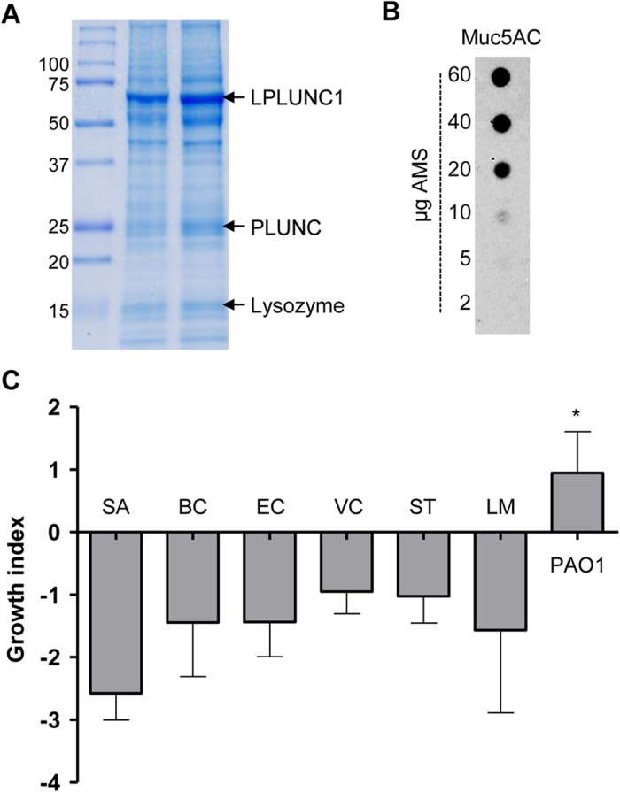
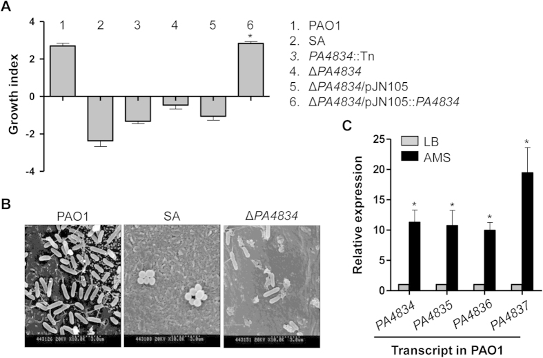
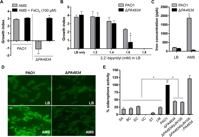
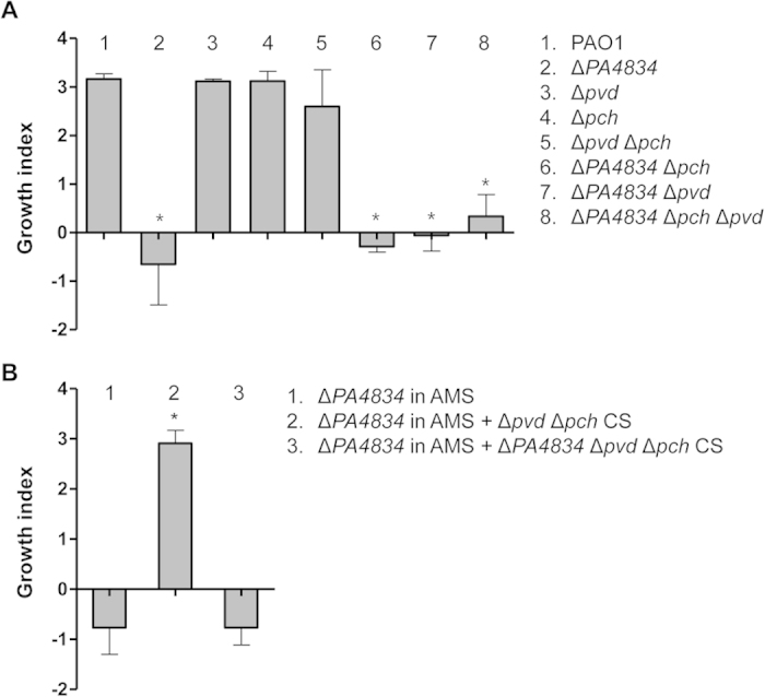
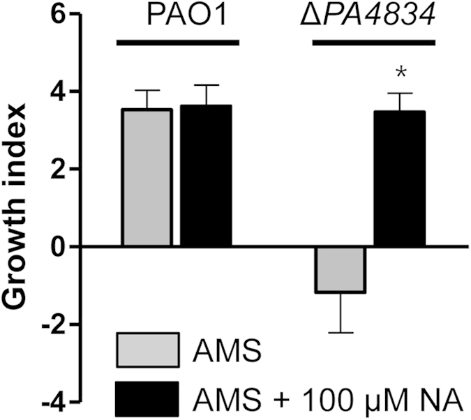
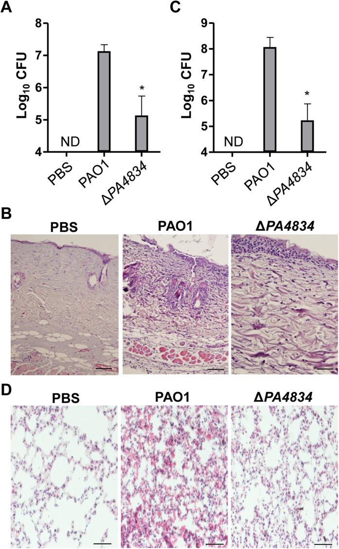
Pseudomonas aeruginosa establishes airway infections in Cystic Fibrosis patients. Here, we investigate the molecular interactions between P. aeruginosa and airway mucus secretions (AMS) derived from the primary cultures of normal human tracheal epithelial (NHTE) cells. PAO1, a prototype strain of P. aeruginosa, was capable of proliferating during incubation with AMS, while all other tested bacterial species perished. A PAO1 mutant lacking PA4834 gene became susceptible to AMS treatment. The ΔPA4834 mutant was grown in AMS supplemented with 100 μM ferric iron, suggesting that the PA4834 gene product is involved in iron metabolism. Consistently, intracellular iron content was decreased in the mutant, but not in PAO1 after the AMS treatment. Importantly, a PAO1 mutant unable to produce both pyoverdine and pyochelin remained viable, suggesting that these two major siderophore molecules are dispensable for maintaining viability during incubation with AMS. The ΔPA4834 mutant was regrown in AMS amended with 100 μM nicotianamine, a phytosiderophore whose production is predicted to be mediated by the PA4836 gene. Infectivity of the ΔPA4834 mutant was also significantly compromised in vivo. Together, our results identify a genetic element encoding a novel iron acquisition system that plays a previously undiscovered role in P. aeruginosa airway infection.


Figures

Figure 1
Figure 1. Preparation of AMS and robust ability of P. aeruginosa to grow in AMS.
(A) SDS-PAGE analysis of two independently prepared AMS samples. Ten μg of each sample was loaded into 12% SDS-PAGE and three distinct bands were identified by Mass Spectrometry. (B) Dot-blot analysis of Muc5AC protein. AMS samples with indicated protein amount were immobilized in the nitrocellulose membrane and probed with anti-Muc5AC antibody. (C) Seven different bacterial strains (SA, Staphylococcus aureus; BC, Bacillus cereus; EC, Escherichia coli; VC, Vibrio cholerae; ST, Salmonella enterica serovar Typhimurium; LM, Listeria monocytogenes; and PAO1, P. aeruginosa) were incubated with AMS prepared from primary cultures of normal human tracheal epithelial cells for 16 h. Changes in bacterial cell viability were monitored by calculating the growth index as described in the Materials and Methods. Three independent experiments were performed, and the mean values ± SD (error bars) are displayed in each bar. *p < 0.05 vs. other species, as determined by ANOVA.
Figure 2
Figure 2. A PAO1 mutant devoid of the PA4834 gene lost its ability to grow in AMS.
(A) Bacterial strains indicated by numbers were grown in AMS, and the growth index for each strain was measured. Experimental conditions were identical to those described in Fig. 1C. Six independent experiments were performed, and mean values ± SD (error bars) are displayed in each bar. SA, S. aureus. *p < 0.05 vs. growth indices of PA4834::Tn, ΔPA4834, and ΔPA4834/pJN105, as determined by students’ t-test. (B) Scanning electron microscope of primary cultured NHTE cells infected with PAO1, SA and ΔPA4834 mutant. The images were acquired at a magnification of 10,000. (C) Quantitative real-time PCR was performed to assess PA4834, PA4835, PA4836, and PA4837 gene transcript levels. PAO1 cells were grown in LB (gray bars) or AMS (black bars) before RNA extraction. Transcript levels of the tested genes indicated at the bottom were normalized with those of the rpoD gene transcript. Three independent experiments were performed, and mean values ± SD are displayed in each bar. *p < 0.05 vs. expression levels of each gene in LB-grown PAO1, as determined by students’ t-test.
Figure 3
Figure 3. The PA4834 gene product plays a role in iron acquisition.
(A) Effect of ferric iron on growth of the ΔPA4834 mutant in AMS. Growth indices of PAO1 and the ΔPA4834 mutant were measured after growth in AMS without (grays bars) or with (black bars) 100 μM FeCl3. Six independent experiments were performed, and mean values ± SD (error bars) are displayed in each bar. *p < 0.05 (students’ t-test) vs. growth of the ΔPA4834 mutant in AMS without FeCl3. (B) Effect of an iron chelator on bacterial growth. Growth indices of PAO1 (gray bars) and the ΔPA4834 mutant (black bars) were calculated after growth in LB supplemented with increasing concentrations of 2, 2′-bipyridyl. Bacterial cells were grown for 12 h. Three independent experiments were performed, and mean values ± SD (error bars) are displayed in each bar. *p < 0.05 (students’ t-test) vs. PAO1 growth in the same media. (C) Intracellular iron concentration of PAO1 (gray bars) and the ΔPA4834 mutant (black bars) after growth in LB or in AMS. Inductively coupled plasma mass spectrometry (ICP-MS) was used for the measurement and iron concentrations are displayed in parts per billion (ppb). Three independent experiments were performed, and mean values ± SD (error bars) are displayed in each bar. *p < 0.05 (students’ t-test) vs. all other values. (D) Fluorescent microscope images of PAO1 and the ΔPA4834 mutant. After growth in LB or AMS, bacterial suspensions were stained with PhenGreen™ SK, diacetate. Experiments were repeated three times and representative images, processed at the same magnification, are shown. (E) Bacterial strains indicated at the bottom were grown in LB for 16 h and cell-free culture supernatants were subject to the iron sequestration assay. The iron sequestration activities of indicated species were normalized with that of PAO1 (black bar). SA, Staphylococcus aureus; BC, Bacillus cereus; EC, Escherichia coli; VC, Vibrio cholerae; ST, Salmonella enterica serovar Typhimurium; LM, Listeria monocytogenes. Three independent experiments were performed, and mean values ± SD (error bars) are displayed in each bar. *p < 0.05 (students’ t-test) vs. all other strains but ΔPA4834/pJN105::PA4834.
Figure 4
Figure 4. Growth of PAO1 pyochelin and pyoverdine mutants in AMS.
(A) Bacterial strains indicated by numbers were grown in AMS for 16 h and the growth index of each strain was measured. Experimental conditions were identical to those described in Fig. 1C. Three independent experiments were performed, and mean values ± SD (error bars) are displayed in each bar. *p < 0.05 vs. growth indices of PAO1, Δpvd, Δpch, and ΔpvdΔpch, as determined by students’ t-test. (B) Effect of bacterial culture supernatant (CS) on ΔPA4834 mutant growth in AMS. The ΔPA4834 mutant was grown in AMS supplemented with the CS from the ΔpvdΔpch double or ΔPA4834ΔpvdΔpch triple mutant. CSs were obtained from bacterial cultures grown in LB and diluted 10-fold in AMS. Three independent experiments were performed, and mean values ± SD (error bars) are displayed in each bar. *p < 0.05 vs. growth of the ΔPA4834 mutant in AMS or AMS amended with the CS from the triple mutant, as determined by students’ t-test.
Figure 5
Figure 5. Effect of the addition of nicotianamine (NA) on ΔPA4834 mutant growth in AMS.
PAO1 and the ΔPA4834 mutant were grown in AMS (gray bars) and AMS amended with 100 μM NA (black bars) and growth indices were measured. Experimental conditions were identical to those described in Fig. 1C. Three independent experiments were performed, and mean values ± SD (error bars) are displayed in each bar. *p < 0.05 vs. growth of the ΔPA4834 mutant in AMS with no NA supplementation, as determined by students’ t-test.
Figure 6
Figure 6. In vivo infectivity of the ΔPA4834 mutant is decreased.
(A,B) Mouse burn wound infection. (A) PAO1 and the ΔPA4834 mutant were infected in the thermally-injured region of the mouse skin. The infection dose was 2 × 108 bacterial cells. After 24 h, the infected area was incised and homogenized to enumerate viable cells. Three independent experiments were performed, and mean values ± SD (error bars) are displayed in each bar. *p < 0.05 vs. CFU of PAO1, as determined by students’ t-test. ND, not detected. (B) Histological images of infected skin sections of mice challenged with PBS (negative control), PAO1, or the ΔPA4834 mutant. (C,D) Mouse airway infection. (C) PAO1 and the ΔPA4834 mutant (1 × 107 CFU) were exposed to the mouse airway via the intranasal route. After 24 h, mouse lungs were removed and homogenized to enumerate viable cells. Three independent experiments were performed, and mean values ± SD (error bars) are displayed in each bar. *p < 0.05 vs. CFU of PAO1, as determined by students’ t-test. ND, not detected. (D) Representative histological images of lung sections of mice challenged with PBS (negative control), PAO1, or the ΔPA4834 mutant.

Similar articles

Cited by

References

    1. Folkesson A. et al. Adaptation of Pseudomonas aeruginosa to the cystic fibrosis airway: an evolutionary perspective. Nat Rev Microbiol 10, 841–851, 10.1038/nrmicro2907 (2012). - DOI - PubMed
    1. Bassi G. L., Ferrer M., Marti J. D., Comaru T. & Torres A. Ventilator-associated pneumonia. Semin. Respir. Crit. Care Med. 35, 469–481, 10.1055/s-0034-1384752 (2014). - DOI - PubMed
    1. Azzopardi E. A. et al. Gram negative wound infection in hospitalised adult burn patients—systematic review and metanalysis. PLoS One 9, e95042, 10.1371/journal.pone.0095042 (2014). - DOI - PMC - PubMed
    1. Smith J. J., Travis S. M., Greenberg E. P. & Welsh M. J. Cystic fibrosis airway epithelia fail to kill bacteria because of abnormal airway surface fluid. Cell 85, 229–236 (1996). - PubMed
    1. Regnis J. A. et al. Mucociliary clearance in patients with cystic fibrosis and in normal subjects. Am. J. Respir. Crit. Care Med. 150, 66–71, 10.1164/ajrccm.150.1.8025774 (1994). - DOI - PubMed

Publication types

MeSH terms