. 2016 Feb;34(2):431-44.
doi: 10.1002/stem.2213. Epub 2015 Oct 23.

Definitive Hematopoiesis in the Yolk Sac Emerges from Wnt-Responsive Hemogenic Endothelium Independently of Circulation and Arterial Identity

Affiliations

Definitive Hematopoiesis in the Yolk Sac Emerges from Wnt-Responsive Hemogenic Endothelium Independently of Circulation and Arterial Identity

Jenna M Frame et al. Stem Cells. 2016 Feb.

Abstract

Adult-repopulating hematopoietic stem cells (HSCs) emerge in low numbers in the midgestation mouse embryo from a subset of arterial endothelium, through an endothelial-to-hematopoietic transition. HSC-producing arterial hemogenic endothelium relies on the establishment of embryonic blood flow and arterial identity, and requires β-catenin signaling. Specified prior to and during the formation of these initial HSCs are thousands of yolk sac-derived erythro-myeloid progenitors (EMPs). EMPs ensure embryonic survival prior to the establishment of a permanent hematopoietic system, and provide subsets of long-lived tissue macrophages. While an endothelial origin for these HSC-independent definitive progenitors is also accepted, the spatial location and temporal output of yolk sac hemogenic endothelium over developmental time remain undefined. We performed a spatiotemporal analysis of EMP emergence, and document the morphological steps of the endothelial-to-hematopoietic transition. Emergence of rounded EMPs from polygonal clusters of Kit(+) cells initiates prior to the establishment of arborized arterial and venous vasculature in the yolk sac. Interestingly, Kit(+) polygonal clusters are detected in both arterial and venous vessels after remodeling. To determine whether there are similar mechanisms regulating the specification of EMPs with other angiogenic signals regulating adult-repopulating HSCs, we investigated the role of embryonic blood flow and Wnt/β-catenin signaling during EMP emergence. In embryos lacking a functional circulation, rounded Kit(+) EMPs still fully emerge from unremodeled yolk sac vasculature. In contrast, canonical Wnt signaling appears to be a common mechanism regulating hematopoietic emergence from hemogenic endothelium. These data illustrate the heterogeneity in hematopoietic output and spatiotemporal regulation of primary embryonic hemogenic endothelium.

Keywords: Embryo; Endothelial cell; Hemangioblast; Hematopoiesis; Hematopoietic progenitors; Hematopoietic stem cells; Vascular development.


Conflict of interest statement

Disclosure of Potential Conflicts of Interest

The authors have no conflicts of interest.

Figures
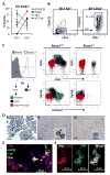
Figure 1
Figure 1. Emerging Kit+ EMPs progress from an endothelial to hematopoietic immunophenotype in a Runx1-dependent manner
A. The majority of E8.5 Kit+CD41+ EMPs are negative for CD16/32 and CD45, and express endothelial markers Flk1 and VE-Cadherin. By E9.5, a higher proportion of the Kit+CD41+ population expresses hematopoietic markers, with reduced endothelial marker expression. B. A significant portion of isolated E8.5 Kit+CD41+CD16/32neg cells upregulate CD16/32 after 16–20 hours of in vitro culture. Plots from one representative experiment are shown; population percentages are the average of 3 experiments +/− SEM. C. Kit+ cells (blue) arise in Runx1+/+ yolk sacs, but very few Kit+ events are detected in Runx1−/− yolk sacs. Kitneg populations of immunophenotypic megakaryocytes (KitnegCD41hiGp1bβ+, black) and macrophages (KitnegCD16/32+CD45hi, green), remain in Runx1−/− yolk sacs, albeit with significant depletion of immunophenotypic macrophages. Plots represent combined flow cytometric data from 4 E10.5 Runx1+/+ yolk sacs and 4 E10.5 Runx1−/− yolk sacs. D. Culture of sorted E9.5 and E10.5 wildtype hematopoietic populations confirm EMP progeny (i, Kit+CD41+CD16/32+), megakaryocyte potential (ii, KitnegCD41+Gp1bβ+), and macrophage potential (iii, KitnegCD16/32+CD45+). Images are of culture dishes (ii, iii) or Wright-Giemsa stained cytospins of cultures (i; ii and iii insets). n=2 pooled litters of yolk sacs. E. Immunohistochemical staining for CD41 demonstrates a significant overlap with Gp1bβ+CD41hi megakaryocytes in the E9.5 yolk sac, in addition to clusters of putative CD41+Gp1bβneg EMPs. EryP, primitive erythroblast autofluorescence. n=3 yolk sacs. F. Kit+ cluster cells are CD41+ and CD41lo/neg by immunohistochemistry. n=5 yolk sacs.
Figure 2
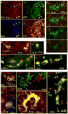
Figure 2. EMPs emerge from morphologic hemogenic endothelium between E8.5-E11
Immunohistochemistry at the stages indicated with indicated markers. A. Flattened and rounded Kit+ cells are detected at E8.5. Arrows indicate flattened hemogenic endothelial cells; arrowheads indicate rounded hematopoietic cells. B. Examples of flattened hemogenic endothelial cells at E9.5-E10. Bottom right has both Runx1+Kit+ rounded cells (arrowheads) and Runx1+Kitlo flattened cells (arrows). C. Examples of polygonal Kit+ clusters (open arrows) and rounded Kit+ cells (closed arrows) at E8.5 (6, 8 somite pairs (sp)), E9.5 (19, 22 sp), and E11 (41 sp). D (top right). Confocal Z-stacks through a polygonal Kit+ cluster at E9.5 indicates Kit+ cells are in the same plane of focus as the endothelium.
Figure 3
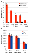
Figure 3. EMPs continually emerge over a broad developmental time and remain in the yolk sac circulation as late as E12
A. Quantification of immunohistochemical Kit+ clusters in the yolk sac (YS) over developmental time. n=3 for all timepoints except 39–40sp (E11) (n=4). Error bars indicate SEM. Significance was determined (10–49 cells; P = 4.81x 10⁻⁶) by one-way Analysis of Variance. All means (10–49 cells) are significantly different from one another (P < 0.05, Tukey-Kramer post-test) except: 27–28sp and 35–36sp; 35–36sp and 39–40sp. B. Flow cytometry indicates that robust numbers of Kit+ cells, many of which are Kit+CD41+CD16/32+ EMPs, remain in the yolk sac circulation through E11, and begin to decline by E12. n=3 20–22sp, n=5 32–40sp, n=4 48–51sp. Error bars indicate SEM. Significant differences within each fraction were determined by one-way Analysis of Variance: P = 0.012 (Kit+), P = 0.001 (EMP). All means are significantly different from one another (P < 0.05, Tukey-Kramer post-test) except 20–22sp and 32–40sp (both Kit+ and EMP).
Figure 4
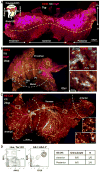
Figure 4. EMPs emerge along a proximal-distal gradient without anterior/posterior or arterial/venous preference
A–C. Immunohistochemistry at the stages indicated with indicated markers. A. Schematic represents an intact E8.5 embryo for clarity of anatomical locations within the flat-mounted yolk sac. EMP clusters are interspersed within the anterior and posterior regions of the blood island region of the proximal yolk sac (above the dotted line), where the primitive erythroblasts (EryP; purple) are also located. See Fig. S2 for images of each separate stain. B. By 14sp (E8.75), EryPs start to appear in the distal portion of the yolk sac (inside dotted circle), while EMP clusters remain in the proximal yolk sac (outside dotted circle). Blue asterisks in both B and C indicate background artifact; see Fig. S2. Boxed regions indicate clusters in venous (V) and arterial (A) regions (B, right; C, insets). C. At 21sp (E9.5), EMP clusters reside in both the arterial and venous regions of the yolk sac (approximated by dotted lines). Scale bar of insets: 50μm. The vitelline artery was identified based on distinct branching morphology[68], which was corroborated both with Sox17 labeling (see Fig. S3), and by identification of Kit+Runx1+ clusters in extensions of the artery (not shown). D. Anterior and posterior regions of the E8.5 yolk sac each produce GR-1+ granulocytes. B-lymphocyte potential is also evident. Flow cytometric analysis of one anterior culture shown at day 12.
Figure 5
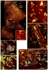
Figure 5. The endothelial-to-hematopoietic transition occurs without circulation, and in larger and smaller vessels after vascular remodeling
A. EMP clusters in the Ncx1−/− yolk sac are localized to the proximal region. Blue asterisks indicate background artifact from folded tissue. Scale bar: 500μm; inset: 50μm. B–C. Examples of polygonal Kit+CD31+Runx1+ clusters (B) and flattened Runx1+ hemogenic endothelial cells (C, arrows) in E9.5 Ncx1−/− yolk sacs. 3 Ncx1−/− yolk sacs were analyzed. D–F. At E9.5-10, flattened and polygonal Kit+ cells are present in larger remodeled vessels and the vascular plexus. (F) Flattened KitloRunx1+ cells are denoted by closed arrows, polygonal cells by open arrows.
Figure 6
Figure 6. Canonical Wnt signaling regulates EMP production in vitro and in vivo
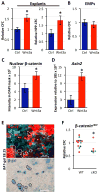
A. Increased production of CFCs and HPP-CFCs in explanted E8.5 yolk sacs with Wnt3a treatment. (Left) Methylcellulose culture (CFCs) of 6–7sp yolk sacs. n=10. (Right) HPP-CFC of 7–9sp yolk sacs (n=7 controls (Ctrl), n=8 Wnt3a treated). Error bars indicate SEM. *P <0.05, 2-tailed student’s t-test. B. No changes in CFC activity were observed with Wnt3a treatment of sorted E9.5-E10 Kit+CD41+CD16/32+ EMPs for 24 hours. n=5, error bars indicate SEM. C. Wnt3a treatment of E9.5 dissociated yolk sacs for 2 hours increased β-catenin staining intensity in the nuclear region of Kit+VEC/AA4.1+CD16/32neg cells. n=3, error bars indicate SEM. *P <0.01, 1-tailed student’s t-test. D. Treatment of sorted VEC/AA4.1+CD16/32neg cells with Wnt3a for 5 hours increased expression of β-catenin target gene Axin2. n=3. *P <0.05, 1-tailed student’s t-test. E. Immunohistochemistry and β-galactosidase (β-gal) staining of E8.25 BAT-gal yolk sac reveals a rare subset of VEC+Runx1+β-gal+ putative hemogenic endothelium (asterisks). 1 example of 4 yolk sacs with VEC+Runx1+β-gal+ staining is shown. Runx1negβ-gal+ cells (dotted lines) are also present. The majority of Runx1+ cells are β-galneg. For whole yolk sac images, see Fig. S4B. F. E9.5-E10.5 Cdh5-Cre; Ctnnb1fl/fl yolk sacs (cKO) have reduced CFCs compared with yolk sacs of Cdh5-Creneg littermates. Colors of data points represent separate litters of embryos (n=4 litters). *P =0.01, 1-tailed student’s t-test.
Figure 7
Figure 7. Summary of EMP specification in the murine yolk sac and comparison with HSC specification
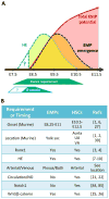
A. Temporal model of yolk sac-derived definitive hematopoiesis. Hematopoietic potential is initiated as a wave of EMP colony-forming activity over developmental time (red arrow with shaded area)[27]. Analysis of hemogenic endothelial-derived Kit+ cluster numbers indicates that EMPs cease to emerge in the yolk sac after E11.5 (black dotted line, orange shaded area). This wave of hematopoietic emergence is preceded by the appearance of flattened hemogenic endothelium (HE; green dotted line) from E8.5 through as late as E10.5-11, as determined in Figure 2. The observed formation of hemogenic endothelium correlates well with the reported requirement of Runx1 in mediating EMP formation through E10.5 (green trapezoid)[79]. Note that presence of hemogenic endothelium at early developmental stages (green triangle) is approximated and remains unknown. Endogenous canonical Wnt signaling in the yolk sac is robust at early stages (blue trapezoid), and increases EMP production from hemogenic endothelium at E8.5 (green dotted line) ex vivo, as determined in Figure 6. Together, these kinetics suggest that canonical Wnt signaling has an early role in regulating hematopoietic potential from hemogenic endothelium. B. Comparison of the timing of and requirements for EMP and HSC emergence. UA, umbilical artery; VA, vitelline artery; HE, hemogenic endothelium, NO; nitric oxide.

Similar articles

Cited by

References

    1. Muller AM, Medvinsky A, Strouboulis J, et al. Development of hematopoietic stem cell activity in the mouse embryo. Immunity. 1994;1:291–301. - PubMed
    1. Bruijn MFTR, de Speck NA, Peeters MCE, et al. Definitive hematopoietic stem cells first develop within the major arterial regions of the mouse embryo. EMBO J. 2000;19(11):2465–2474. - PMC - PubMed
    1. Wang Q, Stacy T, Binder M, et al. Disruption of the Cbfa2 gene causes necrosis and hemorrhaging in the central nervous system and blocks definitive hematopoiesis. Proc Natl Acad Sci. 1996;93(8):3444–3449. - PMC - PubMed
    1. Okuda T, Deursen J, van Hiebert SW, et al. AML1, the target of multiple chromosomal translocations in human leukemia, is essential for normal fetal liver hematopoiesis. Cell. 1996;84:321–330. - PubMed
    1. North T, Gu TL, Stacy T, et al. Cbfa2 is required for the formation of intra-aortic hematopoietic clusters. Development. 1999;126(11):2563–2575. - PubMed

Publication types