

PLZF Controls the Development of Fetal-Derived IL-17+Vγ6+ γδ T Cells
- PMID: 26408661
- PMCID: PMC4610873
- DOI: 10.4049/jimmunol.1500939
PLZF Controls the Development of Fetal-Derived IL-17+Vγ6+ γδ T Cells
Abstract
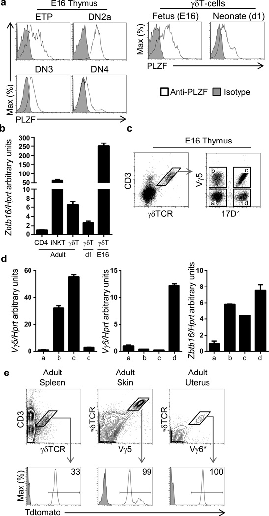
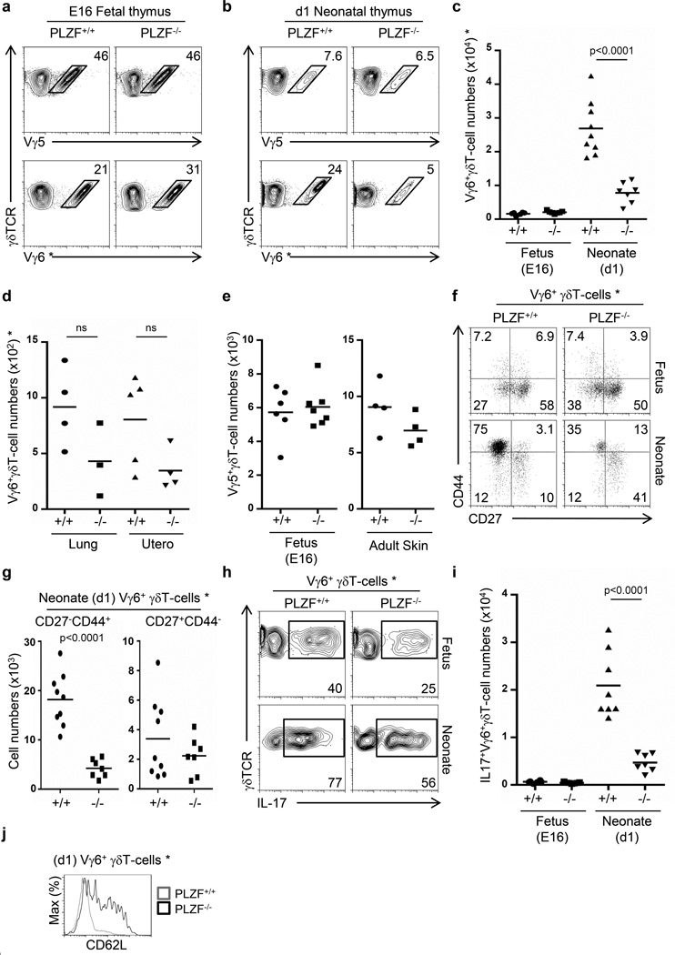
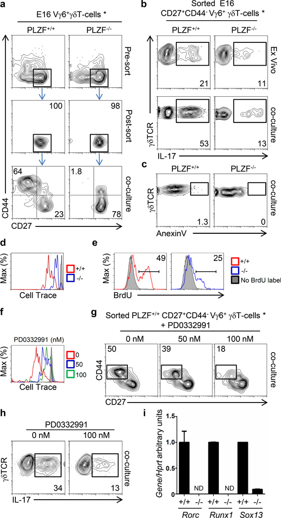
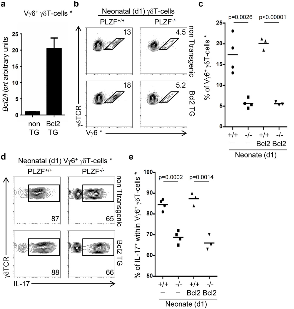
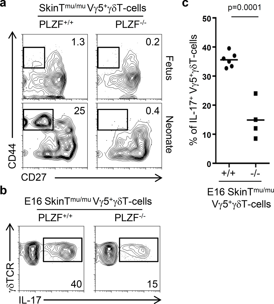
Expression of promyelocytic leukemia zinc finger (PLZF) protein directs the effector differentiation of invariant NKT (iNKT) cells and IL-4(+) γδ NKT cells. In this study, we show that PLZF is also required for the development and function of IL-17(+) γδ T cells. We observed that PLZF is expressed in fetal-derived invariant Vγ5(+) and Vγ6(+) γδ T cells, which secrete IFN-γ and IL-17, respectively. PLZF deficiency specifically affected the effector differentiation of Vγ6(+) cells, leading to reduced numbers of mature CD27(-)CD44(+) phenotype capable of secreting IL-17. Although PLZF was not required for Vγ5(+) γδ T cells to develop, when these cells were reprogrammed into IL-17-secreting cells in Skint-1 mutant mice, they required PLZF for their effector maturation, similarly to Vγ6(+) γδ T cells. The impaired effector differentiation of PLZF-deficient Vγ6(+) γδ T cells was not due to increased apoptosis and it was related to reduced proliferation of immature CD27(+)CD44(-) Vγ6(+) γδ T cells, which was required for their differentiation into mature CD27(-)CD44(+) IL-17-secreting cells. Thus, the present study identifies that PLZF function is not restricted to NKT or IL-4(+) T cells, but it also controls the development of IL-17(+) γδ T cells.
Figures
Similar articles
-
TCR-inducible PLZF transcription factor required for innate phenotype of a subset of gammadelta T cells with restricted TCR diversity.Proc Natl Acad Sci U S A. 2009 Jul 28;106(30):12453-8. doi: 10.1073/pnas.0903895106. Epub 2009 Jul 15. Proc Natl Acad Sci U S A. 2009. PMID: 19617548 Free PMC article.
-
Innate PLZF+CD4+ αβ T cells develop and expand in the absence of Itk.J Immunol. 2014 Jul 15;193(2):673-87. doi: 10.4049/jimmunol.1302058. Epub 2014 Jun 13. J Immunol. 2014. PMID: 24928994 Free PMC article.
-
Identification of CD25+ gamma delta T cells as fetal thymus-derived naturally occurring IL-17 producers.J Immunol. 2008 Nov 1;181(9):5940-7. doi: 10.4049/jimmunol.181.9.5940. J Immunol. 2008. PMID: 18941182
-
Development of PLZF-expressing innate T cells.Curr Opin Immunol. 2011 Apr;23(2):220-7. doi: 10.1016/j.coi.2010.12.016. Epub 2011 Jan 21. Curr Opin Immunol. 2011. PMID: 21257299 Free PMC article. Review.
-
Thymic maturation determines gammadelta T cell function, but not their antigen specificities.Curr Opin Immunol. 2009 Apr;21(2):140-5. doi: 10.1016/j.coi.2009.02.008. Epub 2009 Mar 25. Curr Opin Immunol. 2009. PMID: 19321327 Free PMC article. Review.
Cited by
-
Role of innate T cells in necrotizing enterocolitis.Front Immunol. 2024 Feb 8;15:1357483. doi: 10.3389/fimmu.2024.1357483. eCollection 2024. Front Immunol. 2024. PMID: 38390341 Free PMC article. Review.
-
MAITs and their mates: "Innate-like" behaviors in conventional and unconventional T cells.Clin Exp Immunol. 2023 Jul 5;213(1):1-9. doi: 10.1093/cei/uxad058. Clin Exp Immunol. 2023. PMID: 37256718 Free PMC article.
-
Novel Insight Into the Molecular and Metabolic Mechanisms Orchestrating IL-17 Production in γδ T Cells.Front Immunol. 2019 Dec 3;10:2828. doi: 10.3389/fimmu.2019.02828. eCollection 2019. Front Immunol. 2019. PMID: 31849992 Free PMC article. Review.
-
γδ T, NKT, and MAIT Cells During Evolution: Redundancy or Specialized Functions?J Immunol. 2022 Jul 15;209(2):217-225. doi: 10.4049/jimmunol.2200105. J Immunol. 2022. PMID: 35821101 Free PMC article. Review.
-
Commensal Microbiota Promote Lung Cancer Development via γδ T Cells.Cell. 2019 Feb 21;176(5):998-1013.e16. doi: 10.1016/j.cell.2018.12.040. Epub 2019 Jan 31. Cell. 2019. PMID: 30712876 Free PMC article.
References
-
- Kovalovsky D, Uche OU, Eladad S, Hobbs RM, Yi W, Alonzo E, Chua K, Eidson M, Kim HJ, Im JS, Pandolfi PP, Sant'Angelo DB. The BTB-zinc finger transcriptional regulator PLZF controls the development of invariant natural killer T cell effector functions. Nature immunology. 2008;9:1055–1064. - PMC - PubMed
-
- Kreslavsky T, Savage AK, Hobbs R, Gounari F, Bronson R, Pereira P, Pandolfi PP, Bendelac A, von Boehmer H. TCR-inducible PLZF transcription factor required for innate phenotype of a subset of gammadelta T cells with restricted TCR diversity. Proceedings of the National Academy of Sciences of the United States of America. 2009;106:12453–12458. - PMC - PubMed
Publication types
MeSH terms
Substances
Grants and funding
LinkOut - more resources
Full Text Sources
Other Literature Sources
Molecular Biology Databases
Research Materials
Miscellaneous