. 2015 Sep 24:6:8395.
doi: 10.1038/ncomms9395.

STIM1 dimers undergo unimolecular coupling to activate Orai1 channels

Affiliations

STIM1 dimers undergo unimolecular coupling to activate Orai1 channels

Yandong Zhou et al. Nat Commun. .

Abstract

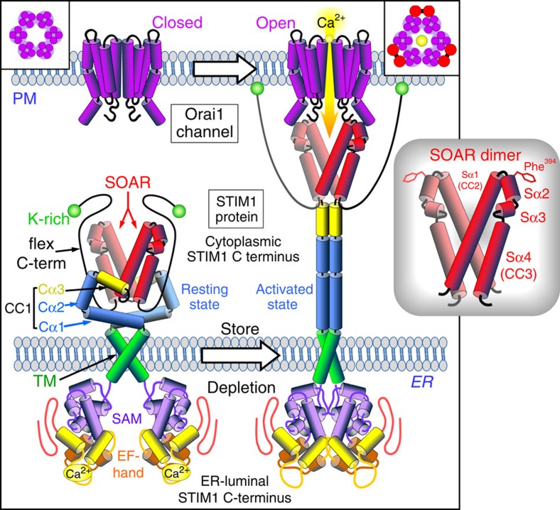
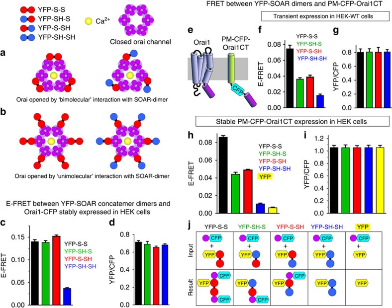
The endoplasmic reticulum (ER) Ca(2+) sensor, STIM1, becomes activated when ER-stored Ca(2+) is depleted and translocates into ER-plasma membrane junctions where it tethers and activates Orai1 Ca(2+) entry channels. The dimeric STIM1 protein contains a small STIM-Orai-activating region (SOAR)--the minimal sequence sufficient to activate Orai1 channels. Since SOAR itself is a dimer, we constructed SOAR concatemer-dimers and introduced mutations at F394, which is critical for Orai1 coupling and activation. The F394H mutation in both SOAR monomers completely blocks dimer function, but F394H introduced in only one of the dimeric SOAR monomers has no effect on Orai1 binding or activation. This reveals an unexpected unimolecular coupling between STIM1 and Orai1 and argues against recent evidence suggesting dimeric interaction between STIM1 and two adjacent Orai1 channel subunits. The model predicts that STIM1 dimers may be involved in crosslinking between Orai1 channels with implications for the kinetics and localization of Orai1 channel opening.


Figures

Figure 1
Figure 1. Molecular model of the activation and coupling of STIM1 to activate the Orai1 channel.
The dimeric, single transmembrane-spanning STIM1 ER protein senses ER luminal Ca2+ change through its N-terminal complex of EF hands and sterile-α motif (SAM). The STIM1 cytoplasmic C terminus is a largely helical complex including the CC1 region, the STIM-Orai-activating region (SOAR), and a flexible C terminus ending with a K-rich region. In the STIM1-resting state, the EF hand/SAM domains are separated, and the SOAR region is occluded within a folded complex of SOAR, CC1 and the flexible C terminus. On luminal Ca2+ store depletion, Ca2+ dissociation from the EF hand/SAM domains allows the N termini to associate forcing a conformational change in the STIM1 C terminus mediated through rearrangement of the TM domains. The STIM1 C terminus undergoes unfolding and elongation through dissociation between the CC1 and SOAR regions. The extended, unfolded C terminus allows the K-rich C termini of STIM1 to bind to the PM, and also exposes SOAR in this extended configuration allowing it to associate with PM Orai1 channels within closely membrane-associated ER–PM junctions. Orai1 is a four transmembrane-spanning PM protein, forming hexameric Ca2+ channels (box, left). Orai1 channels can be tethered within ER–PM junctions by the extended, exposed SOAR unit of activated STIM1. SOAR binding to Orai1 induces gating of the channel to allow Ca2+ entry. On the basis of recent information, the model depicts interaction between the SOAR dimer and two adjacent Orai1 subunits of the channel hexamer (box, top right). A more detailed diagram of the molecular organization of the SOAR dimer (inset, right) is from the crystal structure. Each monomer has four α-helices, interacting together through hydrophobic and hydrogen bonding at three interfaces. The two larger helices, Sα1 and Sα4 (also known as CC2 and CC3, respectively), flank two smaller helices (Sα2 and Sα3). The apical Sα2 helix includes an exposed residue, phenylalanine-394, shown to be critical to the binding and gating of Orai1 channels by STIM1 (ref. 20).
Figure 2
Figure 2. The F394H mutation in SOAR prevents its binding to and gating of Orai1 channels without altering its size or ability to dimerize.
SOAR constructs were transiently expressed in HEK-Orai1-CFP stable cells. The YFP-labelled wild-type SOAR protein (YFP-SOAR) is exclusively PM localized where its distribution is superimposable with Orai1 (ac). In contrast, the YFP-tagged F394H mutant (YFP-SOAR-H) is exclusively cytosolic (df). (g) Expression of YFP-SOAR and YFP-SOAR-H detected with GFP antibody. (h) Average YFP fluorescence intensity (arbitrary fluorescence units, AFUs) of cells shown in i±s.e.m. (i) Fura-2 ratiometric Ca2+ responses in HEK-Orai1-CFP cells transiently expressing similar levels of YFP-SOAR (n=32) or YFP-SOAR-H (n=8). Results are means±s.e.m. and representative of three independent repeats. Constitutive Ca2+ entry was measured in nominally Ca2+-free medium and after addition of 1 mM Ca2+ (arrow). (j) Whole-cell patch-clamp recording of ICRAC in HEK-Orai1-CFP cells transfected with YFP-SOAR or YFP-SOAR-H. Bath solutions contained 20 mM Ca2+ and cytosolic-free Ca2+ was maintained at 90 nM. (k) IV relationship from cells shown in j. (l) Size-exclusion chromatogram of purified wild-type SOAR (red) and F394H SOAR mutant (blue), showing both elute from a Superdex-200 16/60 column as a single peak at ∼245 ml, corresponding to 25 kDa (monomeric SOAR, is 12 kDa). Protein standards (grey) correspond to molecular weights of (i) 670, (ii) 158, (iii) 44, (iv) 17 and (v) 1.3 kDa. (m) Chemical crosslinking of purified SOAR subunits with DSS. Coomassie blue-stained SDS–PAGE of SOAR wild-type (left) and SOAR-F394H (right) after incubation with 0, 0.1 and 1 mM DSS at room temperature for 30 min. M: protein marker.
Figure 3
Figure 3. Expression, distribution and interactions of SOAR monomers and SOAR concatemer–dimers.
(a) Summary of E-FRET values between YFP-SOAR and CFP-SOAR (red, n=21), or YFP-SOAR-H and CFP-SOAR (blue, n=45), or YFP-SOAR-H and CFP (green, n=14), transiently co-expressed in HEK-Orai1-His stable cells. Analysis was restricted to the near-PM area. (b) YFP/CFP ratios of the near-PM-expressed proteins in the cells used for E-FRET in a. Results are means±s.e.m. of three independent repeats. (ce) YFP-SOAR-H and CFP-SOAR co-transfected in HEK-Orai1-His cells. The view shows three cells all expressing YFP-SOAR-H (c), but only two cells co-expressing CFP-SOAR (d). The merged image (e) reveals some YFP-SOAR-H localized to the PM in the two cells co-expressing CFP-SOAR; YFP-SOAR-H remains cytosolic in the cell not co-expressing CFP-SOAR. (f) Western analysis of the four SOAR concatemer–dimers shown, transiently expressed in stable HEK-Orai1-His using an anti-GFP antibody. (gj) Distribution of the same four SOAR YFP-tagged concatemer–dimers transiently expressed in stable HEK-Orai1-CFP cells: (g) wild-type dimer YFP-S-S, (h,i) heterodimer mutants YFP-SH-S and YFP-S-SH, and (j) the homo-mutant dimer YFP-SH-SH; dimers are defined in Supplementary Fig. 1. Distribution of Orai1-CFP in the same cells is shown in Supplementary Fig. 2a–h. (kn) Distribution of the same four concatemer–dimers expressed this time in HEK-WT cells. (oq) Co-transfection of the wild-type YFP-S-S concatemer–dimer with the double-mutant CFP-SH-SH concatemer–dimer in HEK-Orai1-His cells.
Figure 4
Figure 4. The three SOAR concatemer–dimers, YFP-S-S, YFP-SH-S and YFP-S-SH are functionally identical.
Fura-2 ratiometric Ca2+ responses in stable HEK-Orai1-CFP cells transiently expressing the same levels of either: (a) YFP-S-S (43 cells); (b) YFP-SH-S (50 cells); (c) YFP-S-SH (49 cells); (d) YFP-SH-SH (48 cells). Constitutive Ca2+ entry was measured using cells in nominally Ca2+-free solution, replaced with 1 mM Ca2+ solution (Ca2+). (ad) Results are means of the cell numbers indicated±s.e.m. and are representative of three independent repeats. (e) Statistics for average peak of constitutive Ca2+ entry shown in ad ,*P<0.001 from YFP-S-S; (f) statistics for Ca2+ entry rates shown in ad, *P<0.001 from YFP-S-S; (g) average YFP fluorescence intensity (arbitrary fluorescence units; AFUs) of cells used in ad). (eg) Results are means±s.e.m. of three independent experiments. (h) I/V relationship of whole-cell ICRAC measurements for YFP-S-S (black), YFP-SH-S (green), YFP-S-SH (red) or YFP-SH-SH (blue) transiently expressed in HEK-Orai1-CFP stable cells. Quantification of current densities for the three active concatemer–dimers are given in Supplementary Fig. 3.
Figure 5
Figure 5. Evidence for a unimolecular interaction between the SOAR dimer and Orai1.
Two possible models for SOAR–Orai1 interactions: (a) bimolecular binding of a homomeric SOAR dimer (red) to two adjacent Orai1 subunits is required to open the hexameric Orai1 channel (purple). (b) Unimolecular binding of a single monomer within the SOAR dimer (red) to a single Orai1 subunit is sufficient to open the hexameric Orai1 channel (purple). Changes in the association of heterodimers of SOAR comprising one WT and one F394H monomer (red/blue) occurring in the bimolecular (a, right) and unimolecular (b, right) models. (c) Near-PM values of E-FRET between Orai1-CFP in HEK-Orai1-CFP stable cells, and transiently expressed YFP-S-S (black; n=141), YFP-SH-S (green, n=243), YFP-S-SH (red, n=213) or YFP-SH-SH (blue, n=206). (d) YFP/CFP ratios of the near-PM-expressed Orai1-CFP and YFP dimers in the cells used for E-FRET in c. (e) Cartoon of Orai1 protein (left) and PM-CFP-Orai1CT construct (right) comprising C terminus of Orai1 (267–301) attached to CFP and a single PM transmembrane-spanning helix. (f) Near-PM E-FRET between PM-CFP-Orai1CT transiently expressed in HEK-WT cells and transiently expressed SOAR concatemer–dimers: YFP-S-S (black; n=51); YFP-SH-S (green; n=55); YFP-S-SH (red; n=39); and YFP-SH-SH (blue; n=85). (g) YFP/CFP ratios of near-PM-expressed Orai1-CFP and YFP dimers in the cells used (f). (h) Near-PM E-FRET between PM-CFP-Orai1CT stably expressed HEK cells and transiently expressed SOAR concatemer–dimers: YFP-S-S (black; n=141); YFP-SH-S (green; n=137); YFP-S-SH (red; n=389); YFP-SH-SH (blue; n=202); and YFP alone (yellow; n=202). (i) YFP/CFP ratios of near-PM-expressed Orai1-CFP and YFP dimers in the cells used in h. In all cases (c–i), results are means±s.e.m. of three independent experiments. (j) Interpretation of interactions between CFP-tagged Orai1CT (purple) and each of the four YFP-tagged SOAR dimer–concatemers (or YFP alone) used in the E-FRET experiments shown in h. Top row shows input reactants for each condition; bottom row shows resulting interactions. Each WT SOAR monomer (red) of the SOAR dimers is shown to independently bind a single Orai1CT molecule. F394H SOAR monomers (blue) do not bind Orai1CT.

Similar articles

Cited by

References

    1. Soboloff J., Rothberg B. S., Madesh M. & Gill D. L. STIM proteins: dynamic calcium signal transducers. Nat. Rev. Mol. Cell Biol. 13, 549–565 (2012). - PMC - PubMed
    1. Kar P., Nelson C. & Parekh A. B. CRAC Channels drive digital activation and provide analog control and synergy to Ca2+-dependent gene regulation. Curr. Biol. 22, 242–247 (2012). - PubMed
    1. Amcheslavsky A. et al. Molecular biophysics of Orai store-operated Ca channels. Biophys. J. 108, 237–246 (2015). - PMC - PubMed
    1. Shim A. H., Tirado-Lee L. & Prakriya M. Structural and functional mechanisms of CRAC channel regulation. J. Mol. Biol. 427, 77–93 (2015). - PMC - PubMed
    1. Rothberg B. S., Wang Y. & Gill D. L. Orai channel pore properties and gating by STIM: implications from the Orai crystal structure. Sci. Signal. 6, pe9 (2013). - PMC - PubMed

Publication types

MeSH terms

LinkOut - more resources