

Exosome Biogenesis, Regulation, and Function in Viral Infection
- PMID: 26393640
- PMCID: PMC4584306
- DOI: 10.3390/v7092862
Exosome Biogenesis, Regulation, and Function in Viral Infection
Abstract
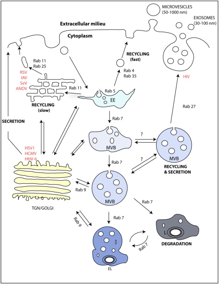
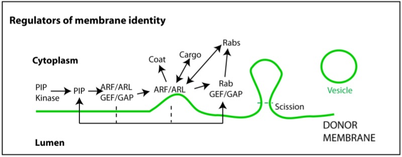
Exosomes are extracellular vesicles released upon fusion of multivesicular bodies(MVBs) with the cellular plasma membrane. They originate as intraluminal vesicles (ILVs) during the process of MVB formation. Exosomes were shown to contain selectively sorted functional proteins, lipids, and RNAs, mediating cell-to-cell communications and hence playing a role in the physiology of the healthy and diseased organism. Challenges in the field include the identification of mechanisms sustaining packaging of membrane-bound and soluble material to these vesicles and the understanding of the underlying processes directing MVBs for degradation or fusion with the plasma membrane. The investigation into the formation and roles of exosomes in viral infection is in its early years. Although still controversial, exosomes can, in principle, incorporate any functional factor, provided they have an appropriate sorting signal, and thus are prone to viral exploitation.This review initially focuses on the composition and biogenesis of exosomes. It then explores the regulatory mechanisms underlying their biogenesis. Exosomes are part of the endocytic system,which is tightly regulated and able to respond to several stimuli that lead to alterations in the composition of its sub-compartments. We discuss the current knowledge of how these changes affect exosomal release. We then summarize how different viruses exploit specific proteins of endocytic sub-compartments and speculate that it could interfere with exosome function, although no direct link between viral usage of the endocytic system and exosome release has yet been reported. Many recent reports have ascribed functions to exosomes released from cells infected with a variety of animal viruses, including viral spread, host immunity, and manipulation of the microenvironment, which are discussed. Given the ever-growing roles and importance of exosomes in viral infections, understanding what regulates their composition and levels, and defining their functions will ultimately provide additional insights into the virulence and persistence of infections.
Keywords: biological sciences; endocytic pathways; exosomes; immunity; mechanisms of viral spread; microbiology; virology.
Figures
Similar articles
-
Mechanisms of RNA loading into exosomes.FEBS Lett. 2015 Jun 4;589(13):1391-8. doi: 10.1016/j.febslet.2015.04.036. Epub 2015 Apr 30. FEBS Lett. 2015. PMID: 25937124 Review.
-
Exosome Biogenesis and Biological Function in Response to Viral Infections.Open Virol J. 2018 Sep 28;12:134-148. doi: 10.2174/1874357901812010134. eCollection 2018. Open Virol J. 2018. PMID: 30416610 Free PMC article. Review.
-
Viral miRNAs exploiting the endosomal-exosomal pathway for intercellular cross-talk and immune evasion.Biochim Biophys Acta. 2011 Nov-Dec;1809(11-12):715-21. doi: 10.1016/j.bbagrm.2011.08.002. Epub 2011 Aug 9. Biochim Biophys Acta. 2011. PMID: 21855666
-
To be or not to be... secreted as exosomes, a balance finely tuned by the mechanisms of biogenesis.Essays Biochem. 2018 May 15;62(2):177-191. doi: 10.1042/EBC20170076. Print 2018 May 15. Essays Biochem. 2018. PMID: 29717057 Review.
-
The Small GTPase Ral orchestrates MVB biogenesis and exosome secretion.Small GTPases. 2018 Nov 2;9(6):445-451. doi: 10.1080/21541248.2016.1251378. Epub 2016 Nov 22. Small GTPases. 2018. PMID: 27875100 Free PMC article.
Cited by
-
Serum proteomics reveals a tolerant immune phenotype across multiple pathogen taxa in wild vampire bats.Front Immunol. 2023 Dec 12;14:1281732. doi: 10.3389/fimmu.2023.1281732. eCollection 2023. Front Immunol. 2023. PMID: 38193073 Free PMC article.
-
Research progress on the mechanism of exosome-mediated virus infection.Front Cell Infect Microbiol. 2024 Jun 26;14:1418168. doi: 10.3389/fcimb.2024.1418168. eCollection 2024. Front Cell Infect Microbiol. 2024. PMID: 38988816 Free PMC article. Review.
-
Exosomes mediate hepatitis B virus (HBV) transmission and NK-cell dysfunction.Cell Mol Immunol. 2017 May;14(5):465-475. doi: 10.1038/cmi.2016.24. Epub 2016 May 30. Cell Mol Immunol. 2017. PMID: 27238466 Free PMC article.
-
Composition, Biogenesis, and Role of Exosomes in Tumor Development.Stem Cells Int. 2022 Sep 9;2022:8392509. doi: 10.1155/2022/8392509. eCollection 2022. Stem Cells Int. 2022. PMID: 36117723 Free PMC article. Review.
-
Extracellular Vesicle Activation of Latent HIV-1 Is Driven by EV-Associated c-Src and Cellular SRC-1 via the PI3K/AKT/mTOR Pathway.Viruses. 2020 Jun 19;12(6):665. doi: 10.3390/v12060665. Viruses. 2020. PMID: 32575590 Free PMC article.
References
Publication types
MeSH terms
LinkOut - more resources
Full Text Sources
Other Literature Sources
Medical
