. 2015 Sep 18;20(9):17288-308.
doi: 10.3390/molecules200917288.

Effect of Amaranthus on Advanced Glycation End-Products Induced Cytotoxicity and Proinflammatory Cytokine Gene Expression in SH-SY5Y Cells

Affiliations

Effect of Amaranthus on Advanced Glycation End-Products Induced Cytotoxicity and Proinflammatory Cytokine Gene Expression in SH-SY5Y Cells

Warisa Amornrit et al. Molecules. .

Abstract

Amaranthus plants, or spinach, are used extensively as a vegetable and are known to possess medicinal properties. Neuroinflammation and oxidative stress play a major role in the pathogenesis of many neurodegenerative diseases, such as Alzheimer's disease and Parkinson's disease. Advanced glycation end-products (AGEs) cause cell toxicity in the human neuronal cell line, SH-SY5Y, through an increase in oxidative stress, as shown by reducing cell viability and increasing cell toxicity in a dose-dependent manner. We found that preincubation of SH-SY5Y cells with either petroleum ether, dichloromethane or methanol extracts of A. lividus and A. tricolor dose-dependently attenuated the neuron toxicity caused by AGEs treatment. Moreover, the results showed that A. lividus and A. tricolor extracts significantly downregulated the gene expression of the pro-inflammatory cytokines, TNF-α, IL-1 and IL-6 genes in AGEs-induced cells. We concluded that A. lividus and A. tricolor extracts not only have a neuroprotective effect against AGEs toxicity, but also have anti-inflammatory activity by reducing pro-inflammatory cytokine gene expression. This suggests that Amaranthus may be useful for treating chronic inflammation associated with neurodegenerative disorders.

Keywords: AGEs; Amaranthus; SH-SY5Y cells; oxidative stress; proinflammatory cytokines.


Conflict of interest statement

The authors declare no conflict of interest.

Figures

Figure 1
Figure 1
Amaranthus lividus L. (A) and Amaranthus tricolor L. (B).
Figure 2
Figure 2
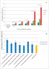
A. lividus L. and A. tricolor L. extracts have an impact on the cell viability of SH-SY5Y cells. SH-SY5Y cells were incubated with different concentrations of A. lividus L. and A. tricolor L. extracts (0–1000 μg/mL) for 24 h. The cell viability of living SH-SY5Y cells was assessed using the MTT assay. (A) A. lividus L. and (B) A. tricolor L. extracts. Values are reported as the means with their standard error of the mean (SEM), depicted by vertical bars. All experiments were performed in triplicate (N = 3). * p < 0.05 for a significant change as compared to untreated control cells.
Figure 2
Figure 2
A. lividus L. and A. tricolor L. extracts have an impact on the cell viability of SH-SY5Y cells. SH-SY5Y cells were incubated with different concentrations of A. lividus L. and A. tricolor L. extracts (0–1000 μg/mL) for 24 h. The cell viability of living SH-SY5Y cells was assessed using the MTT assay. (A) A. lividus L. and (B) A. tricolor L. extracts. Values are reported as the means with their standard error of the mean (SEM), depicted by vertical bars. All experiments were performed in triplicate (N = 3). * p < 0.05 for a significant change as compared to untreated control cells.
Figure 3
Figure 3
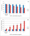
Advanced glycation end-products (AGEs) have an effect on the cell toxicity of SH-SY5Y cells. SH-SY5Y cells were incubated with different concentrations of AGEs (0–4 mg/mL) for 24–48 h. (A) The cell survival of living SH-SY5Y cells was assessed using the trypan blue exclusion assay. (B) The release of LDH of damaged SH-SY5Y cells was assessed using the LDH assay. Values are reported as the means with their standard error of the mean (SEM), depicted by vertical bars. All experiments were performed in triplicate (N = 3). * p < 0.05 for a significant change as compared to untreated control cells.
Figure 4
Figure 4
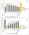
Protective effect of A. lividus L. extracts on AGEs-induced cytotoxicity in SH-SY5Y cells. SH-SY5Y cells were preincubated with A. lividus L. for 24 h and then further incubated with AGEs for 48 h. (A) The cell survival of living SH-SY5Y cells was assessed using the trypan blue exclusion assay. (B) The release of LDH from damaged SH-SY5Y cells was assessed using the LDH assay. Values are reported as the means with their standard error of the mean (SEM), depicted by vertical bars. All experiments were performed in triplicate (N = 3). * p < 0.05 for a significant change as compared to AGEs treatment alone.
Figure 5
Figure 5
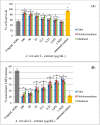
Protective effect of A. tricolor L. extracts on AGEs-induced cytotoxicity in SH-SY5Y cells. SH-SY5Y cells were preincubated with A. tricolor L. for 24 h and then further incubated with AGEs for 48 h. (A) The cell survival of living SH-SY5Y cells was assessed using the trypan blue exclusion assay. (B) The release of LDH of damaged SH-SY5Y cells was assessed using the LDH assay. Values are reported as the means with their standard error of the mean (SEM), depicted by vertical bars. All experiments were performed in triplicate (N = 3). * p < 0.05 for a significant change as compared to AGEs treatment.
Figure 6
Figure 6
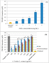
Protective effect of A. lividus L. and A. tricolor L. extracts on AGEs-induced oxidative stress in SH-SY5Y cells. The generation of thiobarbituric acid reactive substances (TBARS) was assessed using the malondialdehyde (MDA) assay. (A) SH-SY5Y cells were incubated with different concentrations of AGEs (0–4 mg/mL) for 24 h. SH-SY5Y cells were preincubated with different concentrations of A. lividus L. (B) and A. tricolor L. (C) extracts (0–100 μg/mL) for 24 h. Values are reported as the means with their standard error of the mean (SEM), depicted by vertical bars. All experiments were performed in triplicate (N = 3). * p < 0.05 for a significant change as compared to AGEs treatment.
Figure 6
Figure 6
Protective effect of A. lividus L. and A. tricolor L. extracts on AGEs-induced oxidative stress in SH-SY5Y cells. The generation of thiobarbituric acid reactive substances (TBARS) was assessed using the malondialdehyde (MDA) assay. (A) SH-SY5Y cells were incubated with different concentrations of AGEs (0–4 mg/mL) for 24 h. SH-SY5Y cells were preincubated with different concentrations of A. lividus L. (B) and A. tricolor L. (C) extracts (0–100 μg/mL) for 24 h. Values are reported as the means with their standard error of the mean (SEM), depicted by vertical bars. All experiments were performed in triplicate (N = 3). * p < 0.05 for a significant change as compared to AGEs treatment.
Figure 7
Figure 7
Effect of A. lividus L. and A. tricolor L. extracts on proinflammatory gene expression in AGEs-stimulated SH-SY5Y cells. (A) Human neuroblastoma SH-SY5Y cells treated with different concentrations of AGEs (0–4 mg/mL) for 24 h and TNF-α, IL-6, and IL-1 gene expression levels were determined by qPCR analysis. SH-SY5Y cells were preincubated with A. lividus L. extracts and A. tricolor L. extracts for 24 h, then further incubated with AGEs for 24 h and qPCR analyses of TNF-α (B), IL-6 (C) and IL-1 (D) were performed. The mRNA expression of GAPDH was used for normalization. Values are reported as the means with their standard error of the mean (SEM), depicted by vertical bars. All experiments were performed in triplicate (N = 3). * p < 0.05 for a significant change on the effect of AGEs on gene expression compared to untreated cells, whereas the effect of A. lividus L. and A. tricolor L. extracts was compared with AGEs-treated cells.
Figure 7
Figure 7
Effect of A. lividus L. and A. tricolor L. extracts on proinflammatory gene expression in AGEs-stimulated SH-SY5Y cells. (A) Human neuroblastoma SH-SY5Y cells treated with different concentrations of AGEs (0–4 mg/mL) for 24 h and TNF-α, IL-6, and IL-1 gene expression levels were determined by qPCR analysis. SH-SY5Y cells were preincubated with A. lividus L. extracts and A. tricolor L. extracts for 24 h, then further incubated with AGEs for 24 h and qPCR analyses of TNF-α (B), IL-6 (C) and IL-1 (D) were performed. The mRNA expression of GAPDH was used for normalization. Values are reported as the means with their standard error of the mean (SEM), depicted by vertical bars. All experiments were performed in triplicate (N = 3). * p < 0.05 for a significant change on the effect of AGEs on gene expression compared to untreated cells, whereas the effect of A. lividus L. and A. tricolor L. extracts was compared with AGEs-treated cells.

Similar articles

Cited by

References

    1. Yong V.W. Inflammation in neurological disorders: A help or a hindrance? Neuroscientist. 2010;16:408–420. doi: 10.1177/1073858410371379. - DOI - PubMed
    1. Emerit J., Edeas M., Bricaire F. Neurodegenerative diseases and oxidative stress. Biomed. Pharmacother. 2004;58:39–46. doi: 10.1016/j.biopha.2003.11.004. - DOI - PubMed
    1. Hsieh H.L., Yang C.M. Role of redox signaling in neuroinflammation and neurodegenerative diseases. BioMed Res. Int. 2013;2013:484613. doi: 10.1155/2013/484613. - DOI - PMC - PubMed
    1. Haddad J.J. Oxygen-sensitive pro-inflammatory cytokines, apoptosis signaling and redox-responsive transcription factors in development and pathophysiology. Cytokines Cell. Mol. Ther. 2002;7:1–14. doi: 10.1080/13684730216401. - DOI - PubMed
    1. Ahmed N. Advanced glycation endproducts—role in pathology of diabetic complications. Diabetes Res. Clin. Pract. 2005;67:3–21. doi: 10.1016/j.diabres.2004.09.004. - DOI - PubMed

Publication types

MeSH terms

LinkOut - more resources