. 2015 Oct;21(10):1154-62.
doi: 10.1038/nm.3951. Epub 2015 Sep 21.

Critical role of acetylation in tau-mediated neurodegeneration and cognitive deficits

Affiliations

Critical role of acetylation in tau-mediated neurodegeneration and cognitive deficits

Sang-Won Min et al. Nat Med. 2015 Oct.

Abstract

Tauopathies, including frontotemporal dementia (FTD) and Alzheimer's disease (AD), are neurodegenerative diseases in which tau fibrils accumulate. Recent evidence supports soluble tau species as the major toxic species. How soluble tau accumulates and causes neurodegeneration remains unclear. Here we identify tau acetylation at Lys174 (K174) as an early change in AD brains and a critical determinant in tau homeostasis and toxicity in mice. The acetyl-mimicking mutant K174Q slows tau turnover and induces cognitive deficits in vivo. Acetyltransferase p300-induced tau acetylation is inhibited by salsalate and salicylate, which enhance tau turnover and reduce tau levels. In the PS19 transgenic mouse model of FTD, administration of salsalate after disease onset inhibited p300 activity, lowered levels of total tau and tau acetylated at K174, rescued tau-induced memory deficits and prevented hippocampal atrophy. The tau-lowering and protective effects of salsalate were diminished in neurons expressing K174Q tau. Targeting tau acetylation could be a new therapeutic strategy against human tauopathies.


Conflict of interest statement

COMPETING FINANCIAL INTERESTS

The authors declare no competing financial interests.

Figures
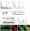
Figure 1
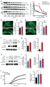
Figure 1. Tau-K174 Is Acetylated at Early Stages of AD
(a) ESI-MS/MS tandem mass spectra of Lys-acetylated peptide IPAKAcTPPAPK (residues 171–180) with a Protein Pilot confidence score of 99 obtained after trypsin digestion of immunoprecipitated Tau from AD brains. K*: Marker ion at 126.1 for acetyllysine immonium ion identification. (b) ESI-MS/MS tandem mass spectrum of acetyl-lysine containing synthetic peptide IPAKacTPPAPK. The corresponding Protein Pilot confidence score was 99. (c) Immunoblot of recombinant tau in the presence or absence of p300 showing acetyl-tau-specificity of AC312. (d) Immunoblot of lysates from HEK293 cells expressing tau mutants. Mutation of K174R diminished p300-induced immunoreactivity of AC312. (e–f) AC312-positive ac-tau (K174) signal was detected in AD brains (Braak stages 1–5). (e) Immunoblot of Tau5 immunoprecipitates with AC312 and PHF-1 antibodies. (f) Levels of ac-K174 were significantly higher at early and late Braak stages than those at Braak stage 0; n = 7 (Braak 0), n = 13 (Braak 1–2), n = 8 (Braak 3–5). * p < 0.05, one-way ANOVA, Tukey-Kramer post hoc analyses. See Supplementary Table-1 for the patient information. (g–j) AC312 immunoreactivity in the hippocampus of PS19 mice. (g) Merged image of AC312 and MC1 immunostaining at low magnification; scale bar 100 μm. (h–j) High-magnification confocal images showing (h) AC312-positive and (i) MC1-positive tau are highly colocalized (j); scale bar 25 μm. Values are means ± SEM (f). WT, wild-type.
Figure 2
Figure 2. Acetyl-Mimic K174Q Leads to Tau Accumulation In Vitro and In Vivo
(a) K174Q tau had longer half-life than WT or K174R tau. Primary neurons were infected with lenti-WT, K174Q or K174R tau for four days before CHX treatment. (left) Representative immunoblot of WT, K174Q or K174R tau in primary neurons treated with CHX for 0–32 hours. (right) Quantification of WT, K174Q, or K174R tau turnover. n=15, n=8, n=8, from 4 independent experiments. Levels of tau were normalized to those of GAPDH at different time points. * p < 0.05, K174Q vs WT at 2, 20, or 24 h. *** p<0.001, at 14 h. One-way ANOVA, Tukey-Kramer post hoc analyses. (b, d) Representative images showing HT7 immunostaining of hippocampus 10 days after injection of equal amounts of AAV-WT vs. K174Q tau (b), or AAV-K174Q vs. AAV-K174R tau (d). Scale bar=500 μm. (c, e) qRT-PCR showing similar levels of human tau mRNA are transduced three months after the infection. GAPDH was used as internal control. (c) n=10 (for WT), 12 (for K174Q). Mann-Whitney non-parametric test. (e) n=9 (for K174R and K174Q), unpaired student t-test. (f,g) K174Q mutation elevated levels of tau monomers and dimers. (f) Representative immunoblot with HT7 showing tau monomers and putative dimers in hippocampus injected with AAV-WT or AAV-K174Q. (g) Quantification of tau monomers (left) and tau dimers (right). n=11 (WT), n=12 (K174Q). *** p < 0.001, unpaired student t-test. (h,i) K174R mutation elevates tau dimers as K174Q, but not monomers. (h) Representative immunoblot with HT7 showing monomeric tau and putative tau dimers in hippocampus injected with AAV-K174Q or AAV-K174R tau. (i) Quantification of tau monomers (left) and dimers (right). n=9/genotype. * p < 0.05, unpaired student t-test. (j) In vitro aggregation analyses of recombinant WT, K174Q and K174R. (left) Tau aggregation kinetics was assayed by Thioflavin T fluorescence following the addition of heparin. (right) Amplitude of curve obtained from parameters fit to the Gompertz equation. ** p < 0.01, one-way ANOVA, Tukey-Kramer post hoc analyses from three independent experiments. KQ=K174Q tau, KR=K174R tau. Values are means ± SEM (a,c,e,g,i,j). WT, wild-type.
Figure 3
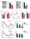
Figure 3. Expression of K174Q Tau Induces Neurodegeneration and Cognitive Deficits
(a,b) Expression of K174Q tau leads to more loss of hippocampal volume than WT or K174R tau. Representative nissl staining and volume quantification of hippocampus from NI (n=7), AAV-WT tau (WT, n=7), or AAV-K174Q tau (KQ, n=7) (a) or NI (n=10), AAV-K174Q tau (KQ, n=9), or AAV-K174R tau (KR, n=9) (b). *** p < 0.001, * p < 0.05, one-way ANOVA, Tukey-Kramer posthoc analyses. Scale bar, 500 μm. (c, e) Hippocampal expression of K174Q tau leads to hyperactivity in the open field test or reduced spontaneous alterations in the Y-maze. (c) Mice injected with K174Q tau exhibited higher accumulative total activity than those injected with WT (left) or with K174R tau (right). (e) Mice injected with K174Q showed significantly lower % alternations in the Y-maze than those injected with WT (left) or K174R tau (right). n=9 (NI, WT, vs. KQ); n=10 (NI), 8 (KQ), 9 (KR). * p<0.05, one-way ANOVA, Dunnett post hoc analyses. (d) The extent of hyperactivity in the open field positively correlated with the levels of tau monomers in mice injected with AAV-K174Q tau (KQ, n=18). Pearson correlation analyses. (f–i) Hippocampal expression of K174Q tau impaired spatial learning in MWM. K174Q tau (f), but not K174R tau (h), impaired spatial learning in both the naive and the reversal learning phases. * p < 0.05, ** p < 0.01, multilevel mixed-effects linear regression model (the naïve and reversal phases were analyzed separately). In probe trials, mice expressing K174Q tau, not WT tau, crossed the new platform location significantly fewer times that NI controls (g). n=9 mice/condition (f,g). n=10 (NI), 9 (KQ), 9 (KR) (h,i). ** p<0.01, *** p<0.001, two-way ANOVA, Bonferroni post-tests. Values are means ± SEM (a,b. c, e, f–i). WT, wild-type.
Figure 4
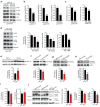
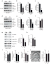
Figure 4. Salicylate Inhibits p300 and Reduces Ac-Tau (K174) In Cultured Neurons and PS19 mice
(a–c) Genetic deletion of p300 lowers levels of ac-K174, p-tau and total tau. (a) Lysates from primary mouse neurons that were derived from p300F/F pups, and infected with lenti-WT tau and lenti-control or lenti-cre virus were immunoblotted with anti-p300, ac-tau (AC312), p-tau (AT8), or t-tau (Tau5) antibody. (b) Quantification of levels of total tau (t-tau) (left), ac-K174 (middle), and AT8-positive p-tau (right). (c) Quantification of the levels of ac-K174 (left) or p-tau (right) relative to t-tau. Levels in non-treated cells were set as 1. n=11 from three independent experiments, *** p < 0.001, ** p < 0.01, unpaired student t-test (ac-tau), Kruskal-Wallis test (p-tau). (d) Rat primary cortical neurons infected with lenti-WT tau were treatment with 0, 5, 10 mM salicylate for 24 hr (DIV 12). Representative immunoblot of p300, ac-tau, p-tau and t-tau. Quantification of the levels of p300 (left), ac-tau (middle) or AT8-positive p-tau (right) relative to t-tau, in primary rat neurons treated with salicylate. Levels in non-treated cells were set as 1. n = 4 from 4 independent experiments.. *, p < 0.05, **, p < 0.01, one –way ANOVA, Tukey-Kramer post-hoc analyses. (e) Representative immunoblot and quantification of ac-H3K18 and H3 in cortical histone extract from 10–11-month-old NTG and PS19 littermates. Levels in NTG group were set as 1. n=9 mice/group, ** p < 0.01, unpaired student t-test. (f–h) Oral gavage of SSA (225 mg/kg) inhibits p300 activity in the brain. Representative immunoblot and quantification of ac-H3K18 and H3 (f, male, n=11/group), ac-H2AK5 and H2A (g, female, n=8/group) or ac-H2BK12K15 and H2B (h, male, n=11/group). Levels in vehicle-treated group were set as 1. * p < 0.05,unpaired student t-test. (i) SSA treatment lowers levels of total hTau protein, but not mRNA. Levels of total hTau protein were measured by ELISA (left). Levels of hTau mRNA were measured by qRT-pCR (right). Levels in vehicle-treated group were set as 1. n=11 mice/condition, 10–11 months old male, * p < 0.05, unpaired student t-test. ns=not significant. (j) SSA treatment lowers levels of ac-tau relative to total tau. Representative immunoblot of ac-tau (AC312), p-tau (AT8), and t-tau (Tau5). Vehicle-treated non-transgenic NTG and tau KO mice were included as controls. Quantification of ac-tau (left) or AT8-positive p-tau relative to t-tau (middle) or GAPDH (right), in the soluble hippocampal lysates from PS19 mice treated with vehicle or SSA. Levels in vehicle-treated group were set as 1. n=8 mice/condition, 10–11 months old female, ** p < 0.01, *, p < 0.05, unpaired student t-test. Values are mean ± SEM (b–j). WT, wild-type. NTG, non-transgenic.
Figure 5
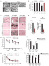
Figure 5. Salsalate Treatment Prevents Hippocampi Atrophy, Reduces Tau Pathology and Rescues Spatial Memory Deficits in PS19 Mice
(a–b) SSA treatment prevented hippocampal volume loss. (a) Representative nissl staining of hippocampus from 8-month-old NTG and PS19 mice before the treatment (non-treated, NT), 10 month-old vehicle-treated NTG, vehicle- or SSA-treated PS19 mice. Scale bar, 500 μm. (b) Quantification of hippocampal volume. n=6 (NTG, NT), 9 (PS19, NT), 8 months old. n=9 (female NTG, veh), 6 (female PS19, veh), 6 (female PS19, SSA), 10–11 months old, ** p < 0.01, one-way ANOVA, Tukey-Kramer post hoc analyses. (c–d) SSA treatment reduced NFTs in PS19 mice. (c) Example of Gallyas staining images of vehicle-treated female NTG mice, and female PS19 mice treated with vehicle or SSA. Scale bar: 250 μm (upper); 25 μm (middle, lower) (d) Quantification of silver-positive cells or neurites in neocortex and hippocampus (CA1+CA3) of vehicle- or SSA treated PS19 mice. n=8 mice/genotype/treatment, 10–11 months old, * p < 0.05, unpaired student t-test. (e–h) SSA treatment ameliorated spatial memory loss in fixed-location dry maze and in MWM. (e) No significant difference in learning rate was observed among the groups in fixed location dry maze; Day 1–2, training with visible hole; Day 3–5, learning with hidden hole. (f) NTG or SSA-treated PS19 mice, but not vehicle-treated PS19 mice, showed preference to the target quadrant. n=10 (female NTG, veh), 8 (female PS19, veh), 8 (female PS19, SSA), 10–11 months old. (g) Learning rate did not differ among the groups in MWM. (h) SSA treatment restored spatial memory deficits in PS19 mice. NTG or SSA-treated PS19 mice, but not vehicle-treated PS19 mice, showed preference to the target quadrant. n=13 (male NTG, veh), 15 (male NTG, SSA), 11 (male PS19, veh), 10 (male PS19, SSA), 9–10 months old. The multilevel mixed-effects linear regression model was used to assess the learning curve (e, g). *** p < 0.001, ** p <0.01, * p <0.05, paired student t-test. Values are means ± SEM (b, d, e–h). NTG, non-transgenic.
Figure 6
Figure 6. Tau-lowering and Protective Effects of SSA/Salicylate Involve Ac-K174 Inhibition
(a–f) SSA treatment lowers levels of WT tau, but not K174Q tau in cultured neurons. Rat primary cortical neurons infected with AAV-WT or AAV-K174Q tau were treated with 5 mM SS for 24 hr (DIV 12). (a, d) Representative immunoblot of p300 and t-tau (human or mouse). Quantification of levels of p300 (b, e; left), mouse t-tau (b, e; right) and human t-tau (c, f) after SS- or veh-treatment. Levels in veh-treated cells were set as 1. n=4 from two independent experiments. *** p < 0.001, ** p <0.01, unpaired student t-test. (g,h) SSA treatment failed to lower t-tau in mice expressing K174Q tau. (g, left) The representative immunoblot of antibodies against ac-H2AK5 and H2A, AT8, PHF1, HT7 or GAPDH. (g, right) Quantification showing that levels of ac-H2AK5/H2A were lower in SSA-treated than veh-treated mice. (h) ELISA assay shows that levels of total K174Q tau were not significantly affected by SSA treatment. n=11 (vehicle), n=10 (SSA), ns = not significant, unpaired student t-test. (i) Levels of PHF1-positive p-tau normalized to HT7 (left), or levels of or AT8-positive p-tau normalized to HT7 (middle) or GAPDH (right) were not affected by SSA treatment. n=11 (vehicle), n=10 (SSA), * p<0.05, ns = not significant, unpaired student t-test. (j) SSA treatment failed to protect against atrophy mice expressing K174Q tau. (left) The representative nissl-stained images of hippocampus treated with either vehicle or SSA. Scale bar: 500 μm. (right) Quantification of hippocampal volume showing no significant difference. n=11 (vehicle), n=10 (SSA), ns = not significant, unpaired student t-test. Values are means ± SEM (b, c, e, f, h–j). WT, wild-type.

Comment in

Similar articles

Cited by

References

    1. Ludolph AC, et al. Tauopathies with parkinsonism: clinical spectrum, neuropathologic basis, biological markers, and treatment options. Eur J Neurol. 2009;16:297–309. - PMC - PubMed
    1. Cairns NJ, et al. Neuropathologic diagnostic and nosologic criteria for frontotemporal lobar degeneration: consensus of the Consortium for Frontotemporal Lobar Degeneration. Acta neuropathologica. 2007;114:5–22. - PMC - PubMed
    1. Braak H, Braak E. Neuropathological stageing of Alzheimer-related changes. Acta Neuropathol Berl. 1991;82:239–259. - PubMed
    1. Lasagna-Reeves CA, et al. Identification of oligomers at early stages of tau aggregation in Alzheimer’s disease. FASEB journal : official publication of the Federation of American Societies for Experimental Biology. 2012;26:1946–1959. - PMC - PubMed
    1. Ren Y, Sahara N. Characteristics of tau oligomers. Frontiers in neurology. 2013;4:102. - PMC - PubMed

Publication types