Recovery of Corneal Endothelial Cells from Periphery after Injury
- PMID: 26378928
- PMCID: PMC4574742
- DOI: 10.1371/journal.pone.0138076
Recovery of Corneal Endothelial Cells from Periphery after Injury
Abstract
Background: Wound healing of the endothelium occurs through cell enlargement and migration. However, the peripheral corneal endothelium may act as a cell resource for the recovery of corneal endothelium in endothelial injury.
Aim: To investigate the recovery process of corneal endothelial cells (CECs) from corneal endothelial injury.
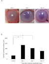
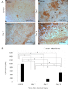
Methods: Three patients with unilateral chemical eye injuries, and 15 rabbit eyes with corneal endothelial chemical injuries were studied. Slit lamp examination, specular microscopy, and ultrasound pachymetry were performed immediately after chemical injury and 1, 3, 6, and 9 months later. The anterior chambers of eyes from New Zealand white rabbits were injected with 0.1 mL of 0.05 N NaOH for 10 min (NaOH group). Corneal edema was evaluated at day 1, 7, and 14. Vital staining was performed using alizarin red and trypan blue.
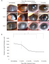
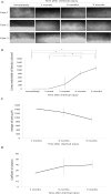
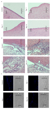
Results: Specular microscopy did not reveal any corneal endothelial cells immediately after injury. Corneal edema subsided from the periphery to the center, CEC density increased, and central corneal thickness decreased over time. In the animal study, corneal edema was greater in the NaOH group compared to the control at both day 1 and day 7. At day 1, no CECs were detected at the center and periphery of the corneas in the NaOH group. Two weeks after injury, small, hexagonal CECs were detected in peripheral cornea, while CECs in mid-periphery were large and non-hexagonal.
Conclusions: CECs migrated from the periphery to the center of the cornea after endothelial injury. The peripheral corneal endothelium may act as a cell resource for the recovery of corneal endothelium.
Conflict of interest statement
Figures
Similar articles
-
Effect of intracameral injection of bisulfite-containing phenylephrine on rabbit corneal endothelium.Cornea. 2015 Apr;34(4):460-3. doi: 10.1097/ICO.0000000000000312. Cornea. 2015. PMID: 25474237
-
Endothelial cell damage following sulfur mustard exposure in rabbits and its association with the delayed-onset ocular lesions.Cutan Ocul Toxicol. 2013 Jun;32(2):115-23. doi: 10.3109/15569527.2012.717571. Epub 2012 Oct 30. Cutan Ocul Toxicol. 2013. PMID: 23106194
-
Effect of Na-hyaluronan on stromal and endothelial healing in experimental corneal alkali wounds.Ophthalmic Res. 1999;31(6):432-9. doi: 10.1159/000055568. Ophthalmic Res. 1999. PMID: 10474072
-
[Transplantation of corneal endothelial cells].Nippon Ganka Gakkai Zasshi. 2002 Dec;106(12):805-35; discussion 836. Nippon Ganka Gakkai Zasshi. 2002. PMID: 12610838 Review. Japanese.
-
The response of the corneal endothelium to intraocular surgery.Refract Corneal Surg. 1991 Jan-Feb;7(1):81-6. Refract Corneal Surg. 1991. PMID: 2043553 Review.
Cited by
-
Analysis of the Effect of Phacoemulsification and Intraocular Lens Implantation Combined With Trabeculectomy on Cataract and Its Influence on Corneal Endothelium.Front Surg. 2022 Feb 18;9:841296. doi: 10.3389/fsurg.2022.841296. eCollection 2022. Front Surg. 2022. PMID: 35252341 Free PMC article.
-
3D in vitro model for human corneal endothelial cell maturation.Exp Eye Res. 2019 Jul;184:183-191. doi: 10.1016/j.exer.2019.04.003. Epub 2019 Apr 10. Exp Eye Res. 2019. PMID: 30980816 Free PMC article.
-
Regulatory Compliant Tissue-Engineered Human Corneal Endothelial Grafts Restore Corneal Function of Rabbits with Bullous Keratopathy.Sci Rep. 2017 Oct 26;7(1):14149. doi: 10.1038/s41598-017-14723-z. Sci Rep. 2017. PMID: 29074873 Free PMC article.
-
New ex vivo model of corneal endothelial phacoemulsification injury and rescue therapy with mesenchymal stromal cell secretome.J Cataract Refract Surg. 2019 Mar;45(3):361-366. doi: 10.1016/j.jcrs.2018.09.030. Epub 2018 Dec 7. J Cataract Refract Surg. 2019. PMID: 30527441 Free PMC article.
-
PAX6, modified by SUMOylation, plays a protective role in corneal endothelial injury.Cell Death Dis. 2020 Aug 12;11(8):683. doi: 10.1038/s41419-020-02848-5. Cell Death Dis. 2020. PMID: 32826860 Free PMC article.
References
-
- Joyce NC. Proliferative capacity of the corneal endothelium. Prog Retin Eye Res. 2003;22:359–89. - PubMed
-
- Iwamoto T, Smelser GK. Electron microscopy of the human corneal endothelium with reference to transport mechanisms. Invest Ophthalmol. 1965;4:270–84. - PubMed
-
- Edelhauser HF. The balance between corneal transparency and edema: the Proctor Lecture. Invest Ophthalmol Vis Sci. 2006;47:1754–67. - PubMed
-
- Engelmann K, Bednarz J, B¨ohnke M. Endothelial cell transplantation and growth behavior of the human corneal endothelium. Ophthalmologe. 1999;96:555–62. - PubMed
Publication types
MeSH terms
Grants and funding
LinkOut - more resources
Full Text Sources
Other Literature Sources
Medical

