Comparative Study
. 2015 Sep 16;35(37):12703-13.
doi: 10.1523/JNEUROSCI.0427-15.2015.

Distinct Functions for Anterograde and Retrograde Sorting of SORLA in Amyloidogenic Processes in the Brain

Affiliations
Comparative Study

Distinct Functions for Anterograde and Retrograde Sorting of SORLA in Amyloidogenic Processes in the Brain

Sonya B Dumanis et al. J Neurosci. .

Abstract

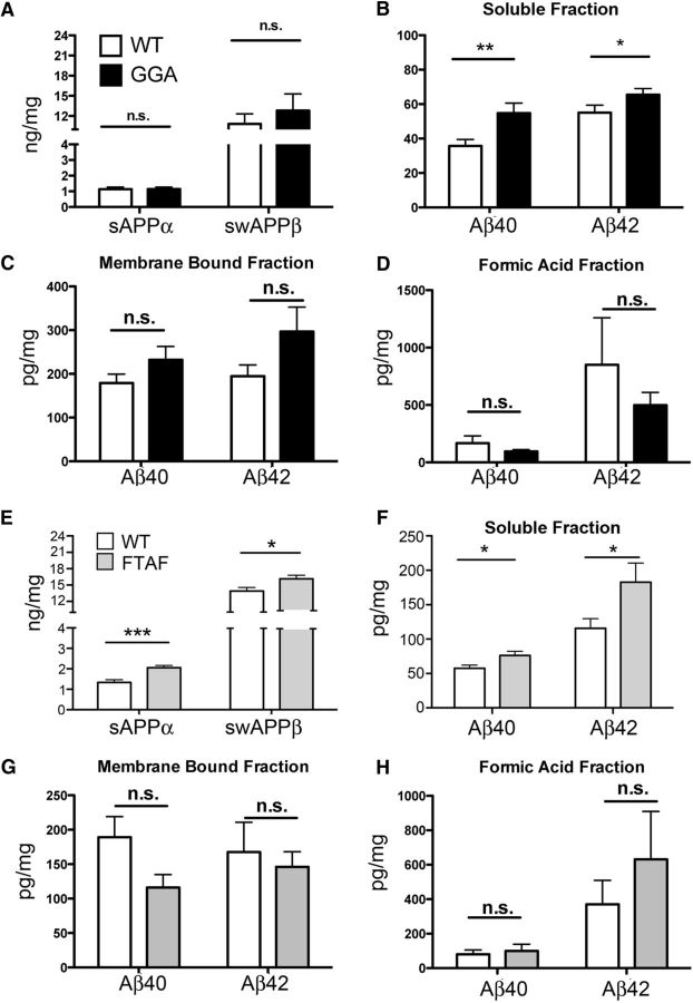
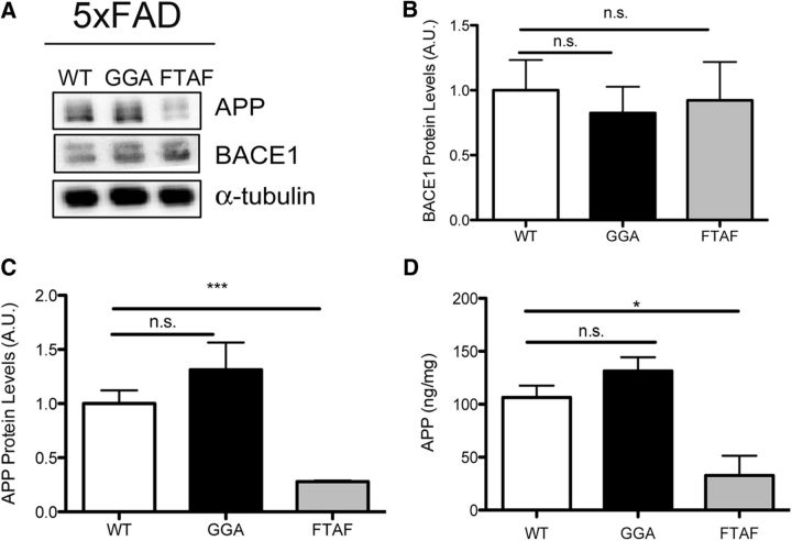
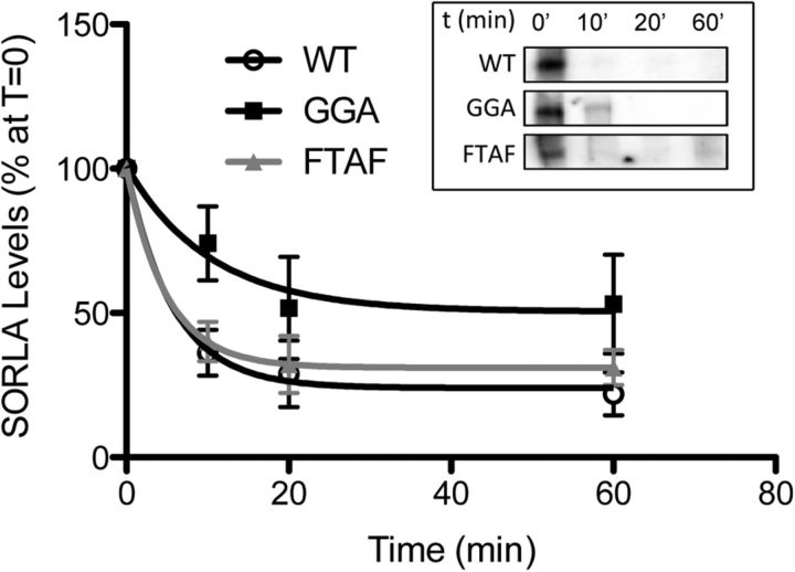
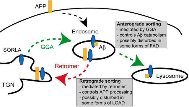
SORLA is a neuronal sorting receptor implicated both in sporadic and familial forms of AD. SORLA reduces the amyloidogenic burden by two mechanisms, either by rerouting internalized APP molecules from endosomes to the trans-Golgi network (TGN) to prevent proteolytic processing or by directing newly produced Aβ to lysosomes for catabolism. Studies in cell lines suggested that the interaction of SORLA with cytosolic adaptors retromer and GGA is required for receptor sorting to and from the TGN. However, the relevance of anterograde or retrograde trafficking for SORLA activity in vivo remained largely unexplored. Here, we generated mouse models expressing SORLA variants lacking binding sites for GGA or retromer to query this concept in the brain. Disruption of retromer binding resulted in a retrograde-sorting defect with accumulation of SORLA in endosomes and depletion from the TGN, and in an overall enhanced APP processing. In contrast, disruption of the GGA interaction did not impact APP processing but caused increased brain Aβ levels, a mechanism attributed to a defect in anterograde lysosomal targeting of Aβ. Our findings substantiated the significance of adaptor-mediated sorting for SORLA activities in vivo, and they uncovered that anterograde and retrograde sorting paths may serve discrete receptor functions in amyloidogenic processes.

Significance statement: SORLA is a sorting receptor that directs target proteins to distinct intracellular compartments in neurons. SORLA has been identified as a genetic risk factor for sporadic, but recently also for familial forms of AD. To confirm the relevance of SORLA sorting for AD processes in the brain, we generated mouse lines, which express trafficking mutants instead of the wild-type form of this receptor. Studying neuronal activities in these mutant mice, we dissected distinct trafficking routes for SORLA guided by two cytosolic adaptors termed GGA and retromer. We show that these sorting pathways serve discrete functions in control of amyloidogenic processes and may represent unique therapeutic targets to interfere with specific aspects of neurodegenerative processes in the diseased brain.

Keywords: APP processing; SORLA; VPS10P domain receptors; adaptors; protein transport; retromer.


Figures

Figure 1.
Figure 1.
Mice expressing human SORLA variants. A, Constructs encoding wild-type SORLA (SORLAWT) or receptor variants lacking the GGA (SORLAGGA) or retromer binding (SORLAFTAF) motifs for targeting the murine Rosa26 locus are shown. Amino acid sequences of the cytoplasmic domains of the receptor variants with residues modified by mutagenesis in the GGA (green) and retromer (red) binding mutants are indicated. The scheme for introducing the targeting vectors between exons 1 and 2 (black squares) of Rosa26 is given below. Removal of the loxP-flanked neomycin resistance cassette (neoR) enables transcription of the SORLA cDNAs from the Rosa26 promoter (activated Rosa26 locus). B, Immunodetection of SORLA (green) in cortices of mice of the indicated genotypes. Sections were costained for the neuronal marker NeuN (blue; left) or astroglial marker GFAP (red) and DAPI (white; right). No immunoreactivity for SORLA is detectable in Sorl1−/− tissue (used as negative control). Scale bar, 10 μm. C, D Western blot analysis (C) and quantification by densitometric scanning of replicate blots (D) documents increased levels for SORLAGGA compared with SORLAWT and SORLAFTAF in brain extracts (n = 3 animals/genotype; *p < 0.05, **p < 0.01, one-way ANOVA, Tukey post hoc analysis). Detection of Na/K ATPase (Na/K) served as loading control in C.
Figure 2.
Figure 2.
Characterization of proteins related to intracellular protein sorting. A, Representative Western blots for proteins related to GGA and retromer activities in the cortices of mice of the various genotypes are shown. Detection of tubulin served as loading control. B, Quantification of the Western blots in A revealed no significant differences in protein expression levels comparing the genotypes (n = 3 animals/genotype). C, Immunodetection of neuronal marker NeuN (blue) and of proteins related to GGA and retromer activities (green) on cortical brain sections of mice of the indicated SORLA genotypes are shown. No discernable differences in neuronal expression patterns in the various genotypes are seen for APP (top row), GGA1 (second row), VPS26 (third row), or CI-MPR (bottom row) are seen. Scale bar, 15 μm.
Figure 3.
Figure 3.
SORLA levels in primary neurons expressing wild-type and mutant receptor variants. A, Representative images for primary hippocampal neurons (DIV 5) of the indicated genotypes stained for SORLA (green) and DAPI (blue; top), or for neuronal marker MAP2 (white) and DAPI (blue; bottom). Analysis of neuronal cultures from Sorl1−/− mice served as negative control. Scale bar, 10 μm. B, C, Levels of SORLA variants were tested by Western blotting of total cell lysates of primary neurons (B) and quantification by densitometric scanning of replicate blots (C). Levels of SORLAGGA were increased compared with SORLAWT and SORLAFTAF (n = 4 per genotype; *p < 0.05, one-way ANOVA, Tukey post hoc analysis).
Figure 4.
Figure 4.
Aberrant trafficking of SORLAGGA and SORLAFTAF in hippocampal neurons. A–C, Primary hippocampal neurons of the indicated genotypes stained for SORLA (green), DAPI (blue), and either VTi1b (red; A), Rab5 (red; B), or Lamp1 (red; C). Pearson's coefficients are given in the merged parts. High-magnification insets indicate colocalization of SORLA with the respective marker. At least 20 neurons per genotype were imaged [*p < 0.05;**p < 0.01; ***p < 0.001; Student's t test (as SORLAWT and SORLAGGA were performed separately from SORLAWT and SORLAFTAF experiments]. D, Model of how SORLA localization changes in mutant receptor variants. SORLAWT is found predominantly in the TGN, but also in endosomes and lysosomes (top). Anterograde sorting from TGN to the endosomal/lysosomal compartments is mediated by GGAs (green arrow). Loss of this interaction results in SORLAGGA accumulation in the TGN and depletion from lysosomes (middle). Retrograde sorting of SORLAWT is mediated by retromer (red arrow). Loss of this interaction causes accumulation of SORLAFTAF in early endosomes but depletion from the TGN (bottom).
Figure 5.
Figure 5.
SORLAGGA and SORLAFTAF differentially impact amyloidogenic processing. SORLAWT, SORLAGGA, and SORLAFTAF mice were crossed with the 5xFAD line. Cortical lysates were collected from females at 8–10 weeks of age and analyzed as indicated. A–D, Quantification by ELISA documents no changes in sAPPα and sAPPβ (swAPPβ). (A), but an increase in Aβ40 and Aβ42 levels (B) in SORLAGGA compared with SORLAWT mice in the soluble brain fraction (n = 12 animals/group). No difference in Aβ40 and Aβ42 levels was seen when the membrane bound (C) or formic acid soluble fractions (D) were analyzed (n = 6–7 animals/group). E–H, ELISA documents increases in sAPPα and sAPPβ (E) and in Aβ40 and Aβ42 (F) in SORLAFTAF compared with SORLAWT animals (n = 14–15 animals/group) in the cytosolic brain fraction. Parallel experiments to E and F were performed, and quantification by ELISA documented no changes in Aβ40 and Aβ42 levels in SORLAFTAF compared with SORLAWT animals in the membrane bound (G) or formic acid soluble (H) fractions (n = 7 animals/group). Statistical analyses were performed using Student's t test (*p < 0.05; **p < 0.01; ***p < 0.001).
Figure 6.
Figure 6.
SORLAFTAF mice have decreased levels of human APP. A, Immunoblot analyses for APP and BACE1 in cortical brain extracts showing decreased levels of APP but not BACE1 in SORLAFTAF mice. Tubulin served as loading control. B, C, Densitometric scanning of replicate Western blots (as exemplified in A) confirms normal levels of BACE1 (B) but decreased levels of APP (C) in SORLAFTAF compared with SORLAWT and SORLAGGA animals (n = 3/group; one-way ANOVA, Tukey post hoc analysis; ***p < 0.001). D, ELISA substantiating decreased levels of APP in cortical extracts of SORLAFTAF compared with SORLAWT and SORLAGGA animals (n = 3/group; one-way ANOVA, Tukey post hoc analysis; ***p < 0.001).
Figure 7.
Figure 7.
Impaired ability of SORLAGGA to mediate lysosomal catabolism of Aβ peptides. A, B, SH-SY5Y cells overexpressing human APP695 together with SORLAWT or SORLAGGA were treated with 5 μm DAPT. At the indicated time points, the levels of intracellular Aβ40 (A) and Aβ42 (B) were determined by ELISA. Data represent the mean ± SEM of nine replicates expressed as percentage of time point 0 (set to 100%). Where no error bars are visible, they are smaller than the actual symbol shown. The inset in A documents expression of SORLA and APP in the cell extracts by Western blotting. C, D, Levels of Aβ40 (C) and Aβ42 (D) are increased in the cell lysate and the cell media of SORLAGGA compared with SORLAWT cells. Cells were treated with 5 μm DAPT for 3 h, and Aβ levels normalized to levels at time point 0 (n = 3 replicates). E, F, SORLAWT cells were treated with 5 μm DAPT in the absence or presence of 100 μm leupeptin, 10 μm pepstatin A, and 50 μm chloroquine (DAPT + Inhibitor). Levels of Aβ40 (E) and Aβ42 (F) in the cell lysate were determined after 3 h and normalized to levels at time point 0 (n = 6 replicates). Application of the lysosomal inhibitor cocktail reduced catabolism of Aβ40 and Aβ42 compared with the DAPT only condition. Student's t test (*p < 0.05; **p < 0.01; ***p < 0.001).
Figure 8.
Figure 8.
Prolonged half-life of SORLAGGA compared with other receptor variants. Pulse-chase experiments were performed in SH-SY5Y cells stably transfected with SORLAWT, SORLAGGA, or SORLAFTAF, and the amount of metabolically labeled SORLA at the chase time points determined by Western blotting (as exemplified in the inset). Following densitometric scanning of replicate blots, values were normalized to time point 0 of each experiment (set to 100%). A best-fit, one-phase exponential decay curve (y = Span*e−K*x + Plateau) was determined using GraphPad Prism software, with t(½) = 0.6932/K. Based on the curve fit, the protein half-life was t(½) = 3.97 min for SORLAWT, 3.43 min for SORLAFTAF, but 7.17 min for SORLAGGA (n = 8 replicates per condition). Data represent the mean ± SEM with the best-fit lines overlaid. Two-way ANOVA analysis (interaction between time and SORLA variant, n.s., time, p < 0.001, SORLA variant, p < 0.05) indicated that the SORLAGGA cohort is significantly different from the other SORLA constructs.
Figure 9.
Figure 9.
Model of SORLA trafficking pathways in amyloidogenic processes. GGAs mediate anterograde sorting of SORLA and impact SORLA-directed Aβ catabolism. An inherited mutation in SORL1 that disrupts Aβ binding, abrogating SORLA-dependent lysosomal-catabolism of Aβ, is implicated in an autosomal-dominant familial form of AD (FAD). Retromer mediates retrograde sorting of SORLA, and disrupting this pathway reduces APP levels in the TGN and results in overall enhanced APP processing rate. Low levels of SORLA or retromer components in the brain are considered risk factors promoting late-onset forms of AD (LOAD).

Similar articles

Cited by

References

    1. Andersen OM, Reiche J, Schmidt V, Gotthardt M, Spoelgen R, Behlke J, von Arnim CA, Breiderhoff T, Jansen P, Wu X, Bales KR, Cappai R, Masters CL, Gliemann J, Mufson EJ, Hyman BT, Paul SM, Nykjaer A, Willnow TE. Neuronal sorting protein-related receptor sorLA/LR11 regulates processing of the amyloid precursor protein. Proc Natl Acad Sci U S A. 2005;28:13461–13466. - PMC - PubMed
    1. Burgert T, Schmidt V, Caglayan S, Lin F, Füchtbauer A, Füchtbauer EM, Nykjaer A, Carlo AS, Willnow TE. SORLA-dependent and -independent functions for PACS1 in control of amyloidogenic processes. Mol Cell Biol. 2013;33:4308–4320. doi: 10.1128/MCB.00628-13. - DOI - PMC - PubMed
    1. Caglayan S, Takagi-Niidome S, Liao F, Carlo AS, Schmidt V, Burgert T, Kitago Y, Füchtbauer EM, Füchtbauer A, Holtzman DM, Takagi J, Willnow TE. Lysosomal sorting of amyloid-beta by the SORLA receptor is impaired by a familial Alzheimer's disease mutation. Sci Transl Med. 2014;6:223ra20. doi: 10.1126/scitranslmed.3007747. - DOI - PubMed
    1. Fjorback AW, Seaman M, Gustafsen C, Mehmedbasic A, Gokool S, Wu C, Militz D, Schmidt V, Madsen P, Nyengaard JR, Willnow TE, Christensen EI, Mobley WB, Nykjaer A, Andersen OM. Retromer binds the FANSHY sorting motif in SorLA to regulate Amyloid Precursor Protein sorting and processing. J Neurosci. 2012;32:1467–1480. doi: 10.1523/JNEUROSCI.2272-11.2012. - DOI - PMC - PubMed
    1. He X, Li F, Chang WP, Tang J. GGA proteins mediate the recycling pathway of memapsin 2 (BACE) J Biol Chem. 2005;280:11696–11703. doi: 10.1074/jbc.M411296200. - DOI - PubMed

Publication types

MeSH terms

Substances

LinkOut - more resources