

'Medusa head ataxia': the expanding spectrum of Purkinje cell antibodies in autoimmune cerebellar ataxia. Part 3: Anti-Yo/CDR2, anti-Nb/AP3B2, PCA-2, anti-Tr/DNER, other antibodies, diagnostic pitfalls, summary and outlook
- PMID: 26377319
- PMCID: PMC4573944
- DOI: 10.1186/s12974-015-0358-9
'Medusa head ataxia': the expanding spectrum of Purkinje cell antibodies in autoimmune cerebellar ataxia. Part 3: Anti-Yo/CDR2, anti-Nb/AP3B2, PCA-2, anti-Tr/DNER, other antibodies, diagnostic pitfalls, summary and outlook
Abstract
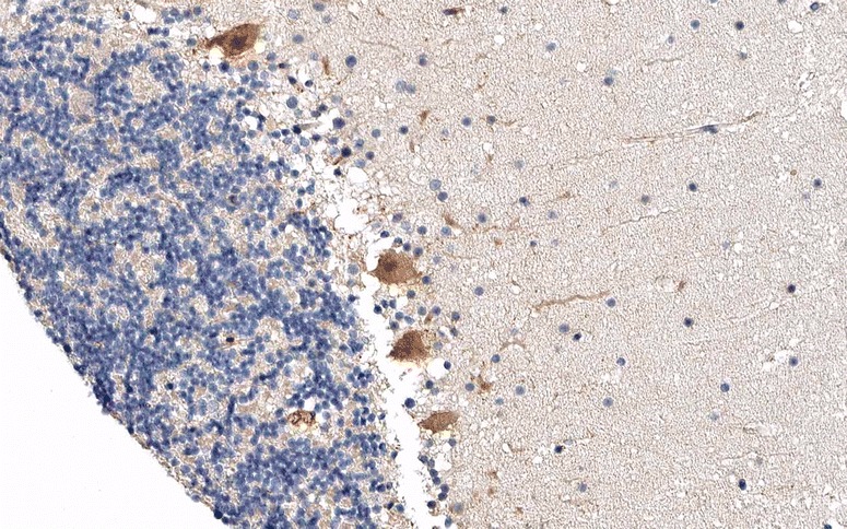
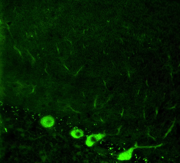
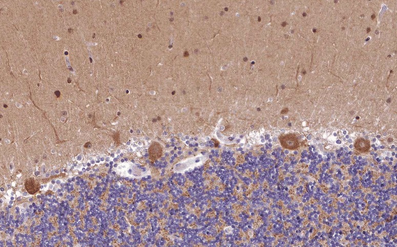
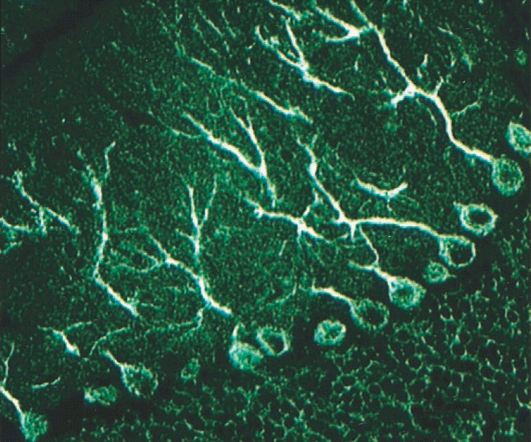
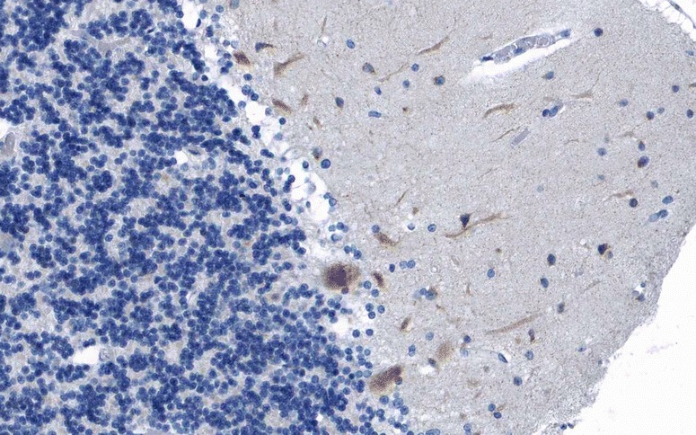
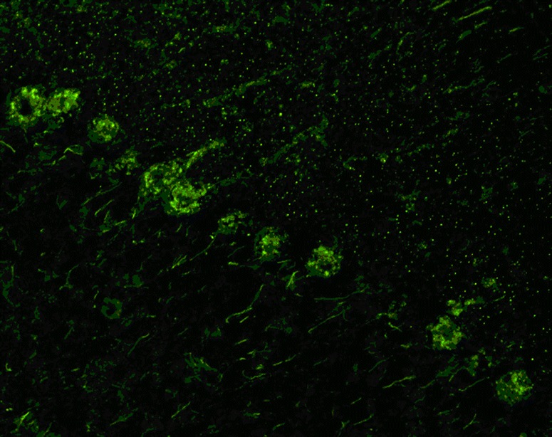
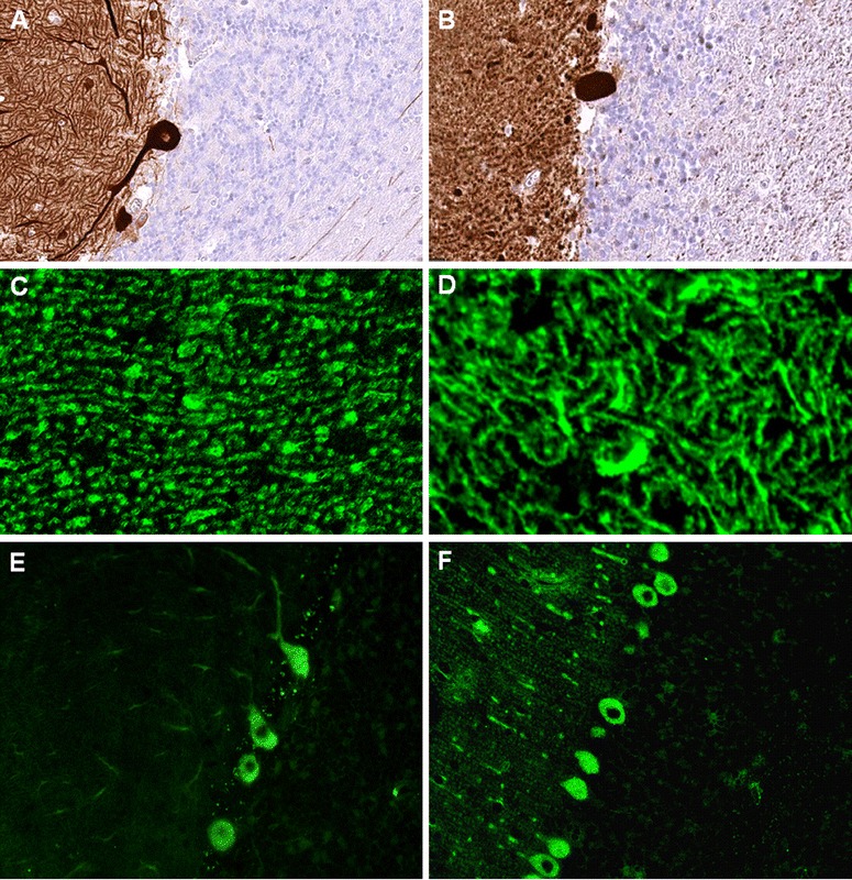
Serological testing for anti-neural autoantibodies is important in patients presenting with idiopathic cerebellar ataxia, since these autoantibodies may indicate cancer, determine treatment and predict prognosis. While some of them target nuclear antigens present in all or most CNS neurons (e.g. anti-Hu, anti-Ri), others more specifically target antigens present in the cytoplasm or plasma membrane of Purkinje cells (PC). In this series of articles, we provide a detailed review of the clinical and paraclinical features, oncological, therapeutic and prognostic implications, pathogenetic relevance, and differential laboratory diagnosis of the 12 most common PC autoantibodies (often referred to as 'Medusa head antibodies' due to their characteristic somatodendritic binding pattern when tested by immunohistochemistry). To assist immunologists and neurologists in diagnosing these disorders, typical high-resolution immunohistochemical images of all 12 reactivities are presented, diagnostic pitfalls discussed and all currently available assays reviewed. Of note, most of these antibodies target antigens involved in the mGluR1/calcium pathway essential for PC function and survival. Many of the antigens also play a role in spinocerebellar ataxia. Part 1 focuses on anti-metabotropic glutamate receptor 1-, anti-Homer protein homolog 3-, anti-Sj/inositol 1,4,5-trisphosphate receptor- and anti-carbonic anhydrase-related protein VIII-associated autoimmune cerebellar ataxia (ACA); part 2 covers anti-protein kinase C gamma-, anti-glutamate receptor delta-2-, anti-Ca/RhoGTPase-activating protein 26- and anti-voltage-gated calcium channel-associated ACA; and part 3 reviews the current knowledge on anti-Tr/delta notch-like epidermal growth factor-related receptor-, anti-Nb/AP3B2-, anti-Yo/cerebellar degeneration-related protein 2- and Purkinje cell antibody 2-associated ACA, discusses differential diagnostic aspects and provides a summary and outlook.
Figures
Similar articles
-
'Medusa head ataxia': the expanding spectrum of Purkinje cell antibodies in autoimmune cerebellar ataxia. Part 2: Anti-PKC-gamma, anti-GluR-delta2, anti-Ca/ARHGAP26 and anti-VGCC.J Neuroinflammation. 2015 Sep 17;12:167. doi: 10.1186/s12974-015-0357-x. J Neuroinflammation. 2015. PMID: 26377184 Free PMC article. Review.
-
'Medusa-head ataxia': the expanding spectrum of Purkinje cell antibodies in autoimmune cerebellar ataxia. Part 1: Anti-mGluR1, anti-Homer-3, anti-Sj/ITPR1 and anti-CARP VIII.J Neuroinflammation. 2015 Sep 17;12:166. doi: 10.1186/s12974-015-0356-y. J Neuroinflammation. 2015. PMID: 26377085 Free PMC article. Review.
-
Autoimmune Vestibulocerebellar Syndromes.Semin Neurol. 2020 Feb;40(1):97-115. doi: 10.1055/s-0039-3402061. Epub 2020 Jan 20. Semin Neurol. 2020. PMID: 31958862 Review.
-
Screening for MOG-IgG and 27 other anti-glial and anti-neuronal autoantibodies in 'pattern II multiple sclerosis' and brain biopsy findings in a MOG-IgG-positive case.Mult Scler. 2016 Oct;22(12):1541-1549. doi: 10.1177/1352458515622986. Epub 2016 Feb 11. Mult Scler. 2016. PMID: 26869529
-
Inositol 1,4,5-trisphosphate receptor type 1 autoantibody (ITPR1-IgG/anti-Sj)-associated autoimmune cerebellar ataxia, encephalitis and peripheral neuropathy: review of the literature.J Neuroinflammation. 2022 Jul 30;19(1):196. doi: 10.1186/s12974-022-02545-4. J Neuroinflammation. 2022. PMID: 35907972 Free PMC article. Review.
Cited by
-
Update on Paraneoplastic Cerebellar Degeneration.Brain Sci. 2021 Oct 26;11(11):1414. doi: 10.3390/brainsci11111414. Brain Sci. 2021. PMID: 34827413 Free PMC article. Review.
-
Therapy response in seronegative versus seropositive autoimmune encephalitis.Front Immunol. 2023 May 31;14:1196110. doi: 10.3389/fimmu.2023.1196110. eCollection 2023. Front Immunol. 2023. PMID: 37325671 Free PMC article.
-
Neurology--the next 10 years.Nat Rev Neurol. 2015 Nov;11(11):658-64. doi: 10.1038/nrneurol.2015.196. Epub 2015 Oct 27. Nat Rev Neurol. 2015. PMID: 26503922 Review.
-
An Autoantigen Atlas From Human Lung HFL1 Cells Offers Clues to Neurological and Diverse Autoimmune Manifestations of COVID-19.Front Immunol. 2022 Mar 24;13:831849. doi: 10.3389/fimmu.2022.831849. eCollection 2022. Front Immunol. 2022. PMID: 35401574 Free PMC article.
-
Phosphodiesterase 10A autoimmunity presenting as cerebellar ataxia responsive to plasma exchange: a case report.J Neurol. 2023 Apr;270(4):2325-2328. doi: 10.1007/s00415-022-11542-9. Epub 2022 Dec 26. J Neurol. 2023. PMID: 36571632 No abstract available.
References
Publication types
MeSH terms
Substances
LinkOut - more resources
Full Text Sources
Other Literature Sources
