. 2015 Nov;89(22):11619-29.
doi: 10.1128/JVI.01739-15. Epub 2015 Sep 9.

Regulation of PACT-Mediated Protein Kinase Activation by the OV20.0 Protein of Orf Virus

Affiliations

Regulation of PACT-Mediated Protein Kinase Activation by the OV20.0 Protein of Orf Virus

Yeu-Yang Tseng et al. J Virol. 2015 Nov.

Abstract

Double-stranded RNA (dsRNA)-activated protein kinase (PKR), a major component of the cellular antiviral system, is activated by the binding of either dsRNA or the cellular PKR activator, the PACT protein. The suppression of PKR activation is one of the main strategies that viruses employ to circumvent interferon signaling. Orf virus (ORFV), a parapoxvirus from the Poxviridae family, causes contagious pustular dermatitis in small ruminants. Previous studies have demonstrated that various OV20.0 isoforms, encoded by the OV20.0L gene, are able to inhibit PKR activation both by sequestering dsRNA and by physically interacting with PKR in vitro. Thus, this gene acts as a virulence factor of ORFV when tested using a mouse infection model. In the present study, the regions within OV20.0 that interact with dsRNA and with PKR have been mapped. Furthermore, this study demonstrates for the first time that OV20.0 is also able to interact with the dsRNA binding domain of PACT and that the presence of dsRNA strengthened the interaction of these two molecules. The presence of OV20.0 diminishes PKR phosphorylation when this is stimulated by PACT. Nevertheless, the association of OV20.0 with PKR, rather than with PACT, was found to be essential for reducing PACT-mediated PKR phosphorylation. These observations elucidate a new strategy whereby innate immunity can be evaded by ORFV.IMPORTANCE Our previous study indicated that ORFV's two OV20.0 isoforms act as a PKR antagonist via sequestering the PKR activator, dsRNA, and by interacting with PKR, leading to an inhibition of PKR activation (Y. Y. Tseng, F. Y. Lin, S. F. Cheng, D. Tscharke, S. Chulakasian, C. C. Chou, Y. F. Liu, W. S. Chang, M. L. Wong, and W. L. Hsu, J Virol 89:4966-4979, 2015, doi:10.1128/JVI.03714-14). In the current study, the possible mechanisms by which OV20.0 protein counteracts PKR activation were studied in depth. OV20.0 is able to bind PKR and its two activators, dsRNA and PACT. In addition, OV20.0 binds directly to the RNA binding domains (RBDs) of PKR, and this interaction does not require dsRNA. Moreover, OV20.0 interacts with or occupies the RBD2 and the kinase domain of PKR, which then prevents PACT binding to PKR. Finally, OV20.0 associates with PACT via the RBDs, which may reduce the ability of PACT to induce PKR activation. The findings in this study provide new concepts in relation to how ORFV modulates PKR activation.


Figures

FIG 1
FIG 1
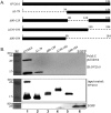
dsRNA binding domain of OV20.0. (A) An illustration showing the constructs expressing the wild type and deletions of ORFV OV20.0 used in this study. The deleted regions are presented as dashes, and the numbers indicate the corresponding amino acids. (B) Plasmids expressing FLAG-tagged wild-type OV20.0 and Δ1-79, Δ80-129, Δ130-184, and Δ79-184 mutants of OV20.0 or EGFP were transfected individually into human 293T cells, which was followed by a poly(I·C) pulldown assay. The collected pellets (upper) as well as 20% of the cell lysate, which was used as the input control (lower), were separately subjected to Western blot analysis using anti-FLAG or anti-EGFP antibodies. The data shown are representative of three independent experiments. Lane M, molecular size marker.
FIG 2
FIG 2
Regions of OV20.0 involved in PKR interaction. (A) Human 293T cells were individually transfected with plasmids expressing the FLAG-tagged wild type and Δ1-79, Δ80-129, Δ130-184, and Δ79-184 mutants of OV20.0. The next day, the cell lysates were harvested for immunoprecipitation (IP) using anti-FLAG agarose beads. (Lower) An aliquot of total cell lysate (20% of IP) was used as the input control. Proteins eluted from beads then were analyzed by Western blotting using either PKR or FLAG antibodies. (B) Full-length (Kozak20) OV20.0 or the Δ1-79 or Δ130-184 mutant fused with EGFP was transiently expressed in 293T cells. At 24 h after transfection, the expression of endogenous PKR was detected by immunofluorescent assay (IFA) using PKR antibody, which was followed by confocal microscopy. The presence of PKR in nuclei in cells expressing full-length OV20.0 is indicated with an arrow. (C) Map of the constructs expressing the wild type and the deletion of the various individual functional domains of PKR. Dashes and the numbers indicate deleted regions and the residues of PKR, respectively. (D) 293T cells were cotransfected with plasmids expressing FLAG-tagged OV20.0 and one of the HA-tagged PKR proteins or EGFP (as a negative control). Immunoprecipitation was conducted with FLAG antibody-conjugated agarose beads followed by detection of PKR in the pellet fraction (upper) or of OV20.0 and EGFP. (Lower) Proteins in whole-cell lysate, which were used as input controls. Three independent experiments were performed.
FIG 3
FIG 3
C terminus of OV20.0 suppresses dsRNA-induced PKR phosphorylation. Human 293T cells were mock transfected (lanes 1 and 7) or transfected with different constructs expressing the various OV20.0 mutants (lanes 2 to 6). At 24 h posttransfection, the cells were left untreated (lane 1, mock) or were transfected with 1 μg poly(I·C) (lanes 2 to 7) for a further 4 h. Total proteins were separated by SDS-PAGE, which was followed by Western blotting using antiserum specific to phosphorylated PKR (PKR-p), basal PKR (PKR), or actin (β-actin), which acted as the loading control. Lane PC indicates cells transfected with poly(I·C) (without OV20.0) for PKR activation. Three independent experiments were performed.
FIG 4
FIG 4
OV20.0 inhibits PACT-mediated PKR phosphorylation. (A) Using 293T cells, 5 μg of PACT plasmid was cotransfected with various amounts (0, 1, 3, 5, or 10 μg) of plasmids expressing OV20.0 (lanes 1 to 5) or were mock transfected (lane 6). At 24 h after transfection, total proteins were collected for Western blot analysis using antibodies specific to phosphorylated PKR (PKR-p), basal PKR (PKR), PACT, OV20.0, or actin (β-actin), which acted as the loading control. (B) The intensity of the phosphorylated PKR band was quantified by ImageJ, and the ratio of the various proteins to the basal level of PKR was calculated. The level of PKR phosphorylation in the mock control (sample in lane 1) was arbitrarily set to 1, and the relative inhibition ability of OV20.0 then was estimated. Results are representative of 3 independent experiments.
FIG 5
FIG 5
OV20.0 interacts with PACT. (A) Plasmids expressing FLAG-tagged wild-type OV20.0 (lane 1), various OV20.0 deletion mutants (lanes 2 to 5), or empty vector (lane 6) were separately transfected in 293T cells; this was followed by immunoprecipitation using FLAG antibody-conjugated agarose beads. The FLAG pulldown fractions then were probed using antiserum specific for either PACT or FLAG. (B) Maps of the PACT deletion constructs are shown. The dashes and the numbers identify the truncated regions and corresponding residues, respectively. (C) Plasmids expressing FLAG-tagged wild-type PACT (lane 1) and the various individual PACT deletion mutants (lanes 2 to 4) were cotransfected with HA-tagged OV20.0 into 293T cells for 24 h. The whole-cell lysate next was immunoprecipitated with anti-FLAG agarose beads. The pellet factions (IP: FLAG; upper) and the input control (input; lower) then were separated by SDS-PAGE, which was followed by immunoblot analysis using antibodies specific to HA (for OV20.0), to FLAG (for the PACTs), or to β-actin. Results are representative of 3 independent experiments.
FIG 6
FIG 6
OV20.0 blocks PACT-mediated PKR phosphorylation by interacting with PKR rather than with PACT. A total of 5 μg of plasmid expressing FLAG-tagged PACTΔ1 was transfected alone (lane 2) or cotransfected with 1, 5, or 10 μg of constructs expressing HA-tagged OV20.0 (lanes 3 to 5) or with empty vectors (lanes 6 to 8) into 293T cells. At 24 h after transfection, the individual cell lysates were harvested and then were subjected to Western blot analysis using antibodies specific to phosphorylated PKR (PKR-p), basal PKR (PKR), HA (OV20.0-HA), FLAG (PACTΔ1-FLAG), or β-actin. Results are representative of 3 independent experiments.
FIG 7
FIG 7
OV20.0 prevents the association of PACT with PKR. Human 293T cells were transfected with 5 μg of construct expressing FLAG-tagged PACT alone (lane 2) or together with 1, 5, or 10 μg of plasmids expressing HA-tagged OV20.0 (lanes 3 to 5) for 24 h. (Lower) Total cell lysates then were harvested individually for immunoprecipitation using anti-FLAG agarose beads; 20% of the cell lysate was kept as the input control. (Upper) The pellet fractions were subjected to Western blot analysis using anti-PKR antibodies. Results are representative of 3 independent experiments.
FIG 8
FIG 8
Double-stranded RNA facilitates the interaction of OV20.0 with PACT but not between OV20.0 and PKR. (A) A construct expressing FLAG-tagged OV20.0 (lanes 2 and 3) or the vector alone (lane 1) were transfected into 293T cells, or (B) goat fibroblast cells (lane 4) were infected with recombinant ORFV OV20.0-FLAG-GFP virus (lanes 5 and 6) at an MOI of 2 for 24 h. Total cell lysates of these transfections then were immunoprecipitated using anti-FLAG agarose beads with (lanes 2 and 5) or without (lanes 3 and 6) the addition of 0.5 U of RNase V1. The proteins bound to the beads were collected and then subjected to Western blot analysis using antibodies against PKR, PACT, or FLAG (OV20.0-FLAG). Results are representative of two independent experiments.
FIG 9
FIG 9
Sequence analysis of RBDs of OV20.0, PACT, and PKR. Sequence of the RBDs of OV20.0, PACT, and PKR were aligned. The dashes represent gaps in the alignment. The asterisks indicate fully conserved residues. The columns (indicated by colons) indicate that one of the following strong residue groups is conserved: FI, FY, KR, KQ, MILV, NQ, or ST. A single dot indicates one of the following groups is conserved: AC, AGS ENS, or HNQR. Boxes mark the conserved residues for dsRNA binding proteins.
FIG 10
FIG 10
Mechanisms by which OV20.0 mediates the inhibition of PKR activation. PKR is activated by dsRNA and PACT protein consisting of domains I, II, and III. Binding of the two activators leads to a change in the conformation of PKR that affects the protein's dimerization and autophosphorylation. ORFV OV20.0 inhibits PKR activation via at least three mechanisms: sequestering dsRNA (1), interaction with PKR (2), and binding to PACT (3). RBD, dsRNA-binding domain; kinase D, kinase domain.

Similar articles

Cited by

References

    1. McInnes CJ, Wood AR, Mercer AA. 1998. Orf virus encodes a homolog of the vaccinia virus interferon-resistance gene E3L. Virus Genes 17:107–115. doi:10.1023/A:1026431704679. - DOI - PubMed
    1. Smith GL, Benfield CT, Maluquer de Motes C, Mazzon M, Ember SW, Ferguson BJ, Sumner RP. 2013. Vaccinia virus immune evasion: mechanisms, virulence and immunogenicity. J Gen Virol 94:2367–2392. doi:10.1099/vir.0.055921-0. - DOI - PubMed
    1. Guerra S, Caceres A, Knobeloch KP, Horak I, Esteban M. 2008. Vaccinia virus E3 protein prevents the antiviral action of ISG15. PLoS Pathog 4:e1000096. doi:10.1371/journal.ppat.1000096. - DOI - PMC - PubMed
    1. Eduardo-Correia B, Martinez-Romero C, Garcia-Sastre A, Guerra S. 2014. ISG15 is counteracted by vaccinia virus E3 protein and controls the proinflammatory response against viral infection. J Virol 88:2312–2318. doi:10.1128/JVI.03293-13. - DOI - PMC - PubMed
    1. Chang HW, Jacobs BL. 1993. Identification of a conserved motif that is necessary for binding of the vaccinia virus E3L gene products to double-stranded RNA. Virology 194:537–547. doi:10.1006/viro.1993.1292. - DOI - PubMed

Publication types

MeSH terms

LinkOut - more resources