. 2015 Sep 22;12(11):1865-75.
doi: 10.1016/j.celrep.2015.08.025. Epub 2015 Sep 3.

Cross-Talk between Carbon Metabolism and the DNA Damage Response in S. cerevisiae

Affiliations

Cross-Talk between Carbon Metabolism and the DNA Damage Response in S. cerevisiae

Kobi J Simpson-Lavy et al. Cell Rep. .

Abstract

Yeast cells with DNA damage avoid respiration, presumably because products of oxidative metabolism can be harmful to DNA. We show that DNA damage inhibits the activity of the Snf1 (AMP-activated) protein kinase (AMPK), which activates expression of genes required for respiration. Glucose and DNA damage upregulate SUMOylation of Snf1, catalyzed by the SUMO E3 ligase Mms21, which inhibits SNF1 activity. The DNA damage checkpoint kinases Mec1/ATR and Tel1/ATM, as well as the nutrient-sensing protein kinase A (PKA), regulate Mms21 activity toward Snf1. Mec1 and Tel1 are required for two SNF1-regulated processes-glucose sensing and ADH2 gene expression-even without exogenous genotoxic stress. Our results imply that inhibition of Snf1 by SUMOylation is a mechanism by which cells lower their respiration in response to DNA damage. This raises the possibility that activation of DNA damage checkpoint mechanisms could contribute to aerobic fermentation (Warburg effect), a hallmark of cancer cells.


Figures

Figure 1
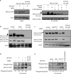
Figure 1. Mms21 is a phospho-protein
a. Glucose to 2% (15 minutes) or 0.03% MMS (1 hour) was added to media containing Mms21-3HA cells (or cells lacking the HA tag). Samples were taken for immunoprecipitations using anti-HA. Left: input. Right: Immunopreciptated Mms21-3HA was run on 15% phos-tag and regular gels, and probed with anti-HA. Phosphorylated Mms21 is indicated with arrows. b, c. Glucose to 2% (15 minutes) (b) or 0.03% MMS (1 hour) (c) was added to media containing Mms21-3HA cells (or cells lacking the HA tag) bearing indicated plasmids or empty vector. Cells were processed for immunoblots, and whole extracts loaded onto 15% gels containing phos-tag. A long and a short exposure for anti-HA are shown. After probing for HA, blots were stripped and reprobed for actin (b) or identical amounts of protein extract loaded onto a new gel and the membrane probed for actin (c). Phosphorylated Mms21 is indicated with arrows - only the upper phospho-Mms21 band is regulated by PKA. d, e. Mms21-3HA (or cells lacking the HA tag) were treated as with 4% glucose for 15 minutes and/or 0.3% MMS for 1 hour, and samples taken for immunoprecipitations using anti-HA beads. Immunoprecipated Mms21-3HA was run on 12% gels, and probed with mouse anti-HA and rabbit anti-RxxS* simultaneously using fluorescent secondary antibodies. Ratios of pRxxS/HA were calculated by quantification using ImageJ.
Figure 2
Figure 2. Phenotypes of Mms21 phospho-mutants
a, b, c. Cells bearing the indicated plasmids were serially diluted tenfold from saturated cultures onto SC-ura plates containing the indicated carbon sources and genotoxins, and grown at the indicated temperatures. Photographs were taken on the indicated days. For the genotoxin and temperature sensitivity assays, similar results were obtained for cells grown on glucose. For the carbon source growth assay, similar results were obtained at 25°C. None of the strains grew on either 3% ethanol or 3% glycerol at 37°C. mec1Δtel1Δ cells are also sml1Δ. d. mms21-11 cells bearing the indicated MMS21 plasmids or empty vector and prHXT3:LacZ were grown overnight in 2% galactose, and glucose added to 2%. HXT3 expression was measured by β-galactosidase activity every half-hour for 90 minutes. The rate of HXT3 expression is normalized to that of wild-type cells. N=3. Error bars are +− 1 standard deviation. e. mms21-11 cells bearing the indicated MMS21 plasmids or empty vector and prADH2:LacZ were grown overnight in 4% glucose. Cells were washed three times with water and resuspended in media containing 3% glycerol. Samples were taken for β-galactosidase assays each hour for four hours. The rate of ADH2 expression is normalized to that of wild-type cells. N=3. Error bars are +− 1 standard deviation.
Figure 3
Figure 3. Alanine scan of the C-terminus of Mms21
a. WT, mms21-11, or cells with the indicated integrated mutations in the MMS21 locus containing a prADH2:LacZ plasmid were grown overnight in 4% glucose. Cells were washed three times with water and resuspended in media containing 3% glycerol. Samples were taken for β-galactosidase assays each hour for four hours. The rate of ADH2 expression is normalized to that of wild-type cells for each experiment. ADH2 expression in snf1Δ cells bearing Snf1K549R is included for comparison. N=3. Error bars are +− 1 standard deviation. b. mms21-11 cells bearing the indicated MMS21 plasmids or empty vector and prADH2:LacZ were grown overnight in 4% glucose. Cells were washed three times with water and resuspended in media containing 3% glycerol. Samples were taken for β-galactosidase assays each hour for four hours. The rate of ADH2 expression is normalized to that of wild-type cells. N=3. Error bars are +− 1 standard deviation. c. WT, mms21-11, or cells with the indicated integrated mutations in the MMS21 locus containing a prHXT3:LacZ plasmid were grown overnight in 2% galactose, and glucose added to 2%. HXT3 expression was measured by β-galactosidase activity each half-hour for 90 minutes. The rate of HXT3 expression is normalized to that of wild-type cells for each experiment. HXT3 expression in snf1Δ cells bearing Snf1K549R is included for comparison. N=3. Error bars are +− 1 standard deviation. d. mms21-CH cells bearing the indicated MMS21 plasmids or empty vector and prHXT3:LacZ were grown overnight in 2% galactose, at 30°C. The temperature was raised to 34°C for one hour, and preheated glucose added to 2%. HXT3 expression was measured by β-galactosidase activity each half-hour for 90 minutes. The rate of HXT3 expression is normalized to that of wild-type cells. N=3. Error bars are +− 1 standard deviation. e, f. Cells bearing the indicated integrated mutations in the MMS21 locus were grown overnight in 2% galactose at 30°C. Glucose was added to 2%, and samples taken for immunoblotting at the indicated times. An extra copy of Snf1 on a CEN plasmid was present which did not affect the experiment. g. mms21-CH cells containing a plasmid with the indicated mutation in MMS21 were grown overnight in 2% galactose at 30°C. The temperature was raised to 34°C for one hour, and preheated glucose added to 2%. Samples were taken for immunoblots at indicated times.
Figure 4
Figure 4. Kinases that regulate Mms21 activity towards Snf1 via phosphorylation of S261
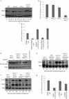
mec1Δ and mec1Δtel1Δ cells are also sml1Δ. a. Cells were grown overnight in 2% galactose at 30°C. Glucose was added to 2% and samples taken for immunoblots at the indicated times. b. Cells were grown overnight in 2% galactose at 30°C. Glucose was added to 2%. HXT3 expression was measured by β-galactosidase activity each half-hour for 90 minutes. The rate of HXT3 expression is normalized to that of wild-type cells. N=3. Error bars are +− 1 standard deviation. c. Indicated cells containing a prADH2:LacZ plasmid and a plasmid with either MMS21WT or MMS21S261D were grown overnight in 4% glucose. Cells were washed three times with water and resuspended in media containing 3% glycerol. Samples were taken for β-galactosidase assays each hour for four hours. The rate of ADH2 expression is normalized to that of wild-type cells. N=3. Error bars are +− 1 standard deviation. d. Indicated cells with an extra copy of plasmid borne Mms21-3HA were grown overnight in 2% galactose at 30°C. Glucose was added to 2% and samples taken after 15 minutes. Cells were processed for immunoblots, and whole extracts loaded onto 15% gels containing phos-tag. After probing for HA, blots were stripped and reprobed for actin. Phosphorylated Mms21 is indicated with arrows. e, f. Indicated cells bearing indicated plasmids were grown overnight in 2% galactose at 30°C. Glucose was added to 2% and samples taken for immunoblots at the indicated times. g. Indicated cells bearing indicated plasmids were grown overnight in 2% galactose at 30°C. Glucose was added to 2%. HXT3 expression was measured by β-galactosidase activity each half-hour for 90 minutes. The rate of HXT3 expression is normalized to that of wild-type cells. N=3. Error bars are +− 1 standard deviation.
Figure 5
Figure 5. MMS downregulates ADH2 expression
a. WT cells were grown overnight in 4% glucose at 30°C. Cells were washed three times with water and resuspended in media containing 3% glycerol and the indicated MMS concentrations. Samples were taken for β-galactosidase assays every hour for four hours. The rate of HXT3 expression is normalized to that of untreated cells. N=3. Error bars are +− 1 standard deviation. mec1Δtel1Δ cells are also sml1Δ. b. ulp1-ts cells (strain 1274 (Wykoff and O'Shea, 2005)) expressing Snf1-8myc (Liu et al., 2011), Snf1K549R-8myc (Simpson-Lavy and Johnston, 2013) or Snf1 (with no tag) (Shirra et al., 2008), together with a plasmid containing GAL:His6-FLAG-SMT3 were grown overnight at 24°C in 2% galactose. The temperature was elevated to 37°C for 4 hours before addition of preheated glucose to 2% for 15 minutes or 0.3% MMS for 1 hour (or both conditions) as indicated. Samples were processed for immunoprecipitations with MYC. c. Cells bearing the indicated mutations were grown overnight in 4% glucose at 30°C. Cells were washed three times with water and resuspended in media containing 3% glycerol with and without 0.03% MMS. Samples were taken for β-galactosidase assays every hour for four hours and the rate of ADH2 expression determined. The rate of ADH2 expression in the presence of 0.03% MMS was normalized against the rate of ADH2 expression for that strain in the absence of MMS (the mutant strains all have dramatically increased ADH2 expression in the absence of MMS (Figures, 2, 3, 4 and (Simpson-Lavy and Johnston, 2013)). Comparing the WT to any of the mutants gave a significant difference at p<0.01. In contrast, the differences between the mutants was not significant (p>0.05). N=3. Error bars are +− 1 standard deviation. d. Wild-type cells were grown overnight in 2% galactose at 30°C. 0.3% MMS was added for two hours. Samples with and without 0.3% MMS treatment for 2 hours were taken for immunoblots.
Figure 6
Figure 6. Snf1 SUMOylation and phosphorylation are independently regulated
a. ulp1-ts cells (strain 1274 (Wykoff and O'Shea, 2005)) expressing Snf1-8myc (Liu et al., 2011) or Snf1 (with no tag) (Shirra et al., 2008), a plasmid with either MMS21 or MMS21S261D, together with a plasmid containing GAL:His6-FLAG-SMT3 were grown overnight at 24°C in 2% galactose. The temperature was elevated to 37°C for 4 hours before harvesting for anti-myc immunoprecipitations with B480. Blots were probed with anti-myc (for Snf1-8myc) and anti-FLAG (for Smt3). b. Cells containing the indicated Snf1-8myc plasmid and MMS21S261D plasmid where applicable were grown with either 2% galactose or 4% glucose as indicated overnight at 30°C. Cells were harvested and Snf1-8myc immunoprecipated. Cells with untagged Snf1 were used as a control. Protein kinase assays were performed using the ADP-glo kinase assay kit with SAMS peptide as a phosphate acceptor. N=3. c. Indicated cells containing a prHXT3:LacZ plasmid and either a 2μ plasmid overexpressing REG1 or empty vector were grown overnight in 2% galactose at 30°C. The temperature was raised to 34°C for one hour, and preheated glucose added to 2%.HXT3 expression was measured by β-galactosidase activity each half-hour for 90 minutes. The rate of HXT3 expression is normalized to that of wild-type cells. N=3. Error bars are +− 1 standard deviation. d. Indicated cells containing a prHXT3:LacZ plasmid and either a plasmid with MMS21WT or MMS21S261D were grown overnight in 2% galactose. HXT3 expression was measured by β-galactosidase activity each half-hour for 90 minutes. The rate of HXT3 expression is normalized to that of wild-type cells. N=3. Error bars are +− 1 standard deviation. e. reg1Δsit4Δsnf1Δubp8Δ cells were transformed with prs314 (TRP1) bearing either SNF1WT, snf1K84R (kinase dead), or snf1K549R (non-SUMOylatable) and with Ycplac33 (URA3) bearing either MMS21WT or MMS21S261D and plated onto –Trp -Ura 2% glucose plates, and photographed after 5 days incubation at 30°C. f. Wild-type ADE2 cells containing plasmids prs314 (TRP1) and prs316 (URA3) (positive control for growth on 5-FOA containing media), wild-type ADE2 cells with integrated TRP1 and URA3 (negative control for growth on 5-FOA containing media), and the three viable strains from figure 6e were serially tenfold diluted onto the indicated media. Plates were photographed after 5 days incubation at 30°C. The 5-FOA promotes loss of the MMS21S261D plasmid from reg1Δsit4Δubp8Δ SNF1 MMS21S261D cells, resulting in Snf1 toxicity on -Trp 5-FOA media.
Figure 7
Figure 7. Model
Proposed signaling pathway based on data presented in this article.
Figure 7
Figure 7. Model
Proposed signaling pathway based on data presented in this article.

Similar articles

Cited by

References

    1. BARRETT L, ORLOVA M, MAZIARZ M, KUCHIN S. Protein kinase A contributes to the negative control of Snf1 protein kinase in Saccharomyces cerevisiae. Eukaryot Cell. 2012;11:119–28. - PMC - PubMed
    1. BOJUNGA N, ENTIAN KD. Cat8p, the activator of gluconeogenic genes in Saccharomyces cerevisiae, regulates carbon source-dependent expression of NADP-dependent cytosolic isocitrate dehydrogenase (Idp2p) and lactate permease (Jen1p). Mol Gen Genet. 1999;262:869–75. - PubMed
    1. BRACHMANN CB, DAVIES A, COST GJ, CAPUTO E, LI J, HIETER P, BOEKE JD. Designer deletion strains derived from Saccharomyces cerevisiae S288C: a useful set of strains and plasmids for PCR-mediated gene disruption and other applications. Yeast. 1998;14:115–32. - PubMed
    1. BUSTI S, COCCETTI P, ALBERGHINA L, VANONI M. Glucose signaling-mediated coordination of cell growth and cell cycle in Saccharomyces cerevisiae. Sensors (Basel) 2010;10:6195–240. - PMC - PubMed
    1. CABA E, DICKINSON DA, WARNES GR, AUBRECHT J. Differentiating mechanisms of toxicity using global gene expression analysis in Saccharomyces cerevisiae. Mutat Res. 2005;575:34–46. - PubMed

Publication types

MeSH terms

Substances