. 2015 Sep 2;10(9):e0135869.
doi: 10.1371/journal.pone.0135869. eCollection 2015.

Design of a Novel Low Cost Point of Care Tampon (POCkeT) Colposcope for Use in Resource Limited Settings

Affiliations

Design of a Novel Low Cost Point of Care Tampon (POCkeT) Colposcope for Use in Resource Limited Settings

Christopher T Lam et al. PLoS One. .

Abstract

Introduction: Current guidelines by WHO for cervical cancer screening in low- and middle-income countries involves visual inspection with acetic acid (VIA) of the cervix, followed by treatment during the same visit or a subsequent visit with cryotherapy if a suspicious lesion is found. Implementation of these guidelines is hampered by a lack of: trained health workers, reliable technology, and access to screening facilities. A low cost ultra-portable Point of Care Tampon based digital colposcope (POCkeT Colposcope) for use at the community level setting, which has the unique form factor of a tampon, can be inserted into the vagina to capture images of the cervix, which are on par with that of a state of the art colposcope, at a fraction of the cost. A repository of images to be compiled that can be used to empower front line workers to become more effective through virtual dynamic training. By task shifting to the community setting, this technology could potentially provide significantly greater cervical screening access to where the most vulnerable women live. The POCkeT Colposcope's concentric LED ring provides comparable white and green field illumination at a fraction of the electrical power required in commercial colposcopes. Evaluation with standard optical imaging targets to assess the POCkeT Colposcope against the state of the art digital colposcope and other VIAM technologies.

Results: Our POCkeT Colposcope has comparable resolving power, color reproduction accuracy, minimal lens distortion, and illumination when compared to commercially available colposcopes. In vitro and pilot in vivo imaging results are promising with our POCkeT Colposcope capturing comparable quality images to commercial systems.

Conclusion: The POCkeT Colposcope is capable of capturing images suitable for cervical lesion analysis. Our portable low cost system could potentially increase access to cervical cancer screening in limited resource settings through task shifting to community health workers.


Conflict of interest statement

Competing Interests: NR has founded a company called Zenalux Biomedical and she and other team members (MSK) have developed technologies related to this work where the investigators or Duke may benefit financially if this system is sold commercially. For the concept of this system a provisional patent application was filed by CTL and NR with the title: COLPOSCOPES HAVING LIGHT EMITTERS AND IMAGE CAPTURE DEVICES AND ASSOCIATED METHODS. Number; PCT/US-14/67038 filed on 11/24/2014. This does not alter the authors' adherence to PLOS ONE policies on sharing data and materials.

Figures

Fig 1
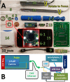
Fig 1. System schematic of our Point of Care Tampon Colposcope (POCkeT Colposcope) with computer aided 3D rendering.
(A) of the assembled device with the essential system components (1) USB cable, (2) manual focus knob, (3) probe handle or body, (4,7) clear sterile disposable sheath, (5) ABS 3D printed shell, (6) linear glass polarizer; and 3D rendering (B) of the exploded schematic view of the device (8) the color CMOS detector, (9) clam shell ABS probe handle, (10) polarizer and illumination ring holder, (11) linear glass polarizer, (12) concentric white and green LED ring, (13) linear film polarizer, (14) clear sterile disposable sheath; and a block diagram (C) of the system interface that combines a single communication cable (Cat5e) to allow for 2-way communication and control of the camera and LED ring through the Smartphone/Tablet and Arduino Microcontroller via a powered USB hub.
Fig 2
Fig 2. A comparison of different colposcopy platforms across resolution, diagonal field of view, and working distance with a representative image of a USAF1951 test target from each.
Representative images of USAF21951 resolution target with the Leisegang Optik 2 at 3.75X magnification (A), 5.0MP POCkeT Colposcope at 4X (B), 2.0MP POCkeT Colposcope at 4X (C), Apple iPhone 5S at 3.5X (D), and Canon SX50HS at 2.5X (E). Our POCkeT Colposcope system (2.0MP as blue circles and 5.0MP as purple circles) have the capability to resolve features as small as 10 micron (F) comparable to the highest magnification of clinical systems. Diagonal field of view (FOV) of Leisegang Optik 2 (G) (Red Diamond) has the largest of any of the digital systems, because of the large image sensor format and 3:2 aspect ratio and would produce images 17% wider than the comparable system with 4:3 aspect ratio. Our POCkeT Colposcope system is still capable of providing a FOV of at least 30 mm, comparable resolving power for identification of potential lesions, while at much closer working distances (H) between 50 to 10 mm. Images have been cropped to better demonstration magnification of ROI (region of interests).
Fig 3
Fig 3. Color accuracy was assessed using NIST calibrated color target (X-Rite Rezchecker) for all digital colposcopy systems.
The X-Rite Rezchecker color target was imaged under D50 or White 5000K illumination, (A) Leisegang Optik 2, (B) 5.0MP POCkeT Colposcope (C) 2.0MP POCkeT Colposcope, (D) Canon SX50HS, and (E) Apple iPhone 5S. Perceptible color difference is therefore the approximately equal to the Euclidean distance between reference and measured L*a*b* values. ΔC ab (F) green bars are for mean and purple bars for max color difference, without accounting for any luminance differences present and ΔE* ab (G) blue bars are for mean and red bars for max color differnece, with weighting for any luminance differences. The best performing is the Leisegang, with mean perceptible color difference ΔC ab of 2.9 (F) and ΔE ab of 5.24 (G), accounting for luminance. Generally, when luminance was included, the error increased 2.5 times.
Fig 4
Fig 4. Performance of a color matching algorithm for use on the POCkeT Colposcope images to the high-end Leisegang Optik 2.
The X-Rite Rezchecker target with NIST calibrated color patches captured in jpeg format by both Leisegang Optik 2 and our 5.0MP TVDC system with on-board white balance and color correction algorithm’s enabled. The pairs are the color matched using Adobe Photoshop CS6, 5.0MP POCkeT Colposcope images on the middle (BC), and 2.0MP POCkeT Colposcope images on the right (DE). In panel FG, we recalculated the ΔC ab (F) green bars for mean and purple bars for max color difference, the darker color bars are the post-color match values, without accounting for any luminance differences present. ΔE* ab (G) blue bars for mean and red bars for max color differnece, again darker color bars are post-CCM,with weighting for any luminance differences. Note, that post color match values were generally lower for both metric values and closer to the Leisegang Optik 2 reference values.
Fig 5
Fig 5. Representative spectra and beam characteristics from white and green field illumination beam characteristics of compared digital colposcopy systems.
(A) Examination of the white spectra of the various digital colposcopy systems: Leisegang Optik 2 (black) with a 5000K white light emitting diode (LED), Wallach Zoomscope halogen source (red), Apple iPhone 5S white LED source (purple), 2.0MP POCkeT Colposcope White LED with 5700K Color Temperature (green), and 5.0MP POCkeT Colposcope with white 5000K LED (blue). (B) Examination of the green field illumination spectra from the Leisegang Optik 2 (LO2) clinical digital colposcope, which has a 5000K White LED source in solid black, the Wallace colposcope using a halogen source (red line), both using broad bass-pass filters. Our 5.0MP POCkeT Colposcope with dedicated green LEDs (dashed blue). (C to G) Luminance contour plots normalized from 0 to 1, the Full Width Half Maximum (FWHM) beam diameter is defined as 50% contour luminance for each illumination systems at their respective working distances. The light blue rings indicate the location of the FWHM and scale bars = 10 mm. The Leisegang Optik 2 (C) had a FWHM beam diameter of 62 mm at Working Distance (WD) of 300 mm, 5.0 MP POCkeT Colposcope (D) had a FHWM beam diameter of 40.2 mm at WD of 40 mm, 2.0MP POCkeT Colposcope (E) had a FWHM beam diameter of 42.8 mm at WD of 40 mm, Wallach Tristar (F) a FWHM beam diameter of 64.2 mm at WD of 300 mm and the Apple iPhone 5S (G) a FWHM beam diameter of 70.2 mm at WD of 70 mm.
Fig 6
Fig 6. Representative images from the percentage of specular reflection as a function of optical power.
The top most panels (ABCD) correspond to White 5000K color temperature LED illumination and bottom panels (EFGH) corresponding to Green LED illumination with the POCkeT Colposcope on a simulated HSIL/CIN2+ cervix. From left to the right is increasing optical power achieved with PWM (pulse width modulation) control and shows the expected increase in specular reflection to above the threshold of failure of 0.5% for white light and 0.05% for green light, set by the commericial system.
Fig 7
Fig 7. Representative cervical images captured the digital colposcopy systems on simulated normal and high grade simulated cervices.
(A-E) Simulated HSIL Cervix in Gaumard Zoe Gynecologic Simulator, (F-J) Simulated Normal cervix in Gaumard Zoe Gynecologic Simulator. Paired images from a Leisegang Optik 2 Digital Colposcope (AF) at 3.75X at a working distance of 300 mm, our 5.0MP POCkeT Colposcope (BG) at 4X at working distance of 30 mm, our 2.0MP POCkeT Colposcope (CH) at 4X at working distance of 30 mm, a Canon SX50HS digital camera (DI) at 2.75X at a working distance of 300 mm, and an Apple iPhone 5S (EJ) at 3.75X at a working distance of 70 mm. There was no color correction performed on the POCkeT Colposcope images. The target illumination intensity was set at the 1% specular reflection threshold for all systems.
Fig 8
Fig 8. Representative cervical images captured with the high-end digital colposcope at 3.75X magnification and our 2.0MP POCkeT Colposcope.
(ABC,GHI) Representative cervical images captured at 3.75 X with Leisegang Optik 2 Digital Colposcope at a working distance of 300 mm and (DEF, JKL) concordant images at 4X with 2.0MP POCkeT Colposcope at a working distance of 40 mm. Normal Cervix (ADGJ), biopsy confirmed LSIL/CIN 1 (BEHK) at 3 o’clock position and normal but suspicious lesion at 7 o’clock, and HSIL/CIN3 confirmed LEEP pathology (CFIL) at 4 to 5 o’clock position. Color correction was performed on the POCkeT Colposcope’s images using color match function in Adobe Photoshop, GHI, JKL and original images in ABC,DEF. The white light illumination power for the POCkeT Colposcope are set at a 1.0% specular reflection threshold.
Fig 9
Fig 9. Representative cervical images captured with the high-end digital colposcope at 3.75X magnification and our 5.0MP POCkeT Colposcope.
(ABC,GHI) Representative cervical images captured at 3.75 X with Leisegang Optik 2 Digital Colposcope at a working distance of 300 mm and (DEF, JKL) concordant images at 4X with 5.0MP POCkeT Colposcope at a working distance of 40 mm. Normal Cervix (ADGJ), biopsy confirmed LSIL/CIN 1 (BEHK) at 1 o’clock position, and biopsy confirmed HSIL/CIN 3 pathology (CFIL) confirmed at the 8 and 11 o’clock. Color correction was performed on the POCkeT Colposcopes images using color match function in Adobe Photoshop, GHI, JKL and original images in ABC, DEF. The white light illumination power for the POCkeT Colposcope was set at the level for 1.0% specular reflection threshold, however we have moved to the 0.5% specular threshold for all subsequent cases.
Fig 10
Fig 10. Comparison of green field images of a Nabothian cyst between the high-end digital colposcope and our POCkeT Colposcope.
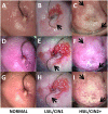
The following image panel is of a biopsy confirmed normal cervix with Nabothian cyst (yellow and clear nodules) taken by the Leisegang Optik 2(AB) at 3.75X magnification and the 5.0MP POCkeT Colposcopes (CD) at 4X magnification. Black arrows indicate prominent vascular features and note the enhanced contrast with the “green field” illumination strategies by both systems. Both systems use White 5000K color temperature LEDs, while a green LED is use by the 5.0MP POCkeT Colposcope when compared to the short-pass Red filter used by the Leisegang Optik 2. Color correction was performed on the POCkeT Colposcope images using the color match function in Adobe Photoshop, GHI, JKL and original images in ABC,DEF. Here the illumination power threshold were set at 1% and 0.5% specular reflection level, respectively for white and green field illumination with the POCkeT Colposcope. We have now moved to a 0.5% and 0.05% specular reflection threshold for illumination power for all subsequent clinical cases.

Similar articles

Cited by

References

    1. Arbyn M, Castellsague X, De Sanjose S, Bruni L, Saraiya M, Bray F, et al. Worldwide burden of cervical cancer in 2008. Annals of oncology. 2011;22(12):2675–86. 10.1093/annonc/mdr015 - DOI - PubMed
    1. Ferlay J, Bray F, Pisani P, Parkin DM. GLOBOCAN 2002: Cancer Incidence, Mortality and Prevalence Worldwide Lyon: IARCPress; 2004.
    1. Sankaranarayanan R, Basu P, Wesley RS, Mahe C, Keita N, Mbalawa CC, et al. Accuracy of visual screening for cervical neoplasia: results from an IARC multicentre study in India and Africa. International Journal of Cancer. 2004;110(6):907–13. - PubMed
    1. Saslow D, Solomon D, Lawson HW, Killackey M, Kulasingam SL, Cain J, et al. American Cancer Society, American Society for Colposcopy and Cervical Pathology, and American Society for Clinical Pathology screening guidelines for the prevention and early detection of cervical cancer. CA: a cancer journal for clinicians. 2012;62(3):147–72. - PMC - PubMed
    1. Ferris DG, Willner WA, Ho JJ. Colposcopes: a critical review. The Journal of family practice. 1991;33(5):506–15. Epub 1991/11/01. . - PubMed

Publication types