

CD1d- and MR1-Restricted T Cells in Sepsis
- PMID: 26322041
- PMCID: PMC4533011
- DOI: 10.3389/fimmu.2015.00401
CD1d- and MR1-Restricted T Cells in Sepsis
Abstract
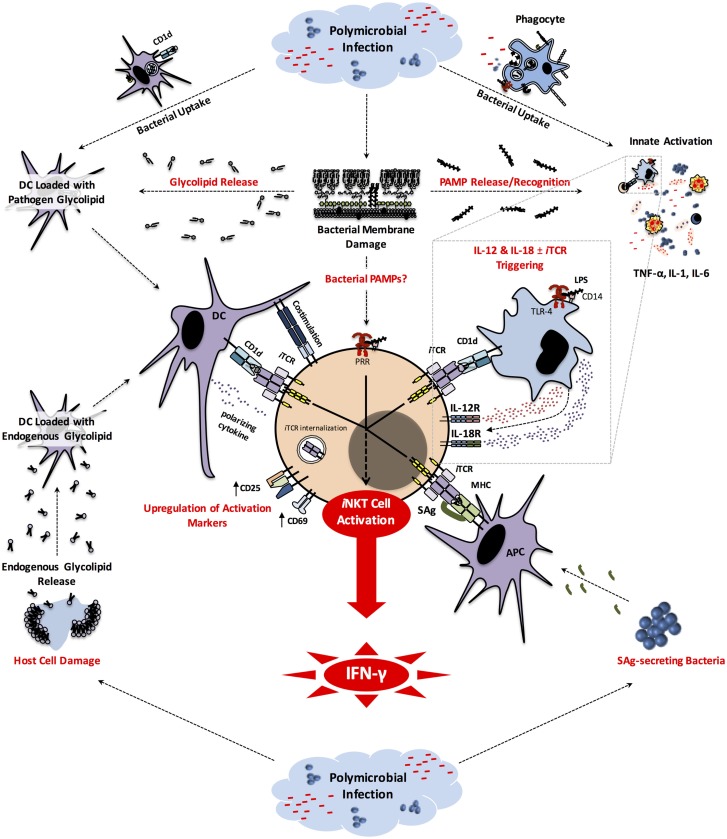
Dysregulated immune responses to infection, such as those encountered in sepsis, can be catastrophic. Sepsis is typically triggered by an overwhelming systemic response to an infectious agent(s) and is associated with high morbidity and mortality even under optimal critical care. Recent studies have implicated unconventional, innate-like T lymphocytes, including CD1d- and MR1-restricted T cells as effectors and/or regulators of inflammatory responses during sepsis. These cell types are typified by invariant natural killer T (iNKT) cells, variant NKT (vNKT) cells, and mucosa-associated invariant T (MAIT) cells. iNKT and vNKT cells are CD1d-restricted, lipid-reactive cells with remarkable immunoregulatory properties. MAIT cells participate in antimicrobial defense, and are restricted by major histocompatibility complex-related protein 1 (MR1), which displays microbe-derived vitamin B metabolites. Importantly, NKT and MAIT cells are rapid and potent producers of immunomodulatory cytokines. Therefore, they may be considered attractive targets during the early hyperinflammatory phase of sepsis when immediate interventions are urgently needed, and also in later phases when adjuvant immunotherapies could potentially reverse the dangerous state of immunosuppression. We will highlight recent findings that point to the significance or the therapeutic potentials of NKT and MAIT cells in sepsis and will also discuss what lies ahead in research in this area.
Keywords: CD1d; LPS; MAIT cell; MR1; NKT cell; infection; sepsis; α-galactosylceramide.
Figures
Similar articles
-
Role of CD1d- and MR1-Restricted T Cells in Asthma.Front Immunol. 2018 Aug 28;9:1942. doi: 10.3389/fimmu.2018.01942. eCollection 2018. Front Immunol. 2018. PMID: 30210497 Free PMC article. Review.
-
Activation and Function of iNKT and MAIT Cells.Adv Immunol. 2015;127:145-201. doi: 10.1016/bs.ai.2015.03.003. Epub 2015 Apr 25. Adv Immunol. 2015. PMID: 26073984 Review.
-
Deficiency of innate-like T lymphocytes in chronic obstructive pulmonary disease.Respir Res. 2017 Nov 28;18(1):197. doi: 10.1186/s12931-017-0671-1. Respir Res. 2017. PMID: 29179729 Free PMC article.
-
Insights Into Mucosal-Associated Invariant T Cell Biology From Studies of Invariant Natural Killer T Cells.Front Immunol. 2018 Jun 28;9:1478. doi: 10.3389/fimmu.2018.01478. eCollection 2018. Front Immunol. 2018. PMID: 30013556 Free PMC article. Review.
-
CD1d- and MR1-restricted invariant T cells: of mice and men.Curr Opin Immunol. 2006 Oct;18(5):519-26. doi: 10.1016/j.coi.2006.07.001. Epub 2006 Jul 25. Curr Opin Immunol. 2006. PMID: 16870416 Review.
Cited by
-
T cell dysregulation in inflammatory diseases in ICU.Intensive Care Med Exp. 2022 Oct 24;10(1):43. doi: 10.1186/s40635-022-00471-6. Intensive Care Med Exp. 2022. PMID: 36279072 Free PMC article. Review.
-
Effect of PD-1: PD-L1 in Invariant Natural Killer T-Cell Emigration and Chemotaxis Following Sepsis.Shock. 2016 May;45(5):534-9. doi: 10.1097/SHK.0000000000000553. Shock. 2016. PMID: 26717105 Free PMC article.
-
Improved MAIT cell functions following fecal microbiota transplantation for metastatic renal cell carcinoma.Cancer Immunol Immunother. 2023 May;72(5):1247-1260. doi: 10.1007/s00262-022-03329-8. Epub 2022 Nov 18. Cancer Immunol Immunother. 2023. PMID: 36396738 Free PMC article.
-
The immune system's role in sepsis progression, resolution, and long-term outcome.Immunol Rev. 2016 Nov;274(1):330-353. doi: 10.1111/imr.12499. Immunol Rev. 2016. PMID: 27782333 Free PMC article. Review.
-
Eomes transcription factor is required for the development and differentiation of invariant NKT cells.Commun Biol. 2019 Apr 29;2:150. doi: 10.1038/s42003-019-0389-3. eCollection 2019. Commun Biol. 2019. PMID: 31044175 Free PMC article.
References
Publication types
LinkOut - more resources
Full Text Sources
Other Literature Sources
Research Materials