. 2015 Aug 19;35(33):11707-18.
doi: 10.1523/JNEUROSCI.0613-15.2015.

Netrin1-DCC-Mediated Attraction Guides Post-Crossing Commissural Axons in the Hindbrain

Affiliations

Netrin1-DCC-Mediated Attraction Guides Post-Crossing Commissural Axons in the Hindbrain

Farnaz Shoja-Taheri et al. J Neurosci. .

Abstract

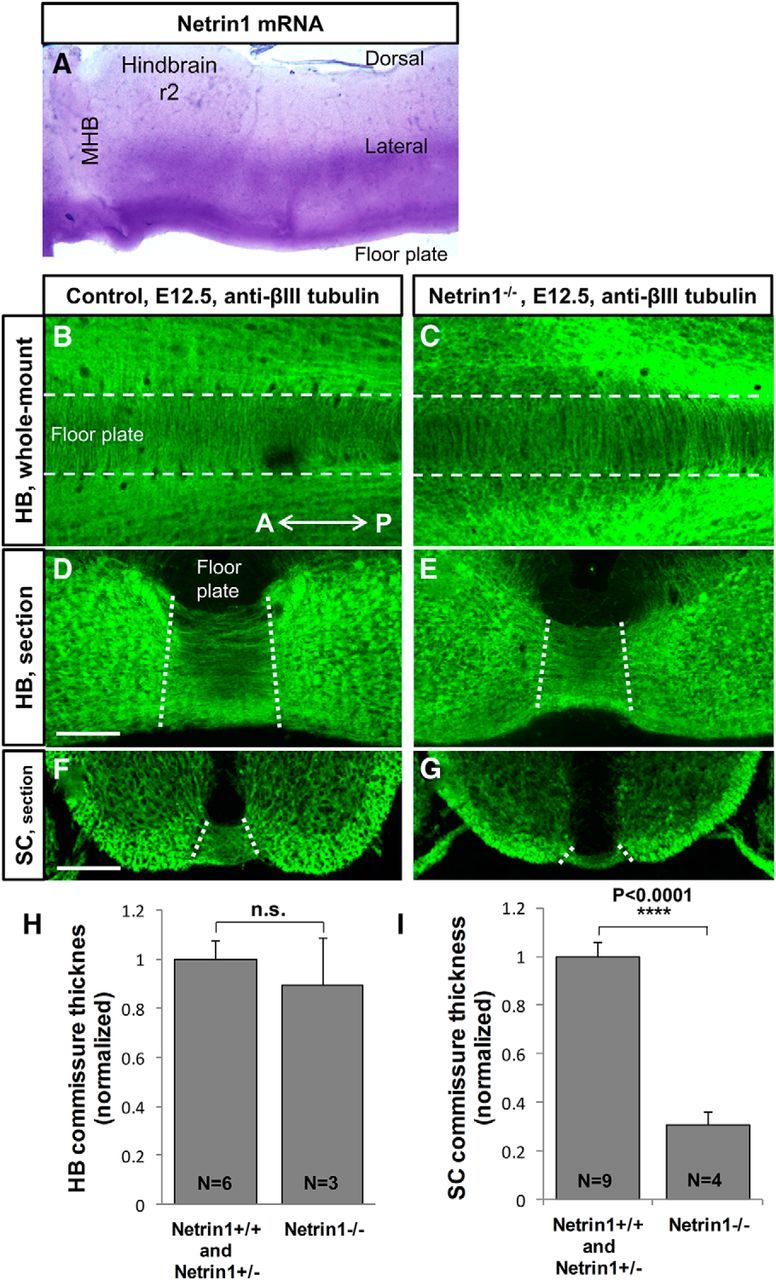
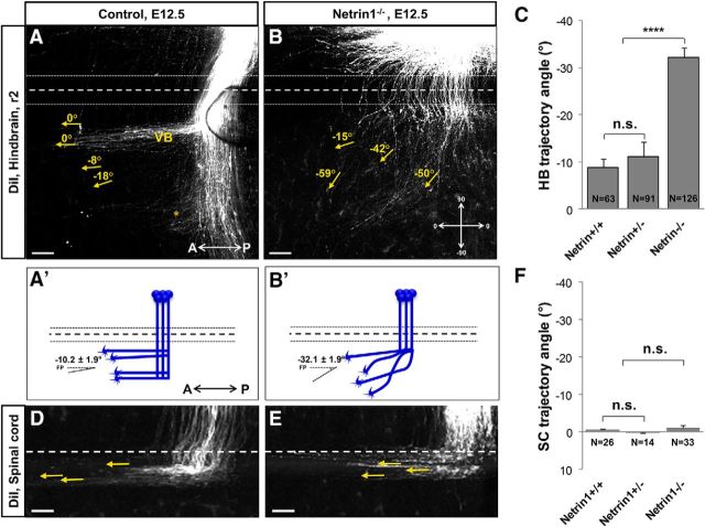
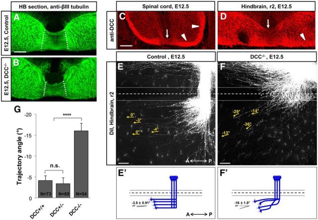
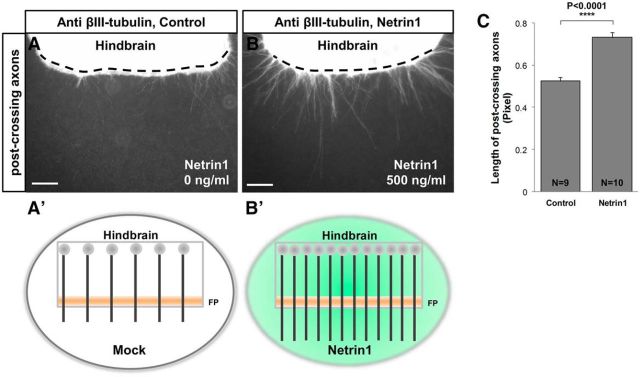
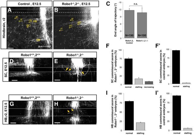
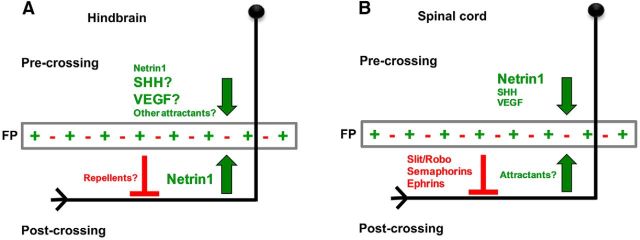
Commissural axons grow along precise trajectories that are guided by several cues secreted from the ventral midline. After initial attraction to the floor plate using Netrin1 activation of its main attractive receptor, DCC (deleted in colorectal cancer), axons cross the ventral midline, and many turn to grow longitudinally on the contralateral side. After crossing the midline, axons are thought to lose their responsiveness to Netrin1 and become sensitive to midline Slit-Robo repulsion. We aimed to address the in vivo significance of Netrin1 in guiding post-crossing axon trajectories in mouse embryos. Surprisingly, in contrast to the spinal cord, Netrin1 and DCC mutants had abundant commissural axons crossing in the hindbrain. In Netrin1 and DCC mutants, many post-crossing axons made normal turns to grow longitudinally, but projected abnormally at angles away from the midline. In addition, exposure of cultured hindbrain explants to ectopic Netrin1 caused attractive deflection of post-crossing axons. Thus, Netrin1-DCC signaling is not required to attract pre-crossing axons toward the hindbrain floor plate, but is active in post-crossing guidance. Also in contrast with spinal cord, analysis of hindbrain post-crossing axons in Robo1/2 mutant embryos showed that Slit-Robo repulsive signaling was not required for post-crossing trajectories. Our findings show that Netrin1-DCC attractive signaling, but not Slit-Robo repulsive signaling, remains active in hindbrain post-crossing commissural axons to guide longitudinal trajectories, suggesting surprising regional diversity in commissural axon guidance mechanisms.

Significance statement: The left and right sides of the brainstem and spinal cord are connected primarily by axon fibers that grow across the ventral midline, and then away on the other side to their targets. Based on spinal cord, axons are initially attracted by diffusible attractive protein signals to approach and cross the midline, and then are thought to switch to repulsive cues to grow away on the opposite side. Our results in the hindbrain show that the major midline attractant, Netrin1, is not required for midline crossing. However, the post-crossing axons depend on Netrin1 attraction to set their proper trajectories on the other side. Overall, these findings suggest that commissural axons use distinct mechanisms to navigate in different CNS regions.

Keywords: DCC; Netrin; Robo; Slit; axon guidance; commissural axon.


Figures

Figure 1.
Figure 1.
Netrin1 mutants have abundant commissural axons crossing the midline in the hindbrain. A, Netrin1 expression pattern revealed by in situ hybridization in E10.5 mouse embryos. Floor plate and lateral expression of Netrin1 mRNA is detected in the hindbrain whole-mount tissues. B–E, To show midline crossing axons, whole-mount and sections of mouse embryos were labeled with neuron-specific βIII-tubulin antibody. B, C, Hindbrain whole-mount preparations of E12.5 control (Netrin1+/+, Netrin1+/−) and Netrin1−/− mutant embryos. White dashed lines represent the borderlines of the floor plate. Abundant commissural axons crossed the midline in both control (B) and Netrin1−/− mutant (C) embryos. DG, Transverse sections of E12.5 at r2 of hindbrain and brachial level of spinal cord. Hindbrain commissures were strongly formed in Netrin1−/− mutants (E) and did not show a significant decrement compared with controls (D). H, I, Quantification of commissures in Netrin1 mutant hindbrain and spinal cord. The thickness of ventral commissure was quantified by measuring the distance between the pial-ventricular boundaries of each commissural bundle, which was then normalized in the spinal cord by the distance between the floor plate and roof plate, and normalized in the hindbrain by the thickness of the neural tube located next to the floor plate. Then, both hindbrain and spinal cord control values were set to 1, to enable comparison between the two regions. Graphs show that hindbrain ventral commissures in Netrin1 mutants were not significantly thinner than in controls (normalized thickness: Net+/+/Net+/−, 1 ± 0.08, n = 6 embryos; Net−/−, 0. 89 ± 0.19, n = 3 embryos; p = 0.53). The spinal cord ventral commissure was significantly thinner in Netrin1−/− (G), compared with controls (F; normalized thickness: Net+/+/Net+/−, 1 ± 0.06, n = 7 embryos; Net−/−, 0.3 ± 0.05, n = 5 embryos; ****p < 0.0001). The commissure thickness was not significantly different between Netrin1+/+ and Netrin1+/− embryos, therefore Netrin1+/+ and Netrin1+/− were pooled as the control group. A, Anterior; SC, spinal cord; HB, hindbrain; MHB, midbrain hindbrain boundary; P, posterior; r2, rhombomere 2. Scale bars, 100 μm.
Figure 2.
Figure 2.
Netrin1 is required for post-crossing longitudinal axon guidance in the hindbrain. A, B, To trace post-crossing axonal trajectories, hindbrain open-book preparations of E12.5 control (Netrin1+/+, Netrin1+/−) and Netrin1−/− mutant embryos were labeled with diI at r2. DiI crystals were placed at a mid-lateral position (data not shown) to label several subpopulations of commissural axons. In control embryos, ventral bundles (VBs) of post-crossing axons turned anteriorly to grow almost parallel to the midline (A). A bundle that turned at a dorsal position (*) and other axons that continued dorsally were also observed, but not further analyzed. In Netrin1−/− mutant embryos, many post-crossing axons turned anteriorly but projected at greater angles away from the midline (B). A′, B′, Schematic diagrams of commissural axon trajectories, as visualized by diI labeling, showing the deviation of post-crossing axons at angles away from the midline in Netrin1−/− mutant embryos (B′) compared with controls (A′). C, Summary graph shows that the end angles of post-crossing axons (yellow arrows) were significantly wider in the ventral bundle of Netrin1−/− mutant embryos compared with their control littermates (normalized angle: Net+/+/Net+/−, −10.17 ± 1.95, n = 154 axons of 10 embryos; Net−/−, −32.15 ± 1.97, n = 126 axons of 9 embryos; ****p < 0.0001). To normalize the angles, they were measured relative to the midline. Quantification of the end angle did not show any significant differences between the wild-type and Netrin1 heterozygous embryos (normalized angle: Net+/+, −8.87 ± 1.76, n = 63 axons of 5 embryos; Net+/−, −11.07 ± 3.08, n = 91 axons of 5 embryos; p = 0.6). D, E, Spinal cord commissural axons were labeled with diI crystals in the forelimb level. These axons turned anteriorly immediately after crossing the midline and made longitudinal trajectories parallel to the midline. Quantification of the end angle of these axons did not show any significant difference between the control embryos and Netrin1−/− mutants (normalized angle: Net+/+/Net+/−, −0.375 ± 0.2489, n = 40 axons of 5 embryos; Net−/−, −1.08 ± 0.684, n = 30 axons of 5 embryos; p = 0.3; F). The end angles of post-crossing axons were not significantly different between Netrin1+/+ and Netrin1+/− embryos, therefore Netrin1+/+ and Netrin1+/− embryos together were considered as the control group. A, Anterior; P, posterior; *, axons at the dorsal position. Scale bars, 100 μm.
Figure 3.
Figure 3.
DCC receptor is required for post-crossing longitudinal axon guidance in the hindbrain. A, B, DCC mutants had abundant commissural axons crossing the midline in the hindbrain. Hindbrain commissure was strongly formed in DCC−/− mutants (B) and did not show a significant decrement compared with controls (A). C, D, To determine DCC expression pattern in the post-crossing axons, hindbrain and spinal cord transverse sections of E12.5 wild-type embryos were labeled with DCC antibody. DCC protein was expressed on midline crossing commissural axons (arrows), and its expression was maintained in longitudinal bundles, which include post-crossing axons (arrowheads) in both spinal cord (A) and hindbrain (B). E, F, To trace post-crossing commissural axon trajectories, hindbrain open books of E12.5 control (wild-type and DCC+/−) and DCC−/− embryos were labeled with diI crystals at intermediate positions of r2. Post-crossing commissural axons turned at angles away from the midline in DCC−/− (F) compared with controls (E). Note that the length of post-crossing trajectories varied from embryo to embryo due to slightly different placements of label sites; DCC mutant labels tended to have post-crossing trajectories similar in length and angle to Netrin1 mutants (Fig. 2). E′, F′, Schematic drawing of the post-crossing axonal behavior showing deviation of axons at angles away from the midline in DCC−/− (F′), but not in the control embryos (E′). G, Summary graph showing that the distal ends of post-crossing axons (yellow arrows) significantly diverged dorsally in DCC mutants compared with wild-type (normalized angle: DCC+/+/DCC+/−, −3.84 ± 0.91, n = 142 axons of 8 embryos; DCC−/−, −16.1 ± 1.81, n = 54 axons of 4 embryos; ****p < 0.0001). The end angles of post-crossing axons were not significantly different between DCC+/+ and DCC+/− embryos, therefore DCC+/+ and DCC+/− together were considered as the control group (normalized angle: DCC+/+, −4.19 ± 1.16, n = 73 axons of 3 embryos; DCC+/−, −3.46 ± 1.42, n = 69 axons of 5 embryos; p = 0.69). A, Anterior; P, posterior. Scale bars, 100 μm.
Figure 4.
Figure 4.
Netrin1 is sufficient to attract hindbrain post-crossing commissural axons. To determine the effect of ectopic Netrin1 on post-crossing axon guidance, hindbrain open-book preparations of E12.5 wild-type embryos were cultured in vitro with aggregates of Netrin1-transfected or mock-transfected COS cells. A, B, Hindbrain explant tissues were fixed after 24 h incubation, followed by labeling with diI crystals to trace the trajectory of r2 post-crossing axons projecting next to the mock (A) and Netrin1 (B) expressing cells. White dashed lines represent the midline. C, Schematic diagram illustrating the post-crossing trajectory of commissural axons in response to mock (dotted line) and Netrin1 (solid arrow) expressing cells. Aggregates of Netrin1-expressing cells were placed lateral to the post-crossing axons. In Netrin1 explants, post-crossing axons deflected away from their longitudinal trajectories toward the Netrin1 source (B, arrows). D, Graph of the proportion of explants that showed any axons with deflections away from the floor plate (i.e., angles of growth toward the ectopic Netrin1 source). Deflections were significantly more frequent in Netrin1 explants (almost 80% of explants had deflected axons) compared with the control group (∼35% of explants had deflected axons; Netrin1, 0.7714 ± 0.07, n = 35 explants; control, 0.359 ± 0.78, n = 39 explants; ****p < 0.0001). E, Quantification of the total number of deflected post-crossing axons within each explant. The number of axons that abnormally turned (asterisk) toward the Netrin1+ cell aggregates was also significantly higher in Netrin1 explants compared with the control experiments (Net, 1.7714 ± 0.29; control, 0.487 ± 0.115; ****p < 0.0001). FP, Floor plate. Scale bars, 100 μm.
Figure 5.
Figure 5.
Netrin1 is sufficient in hindbrain explants to increase the number and length of post-crossing commissural axons. Explants of bisected hindbrain plus floor plate of HH stage 24–26 chicken hindbrain tissues were cultured with recombinant chick Netrin1 protein (0 and 500 ng/ml) to test the effect of Netrin1 on post-crossing axons. A, B, Embryos were stained with βIII-tubulin antibody to label post-crossing axons. Netrin1 increases the number of post-crossing axons emerging from the explants. Control explants showed little outgrowth of post-crossing axons (A), whereas explants incubated with 500 ng/μl Netrin1 showed a significant increase compared with the control (B, C). White dashed lines represent the contralateral borderline of the floor plate. A′, B′. Schematic diagram illustrating the post-crossing outgrowth of commissural axons in the presence and absence of recombinant chicken Netrin1 protein. C, Quantification of the number of axons emerging from the hindbrain explants shows a significant increase in post-crossing axon length at higher concentrations of Netrin1 (post-crossing: Net, 0.7313 ± 0.02, n = 10 hindbrain tissues; Control, 0.5247 ± 0.02, n = 9 hindbrain tissues; ****p < 0.0001). FP, Floor plate. Scale bar, 100 μm.
Figure 6.
Figure 6.
Robo1 and Robo2 are not required for the guidance of post-crossing longitudinal trajectories in the hindbrain. To trace post-crossing axonal trajectories, hindbrain and spinal cord open-book preparations of E12.5 wild-type (or Robo1+/−,2+/−) and Robo1−/−,2−/− embryos were labeled with diI crystals. A, B, In the hindbrain of control embryos, post-crossing axons turned anteriorly to form two distinct ventral and dorsal bundles parallel to the midline (A). Post-crossing axons in Robo1−/−,2−/− double-mutant embryos exhibited the same axon pattern. Midline crossing appeared normal, although a low level of midline stalling was noted in these mutants (B). C, Quantification of the end angle of post-crossing trajectories (yellow arrows) showed no significant differences between the control and mutant embryos (normalized angle: Robo1+/+Robo2+/+ and Robo1+/− Robo2+/−, −11.64 ± 0.81, n = 190 axons in 10 embryos; Robo1−/−Robo2−/−, −10.71 ± 1.99, n = 100 axons in 5 embryos; p = 0.6; C). DF′. Post-crossing axons recrossed the midline in the spinal cord of Robo1−/−,2−/− double-mutant embryos (E, arrowheads). In addition to recrossing, crossing axons stalled in the FP of these mutants (E, arrows). G–I, Hindbrain commissural axons also showed increased stalling in the floor plate of Robo1−/−,2−/− double-mutant embryos (H, arrows) in comparison with their control littermates (G). To see trajectories in the floor plate, these embryos were labeled more sparsely than for A and B. However, there were no detectable recrossing axons in the hindbrain of Robo1−/−, 2−/− mutant embryos (H, I). FF′, II′. Summary graphs showing the percentage of stalling and recrossing axons in the spinal cord and hindbrain of Robo1−/−,2−/− double-mutant (F, I) and control embryos (F′, I′). A, Anterior; DB, dorsal bundle; FP, floor plate; P, posterior; VB, ventral bundle. Scale bars, 100 μm.
Figure 7.
Figure 7.
Molecular mechanisms regulating post-crossing longitudinal trajectories in the hindbrain and spinal cord. A, B, Schematic diagrams of two different proposed models regulating the proper pathfinding of growing commissural axons in the hindbrain and the spinal cord. A, Hindbrain pre-crossing axons: growing hindbrain commissural axons reach the midline by responding to floor plate-derived attractive cues. Netrin1 is not predominantly required, but other floor plate-derived attractants may be more important for the attraction of pre-crossing axons toward the floor plate. Hindbrain post-crossing axons: commissural axons leave the floor plate, and turn to make longitudinal trajectories. Netrin1 is required to attract the axons into trajectories parallel to the floor plate. This midline attraction may be balanced by unknown midline repellents, acting independently of Slit/Robo repulsion. B, Spinal cord pre-crossing axons: Netrin1-DCC signaling predominantly attracts spinal cord commissural axons toward the floor plate, with parallel but weaker attraction by SHH and VEGF. Spinal cord post-crossing axons: Slit-Robo signaling, but not Netrin1-DCC signaling, is required to guide spinal cord post-crossing longitudinal trajectories. Netrin1 attraction may be active, but redundant to other midline attractants, or it may be silenced by activated Robo receptors.

Similar articles

Cited by

References

    1. Andrews GL, Tanglao S, Farmer WT, Morin S, Brotman S, Berberoglu MA, Price H, Fernandez GC, Mastick GS, Charron F, Kidd T. Dscam guides embryonic axons by Netrin-dependent and -independent functions. Development. 2008;135:3839–3848. doi: 10.1242/dev.023739. - DOI - PMC - PubMed
    1. Bai G, Chivatakarn O, Bonanomi D, Lettieri K, Franco L, Xia C, Stein E, Ma L, Lewcock JW, Pfaff SL. Presenilin-dependent receptor processing is required for axon guidance. Cell. 2011;144:106–118. doi: 10.1016/j.cell.2010.11.053. - DOI - PMC - PubMed
    1. Bielle F, Marcos-Mondéjar P, Leyva-Díaz E, Lokmane L, Mire E, Mailhes C, Keita M, García N, Tessier-Lavigne M, Garel S, López-Bendito G. Emergent growth cone responses to combinations of Slit1 and Netrin 1 in thalamocortical axon topography. Curr Biol. 2011;21:1748–1755. doi: 10.1016/j.cub.2011.09.008. - DOI - PubMed
    1. Bourikas D, Pekarik V, Baeriswyl T, Grunditz A, Sadhu R, Nardó M, Stoeckli ET. Sonic hedgehog guides commissural axons along the longitudinal axis of the spinal cord. Nat Neurosci. 2005;8:297–304. doi: 10.1038/nn1396. - DOI - PubMed
    1. Causeret F, Danne F, Ezan F, Sotelo C, Bloch-Gallego E. Slit antagonizes netrin-1 attractive effects during the migration of inferior olivary neurons. Dev Biol. 2002;246:429–440. doi: 10.1006/dbio.2002.0681. - DOI - PubMed

Publication types

LinkOut - more resources