. 2015 Oct 2;290(40):24255-66.
doi: 10.1074/jbc.M115.658559. Epub 2015 Aug 14.

Prolyl isomerase Pin1 negatively regulates AMP-activated protein kinase (AMPK) by associating with the CBS domain in the γ subunit

Affiliations

Prolyl isomerase Pin1 negatively regulates AMP-activated protein kinase (AMPK) by associating with the CBS domain in the γ subunit

Yusuke Nakatsu et al. J Biol Chem. .

Abstract

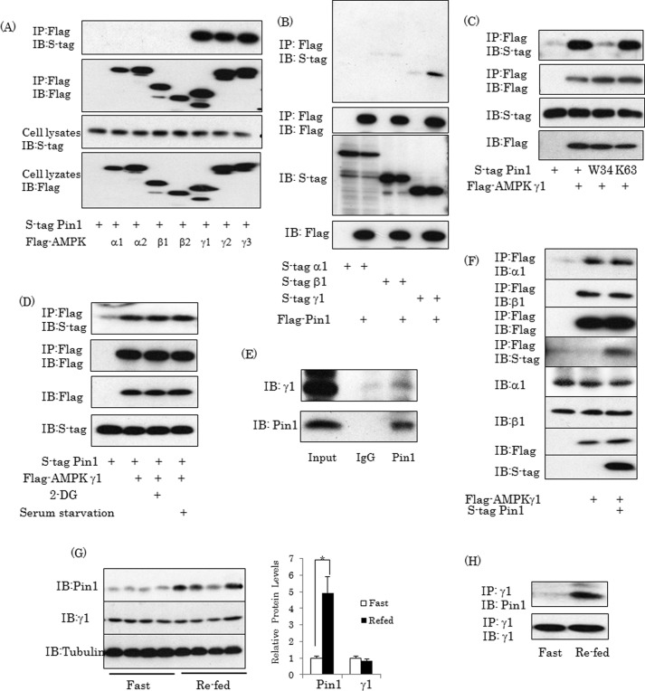
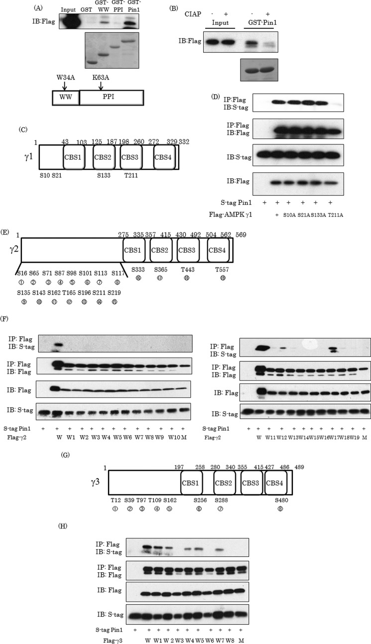
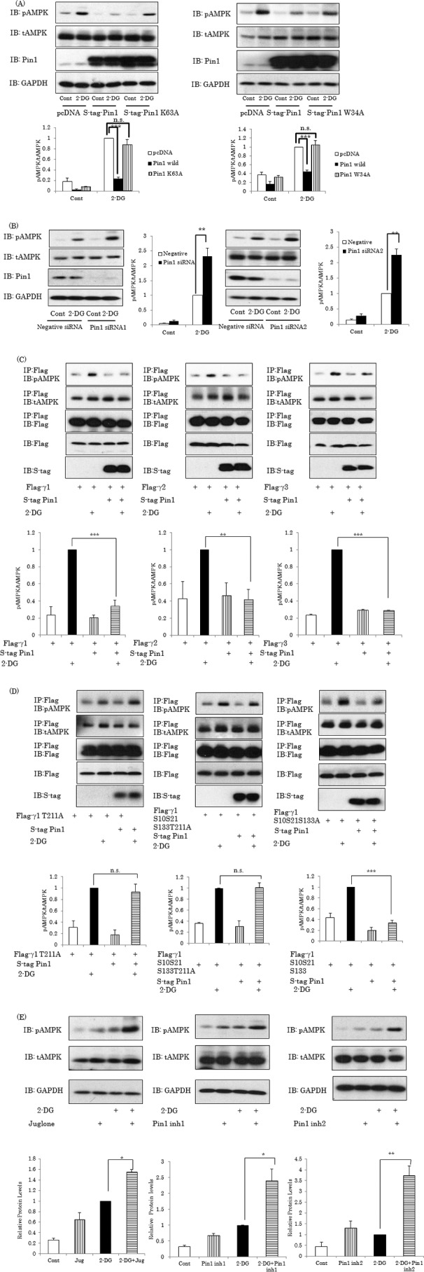
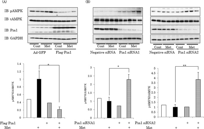
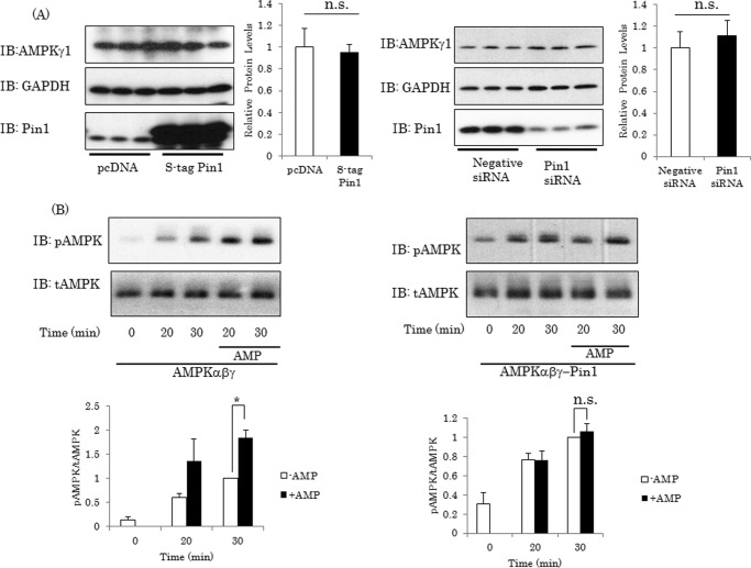
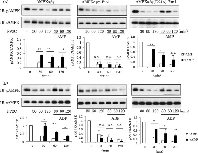
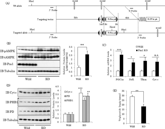
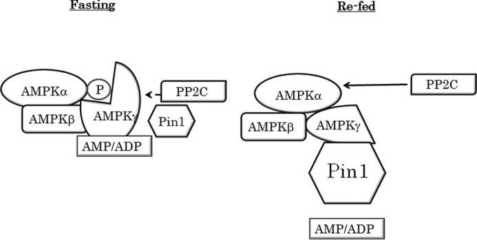
AMP-activated protein kinase (AMPK) plays a critical role in metabolic regulation. In this study, first, it was revealed that Pin1 associates with any isoform of γ, but not with either the α or the β subunit, of AMPK. The association between Pin1 and the AMPK γ1 subunit is mediated by the WW domain of Pin1 and the Thr(211)-Pro-containing motif located in the CBS domain of the γ1 subunit. Importantly, overexpression of Pin1 suppressed AMPK phosphorylation in response to either 2-deoxyglucose or biguanide stimulation, whereas Pin1 knockdown by siRNAs or treatment with Pin1 inhibitors enhanced it. The experiments using recombinant Pin1, AMPK, LKB1, and PP2C proteins revealed that the protective effect of AMP against PP2C-induced AMPKα subunit dephosphorylation was markedly suppressed by the addition of Pin1. In good agreement with the in vitro data, the level of AMPK phosphorylation as well as the expressions of mitochondria-related genes, such as PGC-1α, which are known to be positively regulated by AMPK, were markedly higher with reduced triglyceride accumulation in the muscles of Pin1 KO mice as compared with controls. These findings suggest that Pin1 plays an important role in the pathogenic mechanisms underlying impaired glucose and lipid metabolism, functioning as a negative regulator of AMPK.

Keywords: AMP-activated kinase (AMPK); diabetes; energy metabolism; lipid metabolism; metabolic syndrome; muscle; prolyl isomerase.


Figures

FIGURE 1.
FIGURE 1.
Pin1 associates with AMPK γ subunit. A and B, Pin1 interacts with the AMPK γ subunit in 293T cells. The 293T cells were transfected with S tag Pin1 (wild type, W34A, and K63A) and the FLAG-tagged α1, α2, β1, β2, γ1, γ2, or γ3 subunit of AMPK. Then cell lysates were immunoprecipitated (IP) with anti-FLAG tag antibody beads, followed by immunoblotting (IB) with anti-S tag or anti-FLAG tag antibody. C, Pin1 associates with the γ1 subunit independently of 2-DG stimulation or serum starvation. The 293T cells overexpressing Pin1 and the γ1 subunit were treated with 2-DG for 1 h or serum-starved overnight. Then cell lysates were immunoprecipitated with anti-FLAG tag antibody, followed by immunoblotting with anti-S tag or anti-FLAG tag antibody. D, Pin1 endogenously binds to the γ1 subunit in muscle. Protein extracts from mouse muscle were immunoprecipitated with anti-Pin1 antibody, followed by immunoblotting with anti-γ1 subunit antibody. E, Pin1 had no effect on formation of the AMPK complex. FLAG-tagged γ1 subunits alone or with S-tagged Pin1 were transfected into 293T cells, followed by immunoprecipitation with anti-FLAG tag antibody and immunoblotting with the indicated antibodies. F, Pin1 expressions changed in response to nutrient conditions. C57BL/6J mice were fasted for 16 h and then re-fed for 4 h. The mice were then sacrificed, and muscles were harvested. The indicated proteins were detected using the corresponding antibody. G, the association between Pin1 and γ1 was increased in the re-fed state. Tissue lysates from muscle were immunoprecipitated with γ1 antibody, followed by immunoblotting. Shown are representative data from three independent experiments. Error bars, S.E.; *, p < 0.05.
FIGURE 2.
FIGURE 2.
Thr211-Pro motif in γ1 subunit is required for the association with Pin1. A, the AMPK γ1 subunit interacts with the WW domain in Pin1. The cell lysates containing the overexpressed γ1 subunit were incubated with glutathione beads conjugated with GST alone, the GST-WW domain, the GST-PPIase domain, or GST-full-length Pin1. The middle panel shows the Coomassie blue staining of these GST or GST fusion proteins, and the top panel shows the Pin1 associated with each protein. B, phosphorylation of the γ1 subunit is required for the association with Pin1. The cell lysates containing the overexpressed γ1 subunit were incubated with 50 units of calf intestinal alkaline phosphatase for 2 h at 30 °C and subjected to a pull-down assay using GST-Pin1. C, scheme of Ser/Thr-Pro motif in AMPK γ1. D, substitution of Thr211 in the γ1 subunit by alanine abolished the association with Pin1. The 293T cells were transfected with S-tagged Pin1 and the γ1 subunit in which Ser10, Ser21, Ser133, or Thr211 had been substituted with alanine, and their associations were examined employing immunoprecipitation (IP) and immunoblotting (IB). E–H, the AMPK γ2 and γ3 subunits possess 15 and 8 Ser/Thr-Pro motifs, respectively. To identify the Ser/Thr-Pro-containing domains involved in the association with Pin1, the mutant γ2 and γ3 subunits, in which all or all but one of the Ser/Thr-Pro motifs had been generated by substituting Ser/Thr with alanine, were prepared. Their associations with Pin1 were then examined. Next, the 293T cells were transfected with S-tagged Pin1 and mutated AMPK γ2 or γ3, followed by immunoprecipitation with FLAG beads. W, wild type; W+number, only one Ser/Pro or Thr/Pro site is wild type, and other sites have been substituted with alanine; M, all Ser/Pro or Thr/Pro sites substituted with alanine. Shown are representative data from three independent experiments.
FIGURE 3.
FIGURE 3.
Pin1 suppresses AMPK α subunit phosphorylation by 2-DG. A, Pin1 overexpression suppresses AMPK α subunit phosphorylation. 293T cells were transfected with S-tagged wild-type, W34A, or W63A Pin1 and stimulated with or without 10 mm 2-DG for 1 h. B, knockdown of Pin1 enhances AMPK phosphorylation. The 293T cells were treated with each of the two Pin1 siRNAs. After 72 h, the protein expression levels of Pin1 and the α subunit were determined. After stimulation with or without 2-DG for 1 h, phosphorylation levels of the α subunit were examined. C, Pin1 suppresses α subunit phosphorylation, regardless of whether the γ subunit is γ1, γ2, or γ3. The FLAG-tagged γ1, γ2, or γ3 subunit and S-tagged Pin1 were overexpressed, followed by stimulation with or without 2-DG. The phosphorylation levels and amount of the α subunit in the FLAG tag antibody immunoprecipitates were examined. D, the α subunit phosphorylation level of the AMPK complex containing the γ1 subunit with T211A replacement, unable to associate with Pin1, was not affected by Pin1 overexpression. E, treatment with Pin1 inhibitors promotes AMPK phosphorylation. Three Pin1 inhibitors, including juglone (30 μm), were independently added to the cells, which were then treated with 2-DG. The bar graphs indicate the relative pAMPK levels normalized by tAMPK. Shown are representative data from three independent experiments. IB, immunoblotting; IP, immunoprecipitation. Error bars, S.E. **, p < 0.01; ***, p < 0.001; n.s., not significant.
FIGURE 4.
FIGURE 4.
Pin1 suppresses AMPK α subunit phosphorylation by metformin. A, FLAG-tagged Pin1 or control GFP was overexpressed in HepG2 cells employing adenoviral gene transfer. B, HepG2 cells were treated with each of the two Pin1 siRNAs. Then the cells were treated with or without 1 mm metformin for 1 h, and phosphorylation of the α subunit was examined. The bar graph indicates the relative pAMPK levels normalized by tAMPK. Shown are representative data from three independent experiments. Error bars, S.E.; *, p < 0.05; **, p < 0.01.
FIGURE 5.
FIGURE 5.
Pin1 does not affect AMPK γ1 stability but inhibits the enhancing effect of AMP on LKB1-induced AMPK α subunit phosphorylation. A, expression of AMPK γ1 was not affected by Pin1 overexpression or Pin1 gene silencing. B and C, Pin1 inhibited the enhancing effect of AMP on LKB1-induced AMPK phosphorylation in vitro. AMPK complexes were reacted with recombinant LKB1 in the presence or absence of 300 μm AMP for 20 or 30 min. Then the phosphorylation level of the α subunit was determined by immunoblotting (IB). Shown are representative data from three independent experiments. Error bars, S.E. n.s., not significant; *, p < 0.05.
FIGURE 6.
FIGURE 6.
Pin1 abolishes the protective effect of AMP or ADP against PP2C. A and B, the AMPK complex with or without Pin1 was reacted with recombinant PP2C in the presence or the absence of AMP or ADP for the indicated periods, and the phosphorylation level and amount of each subunit were then determined by immunoblotting (IB). The bar graphs indicate the relative protein levels of pAMPK normalized by tAMPK. Shown are representative data from three independent experiments. Error bars, S.E.; *, p < 0.05; **, p < 0.01; n.s., not significant.
FIGURE 7.
FIGURE 7.
AMPK phosphorylation is elevated in Pin1 KO mouse muscle. A, scheme of the genomic construct of Pin1 flox mice. B, loss of Pin1 in muscle promotes AMPK phosphorylation. After the animals had been fasted for 16 h, muscles were harvested from Pin1 KO and wild-type mice, and the indicated protein levels were detected, using the corresponding antibody. C and D, expressions of mitochondria-related genes and proteins were elevated in Pin1 KO mice. E, muscle triglyceride content was lower in Pin1 KO mice. IB, immunoblotting. Error bars, S.E.; *, p < 0.05; **, p < 0.01; ***, p < 0.001; n.s., not significant.
FIGURE 8.
FIGURE 8.
Proposed mechanism of AMPK regulation by Pin1. As the Pin1 expression level is low in the fasted state, AMP can protect the α subunit from dephosphorylation. In the re-fed state, however, Pin1 impairs AMP binding to the g subunit and thereby abolishes the protective effect of AMP.

Similar articles

Cited by

References

    1. Hardie D. G., Ross F. A., Hawley S. A. (2012) AMPK: a nutrient and energy sensor that maintains energy homeostasis. Nat. Rev. Mol. Cell Biol. 13, 251–262 - PMC - PubMed
    1. Kahn B. B., Alquier T., Carling D., Hardie D. G. (2005) AMP-activated protein kinase: ancient energy gauge provides clues to modern understanding of metabolism. Cell Metab. 1, 15–25 - PubMed
    1. Hardie D. G. (2007) AMP-activated/SNF1 protein kinases: conserved guardians of cellular energy. Nat. Rev. Mol. Cell Biol. 8, 774–785 - PubMed
    1. Hardie D. G., Carling D. (1997) The AMP-activated protein kinase: fuel gauge of the mammalian cell? Eur. J. Biochem. 246, 259–273 - PubMed
    1. Andersson U., Filipsson K., Abbott C. R., Woods A., Smith K., Bloom S. R., Carling D., Small C. J. (2004) AMP-activated protein kinase plays a role in the control of food intake. J. Biol. Chem. 279, 12005–12008 - PubMed

Publication types

MeSH terms