. 2015 Aug 14;10(8):e0135845.
doi: 10.1371/journal.pone.0135845. eCollection 2015.

GNL3L Is a Nucleo-Cytoplasmic Shuttling Protein: Role in Cell Cycle Regulation

Affiliations

GNL3L Is a Nucleo-Cytoplasmic Shuttling Protein: Role in Cell Cycle Regulation

Indu Jose Thoompumkal et al. PLoS One. .

Abstract

GNL3L is an evolutionarily conserved high molecular weight GTP binding nucleolar protein belonging to HSR1-MMR1 subfamily of GTPases. The present investigation reveals that GNL3L is a nucleo-cytoplasmic shuttling protein and its export from the nucleus is sensitive to Leptomycin B. Deletion mutagenesis reveals that the C-terminal domain (amino acids 501-582) is necessary and sufficient for the export of GNL3L from the nucleus and the exchange of hydrophobic residues (M567, L570 and 572) within the C-terminal domain impairs this process. Results from the protein-protein interaction analysis indicate that GNL3L interaction with CRM1 is critical for its export from the nucleus. Ectopic expression of GNL3L leads to lesser accumulation of cells in the 'G2/M' phase of cell cycle whereas depletion of endogenous GNL3L results in 'G2/M' arrest. Interestingly, cell cycle analysis followed by BrdU labeling assay indicates that significantly increased DNA synthesis occurs in cells expressing nuclear export defective mutant (GNL3L∆NES) compared to the wild type or nuclear import defective GNL3L. Furthermore, increased hyperphosphorylation of Rb at Serine 780 and the upregulation of E2F1, cyclins A2 and E1 upon ectopic expression of GNL3L∆NES results in faster 'S' phase progression. Collectively, the present study provides evidence that GNL3L is exported from the nucleus in CRM1 dependent manner and the nuclear localization of GNL3L is important to promote 'S' phase progression during cell proliferation.


Conflict of interest statement

Competing Interests: The authors have declared that no competing interests exist.

Figures

Fig 1
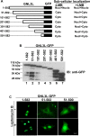
Fig 1. GNL3L localizes to distinct subcellular compartments.
COS-7 cells were transfected with GFP tagged wild type or deletion mutants of GNL3L (GNL3L1–50, GNL3L51–100 and GNL3L51–582) and their sub-cellular distribution was analyzed using fluorescence microscopy. The scale bar represents 10 μm.
Fig 2
Fig 2. GNL3L shuttles between nucleus and cytoplasm and the export from the nucleus is sensitive to Leptomycin-B.
HepG2 cells were transiently transfected with full length or indicated N-terminal deletion mutants of GNL3L and subcellular localization was determined in the presence (B) or the absence (A) of leptomycin-B (LMB). Nuclei were stained with DAPI. The scale bar represents 20μm.
Fig 3
Fig 3. The C-terminal domain of GNL3L harbours a nuclear export signal.
(A)Expression of wild type and indicated deletion mutants of GNL3L was determined by western blot using anti-GFP antibody. The indicated variants of GNL3L were expressed correct sized polypeptides. (B) Schematic diagram of GNL3L deletion mutants and summary of their subcellular distribution. (C) Cells expressing full length and deletion mutants of GNL3L were treated with (+LMB) or without (-LMB) and subcellular distributions were analyzed by fluorescence microscopy.
Fig 4
Fig 4. GNL3L is a nucleo-cytoplasmic shuttling protein.
Heterokaryon assay was performed by transfecting GNL3L1–582-GFP, GNL3L51–582-GFP or GNL3L501–582-GFP into HeLa cells and fusing with NIH3T3 cells as described in Materials and Methods. The punctate pattern of murine nucleus (‘M’) upon staining with Hoechst 33342 distinguished it from the human HeLa nucleus (‘H’). In the presence of LMB, the nuclear export of GNL3L51–582-GFP or GNL3L501–582-GFP was prevented and the protein was localized in both HeLa and NIH3T3 nucleus.
Fig 5
Fig 5. GNL3L interacts with the export receptor, CRM1 via C-terminal domain.
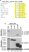
(A) Comparison of GNL3L Nuclear Export Signals (NES) with known NESs from various nucleo-cytoplasmic shuttling proteins. Conserved hydrophobic residues are highlighted in yellow colour. (B) Wild type or indicated variants of GNL3L were transiently transfected into HeLa cells and the cell lysates were subjected to co-immunoprecipitation with anti-CRM1 antibody followed by western blot using anti-GFP antibody (Top panel). The expression of GNL3L and CRM1 (endogenous) was confirmed prior to immunoprecipitation by western blot using anti-GFP (lower panel) and anti-CRM1 antibodies (bottom panel).
Fig 6
Fig 6. The hydrophobic residues within the C-terminal domain of GNL3L constitute a functional nuclear export signal.
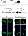
(A) Indicated variants of GNL3L 51–582-GFP and GNL3L 501–582-GFP were generated using QuickChange site-directed mutagenesis as described in Materials and Methods. (B) All the indicated GNL3L mutants were expressed correct size polypeptides as evident from the western blot analysis using anti-GFP antibody. HeLa cells were transfected with wild type GNL3L 51–582-GFP or GNL3L 501–582-GFP (C) and their variants (D). Transfected cells were fixed in 3% paraformaldehyde and nuclei were stained using DAPI. Subcellular localization GNL3L variants were analyzed using confocal microscope. The scale bar represents 20 μm.
Fig 7
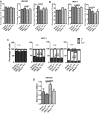
Fig 7. GNL3L modulates ‘G2/M’ phase progression.
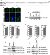
(A) Schematic representation of wild type, nuclear import (GNL3LΔNLS) and nuclear export (GNL3LΔNES) defective mutants of GNL3L. (B) HeLa cells were transfected with Flag-tagged GNL3LWT, GNL3LΔNES and GNL3LΔNLS expression plasmids and the subcellular localization was analyzed using confocal microscopy. The scale bar represents 20μm. (C) HEK293 cells were transfected with Flag-tagged GNL3LWT, GNL3LΔNES and GNL3LΔNLS and the expression was determined by western blot analysis using anti-Flag antibody. (D) Ectopic expression of GNL3L results in decreased accumulation of ‘G2/M’ population. GNL3LWT was overexpressed in HEK293 cells and the asynchronous cell cycle pattern was analyzed using flow cytometry as described in Materials and Methods. (E) Western blot was performed to analyze the protein levels of endogenous cyclins A2 and B1 upon GNL3LWT expression in HEK293 cells. Beta actin served as loading control. (F) GNL3L knockdown leads to increased accumulation of cells in G2/M phase of the cell cycle. Transient knockdown of GNL3L was performed in HEK293 cells using specific siRNA and the cell cycle profile was analyzed using flow cytometry. (NC: Negative control). (G) Endogenous cyclins A2 and B1 levels were analyzed using western blot upon GNL3L knockdown in HEK293.
Fig 8
Fig 8. Nuclear localization of GNL3L promotes ‘S’ phase progression.
HEK293 (A) and MCF-7 (B) cells were transfected with wild type or indicated variants of GNL3L and the asynchronous cell cycle profiles were analyzed. (C) MCF-7 cells transfected with wild type or variants of GNL3L were synchronized by single thymidine block as described in Materials and Methods. Cell cycle profiles were analyzed at 0, 4, 6 and 9 h post thymidine release by flow cytometry. Propidium Iodide (PI) was used to label the nuclei. (D) HEK293 cells were transfected with wild type or indicated variants of GNL3L and the DNA synthesis was measured by incubating the cells with 1X BrdU for 4 h followed by staining with anti-BrdU primary antibody and HRP-conjugated secondary antibody. Stained cells were incubated with TMB substrate and the absorbance was read at 450 nm.
Fig 9
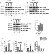
Fig 9. Nuclear localization of GNL3L modulates the levels of E2F1, cyclins A2 and E1 to promote ‘S’ phase progression.
(A) Ectopic expression of GNL3LΔNES upregulates endogenous cyclin A2 and cyclin E1 protein levels. HEK293 cells were transfected with wild type or indicated variants of GNL3L and the cyclin levels were analyzed by western blot using anti-cyclin A and anti-cyclin E1 antibodies. Real-time qPCR analysis suggests that ectopic expression of GNL3LΔNES upregulates E2F1, cyclin A2 and cyclin E1 transcription in HEK293 (B) and MCF-7 (C) cells. (D) Schematic diagram showing the cyclins and cyclin dependent kinases mediated cell cycle regulation.
Fig 10
Fig 10. Nuclear localization of GNL3L modulates Rb-E2F1 pathway.
Ectopic expression of GNL3LΔNES leads to hyperphosphorylation of Retinoblastoma protein (Rb) at serine 780. HEK293 (A) and MCF-7 (B) cells were transfected with wild type or variants of GNL3L. Anti-phospho Rb (S780) antibody and anti-Rb antibodies were used to determine the status of Rb phosphorylation and total Rb protein levels, respectively, by western blot analysis. Beta actin served as loading control. (C) GNL3LΔNES expression leads to increased association between cyclin D1 and cdk4. HEK293 cell lysates containing wild type or variants of GNL3L were subjected to co-immunoprecipitation with anti-cyclin D1 antibody followed by western blot using anti-cdk4 antibody. Expression levels of the indicated variants of GNL3L were normalized to that of GNL3LWT and used as the reference for these analyses. (D) GNL3LΔNES displays Rb dependency in the upregulation of E2F1, cyclins A2 and E1 levels. The Rb null cell line, Hep3B was transfected with wild type or variants GNL3L, alone or in combination with Rb expression plasmid. RT-qPCR was performed for the respective genes using specific primers (S1 Table).
Fig 11
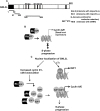
Fig 11. Proposed model for GNL3L function during cell proliferation.
(A) GNL3L harbors multiple functional domains. Amino terminus of GNL3L encodes a functional nucleolar targeting signal (NoLS; amino acids 1–50) and a nuclear localization signal (NLS; amino acids 51–100). A functional nuclear export signal is mapped towards the carboxyl terminal domain (501–582) and the combination of mutagenesis and subcellular localization studies suggest that residues M567, L570 and 572 are critical for GNL3L export from the nucleus. (B) Interaction of Rb with E2F1 is essential to regulate cell proliferation by controlling E2F1 transcriptional activity. (C) The binding of Retinoblastoma protein (Rb) with E2F1 transcription factor inhibits transcription of S-phase regulatory genes as cyclin A2 and cyclin E1. Nuclear localization of GNL3L leads to phosphorylation of Rb at S780, which is critical to release E2F1 to activate transcription of cyclin A and cyclin E, resulting in ‘S’ phase progression. These data suggest the possibility that nuclear localization of GNL3L promotes ‘S’ phase progression during cell proliferation by modulating the Rb-E2F1 pathway.

Similar articles

Cited by

References

    1. Caldon CE, Yoong P, March PE. Evolution of a molecular switch: universal bacterial GTPases regulate ribosome function. Mol. Microbiol. 2001; 41: 289–297. - PubMed
    1. Bourne HR, Sanders DA, McCormick F. The GTPase superfamily: conserved structure and molecular mechanism. Nature. 1991; 349: 117–127. - PubMed
    1. Leipe DD, Wolf YI, Koonin EV, Aravind L. Classification and Evolution of P-loop GTPases and Related ATPases. J. Mol.Biol. 2002; 317: 41–72. - PubMed
    1. Reynaud GE, Andrade AM, Bonneau F, Ly NBT, Knop M, Scheffzek K, et al. Human Lsg1 defines a family of essential GTPases that correlates with the evolution of compartmentalization. BMC Biology. 2005; 3: 21 - PMC - PubMed
    1. Du X, Subba Rao MRK, Chen XQ, Wu W, Mahalingam S, Balasundaram D. The Homologous Putative GTPases Grn1p from Fission Yeast and the Human GNL3L Are Required for Growth and Play a Role in Processing of Nucleolar Pre-rRNA. Mol. Biol. Cell. 2006; 17: 460–474. - PMC - PubMed

Publication types

MeSH terms

Grants and funding

This work was supported by grant VI-D&P/411/2012-2013/TDT(G) from the Department of Science and Technology, Govt. of India (http://www.dst.gov.in/) to SM, and a graduate fellowship (09/084(05000/2009-EMR-I) from the Council of Scientific and Industrial Research (CSIR) (http://www.csirhrdg.res.in/) to IJT. The funders had no role in study design, data collection and analysis, decision to publish, or preparation of the manuscript.