Review
. 2015 Nov;91(6):1263-90.
doi: 10.1111/php.12506. Epub 2015 Sep 20.

Photochemistry and Photobiology of the Spore Photoproduct: A 50-Year Journey

Affiliations
Review

Photochemistry and Photobiology of the Spore Photoproduct: A 50-Year Journey

Peter Setlow et al. Photochem Photobiol. 2015 Nov.

Abstract

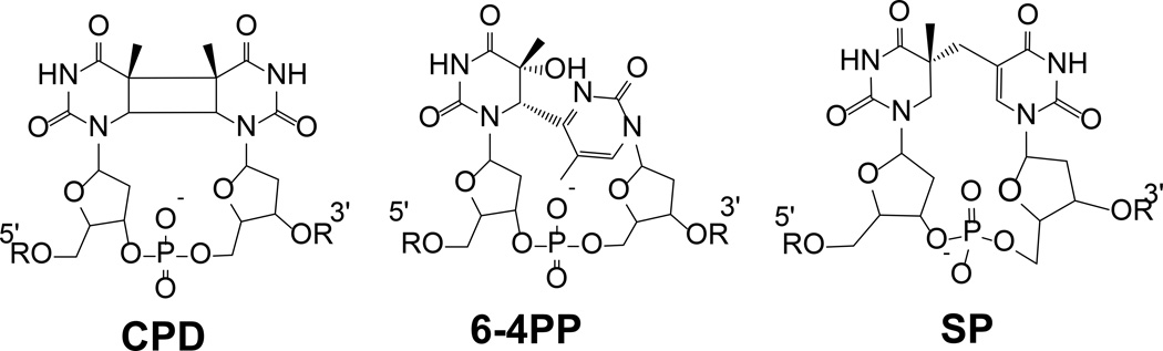
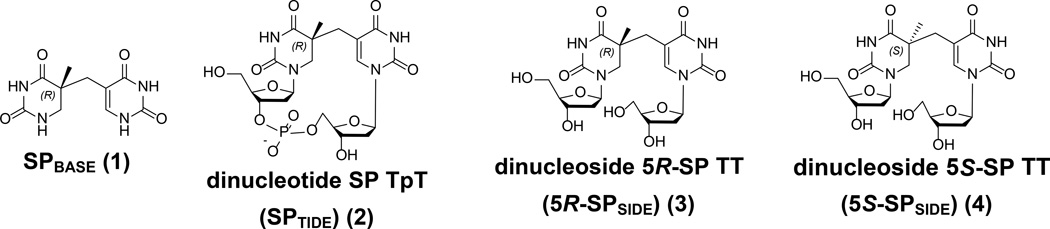
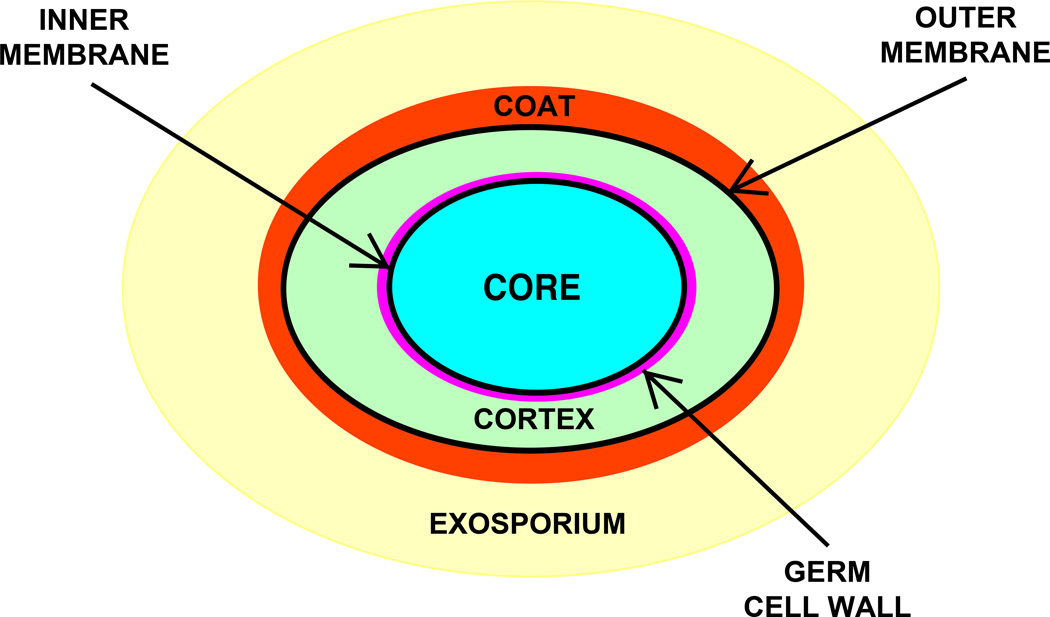
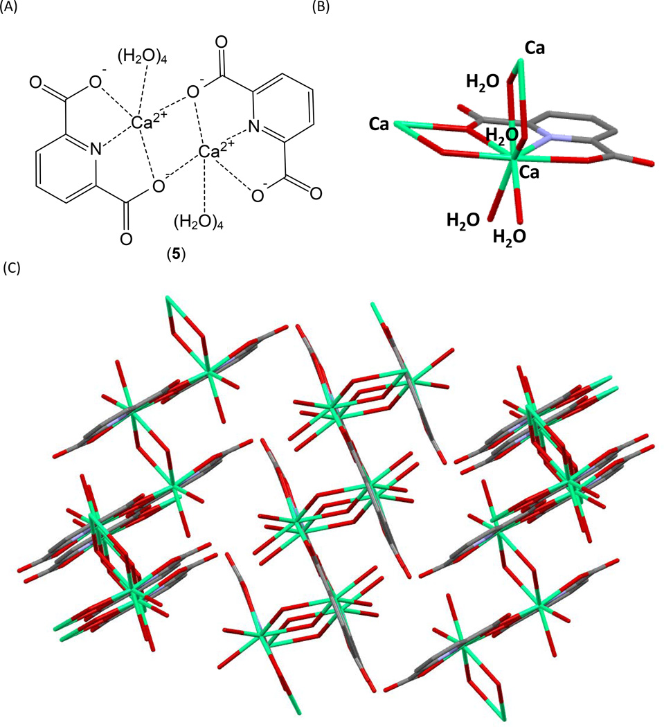
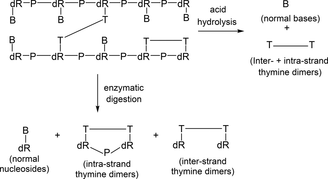
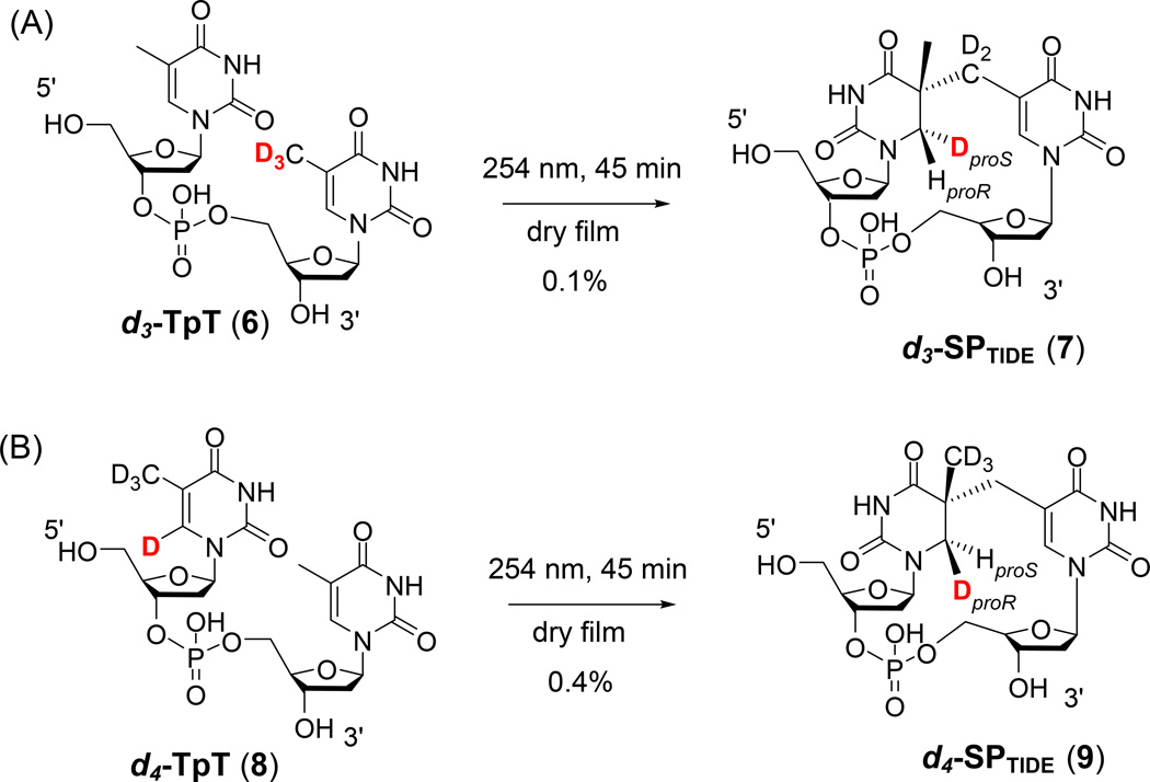
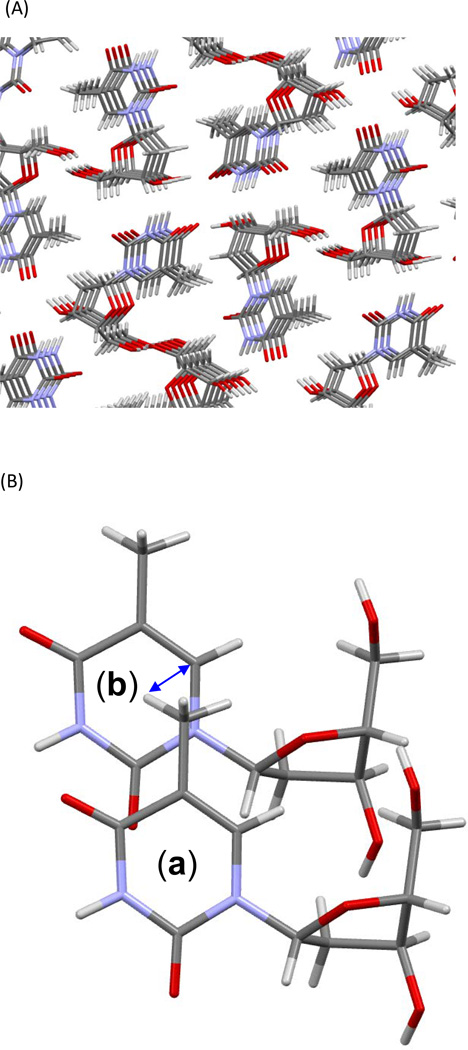
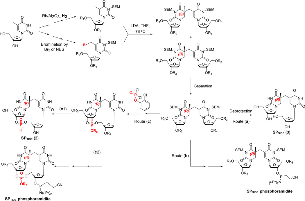
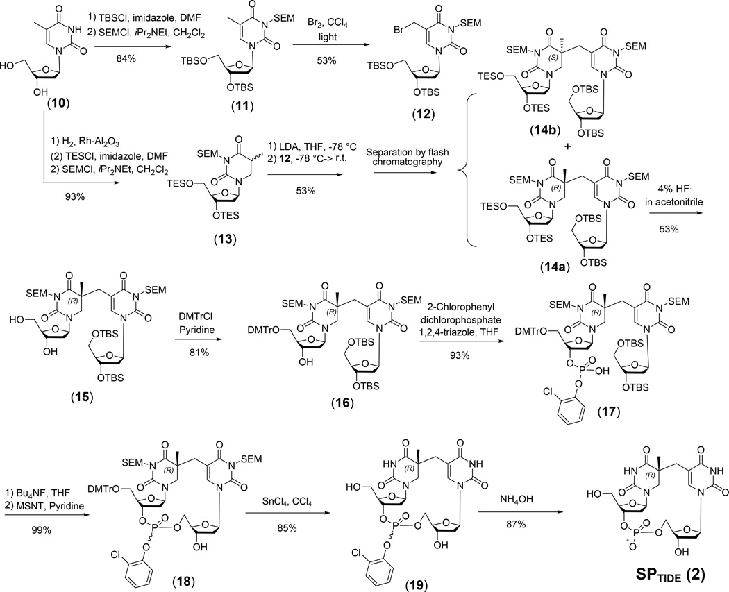
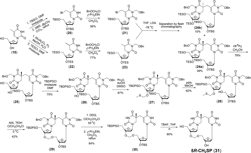
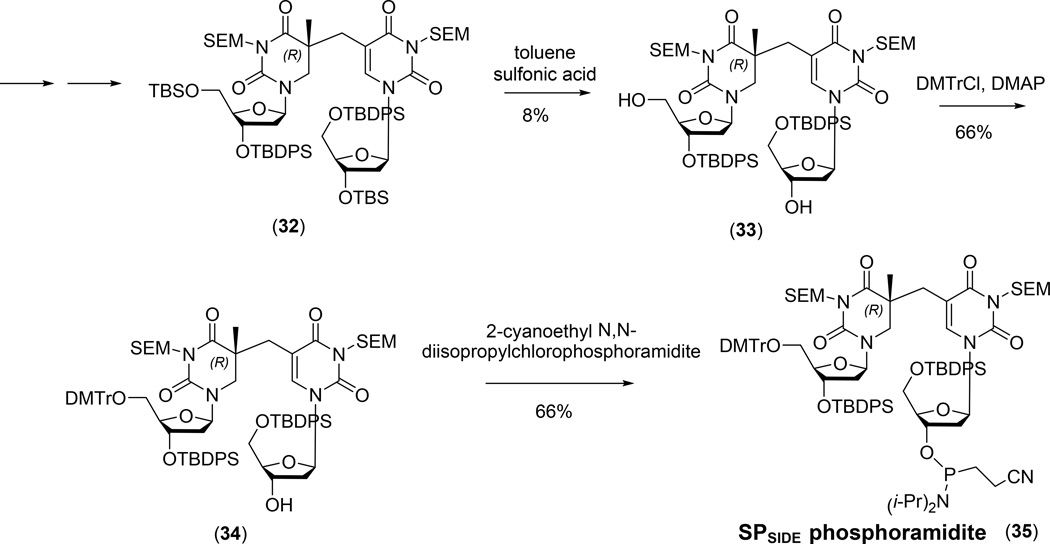
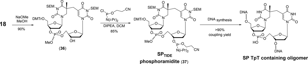
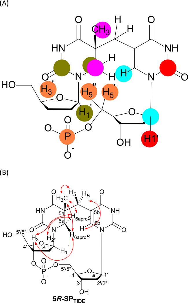
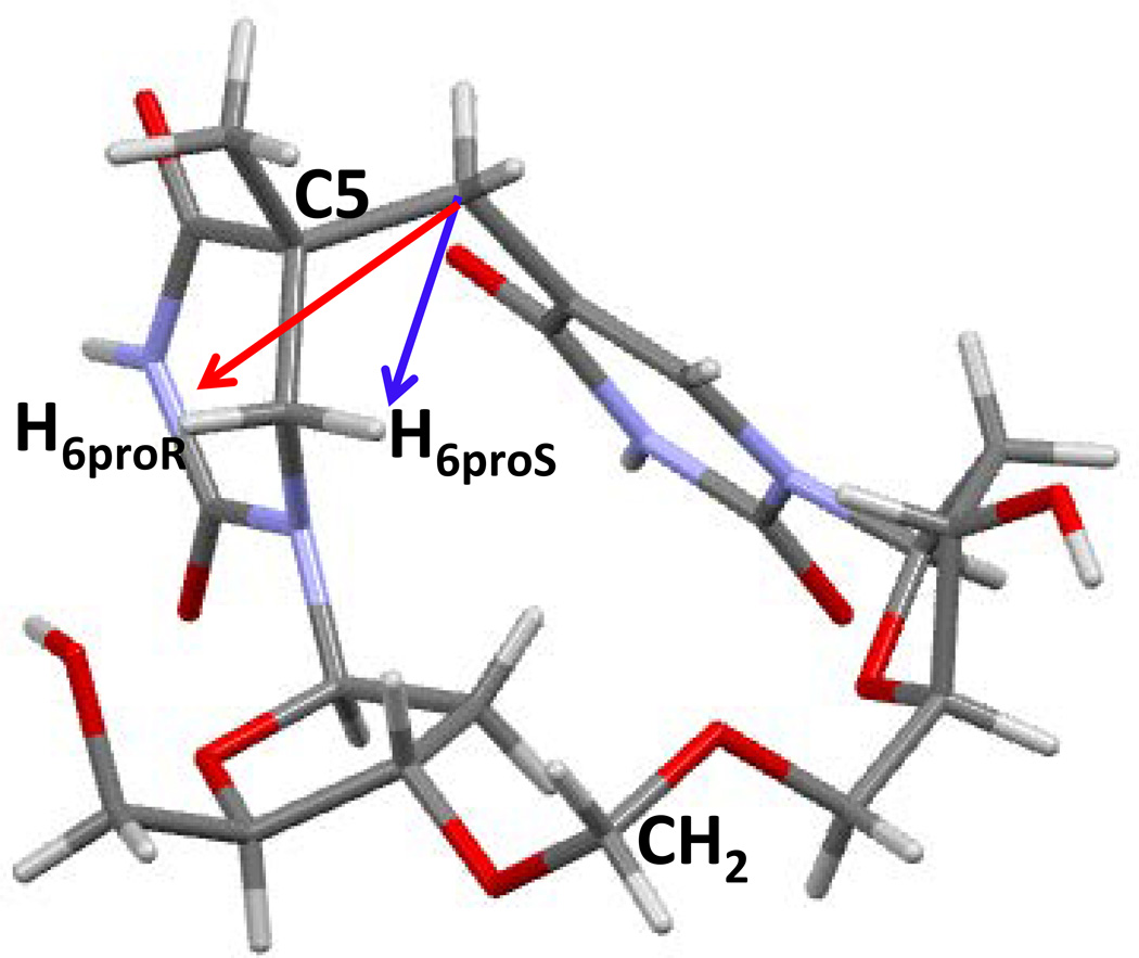
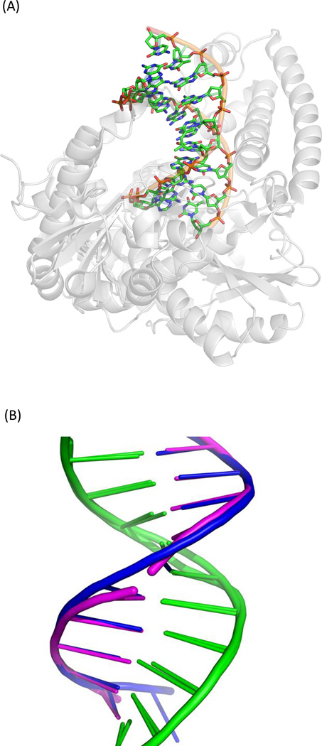
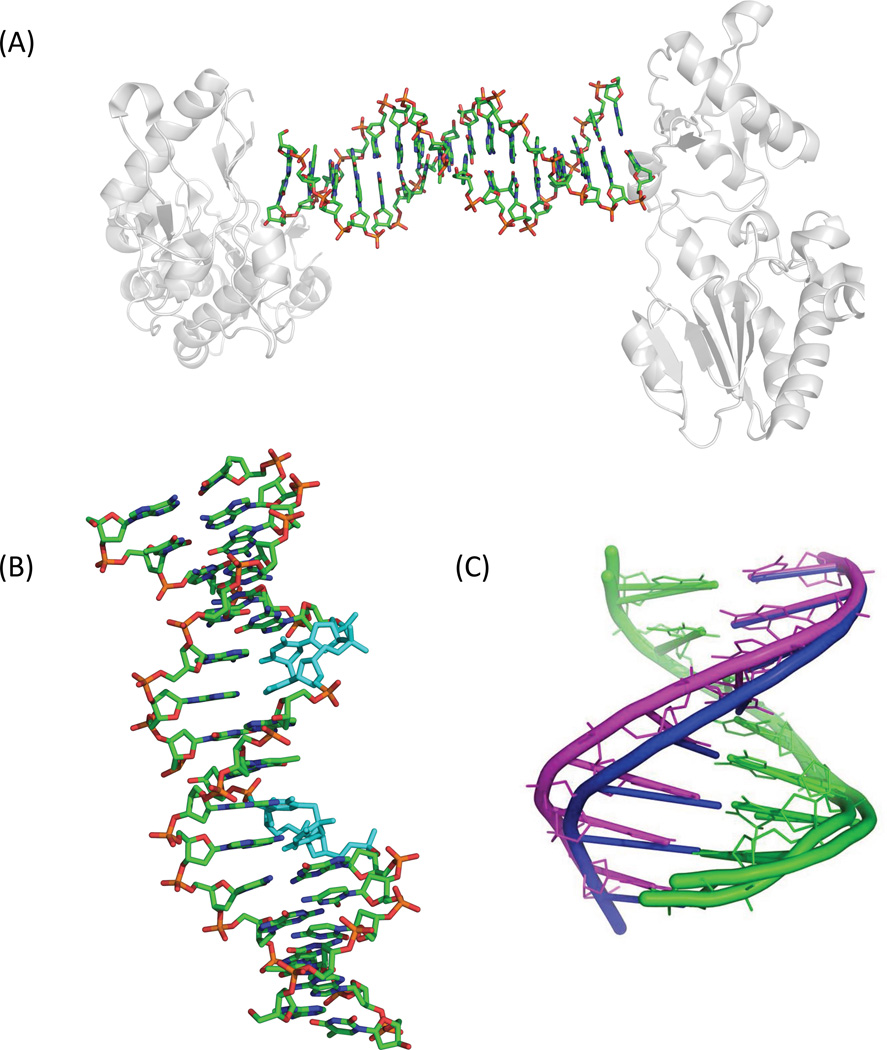
Fifty years ago, a new thymine dimer was discovered as the dominant DNA photolesion in UV-irradiated bacterial spores [Donnellan, J. E. & Setlow R. B. (1965) Science, 149, 308-310], which was later named the spore photoproduct (SP). Formation of SP is due to the unique environment in the spore core that features low hydration levels favoring an A-DNA conformation, high levels of calcium dipicolinate that acts as a photosensitizer, and DNA saturation with small, acid-soluble proteins that alters DNA structure and reduces side reactions. In vitro studies reveal that any of these factors alone can promote SP formation; however, SP formation is usually accompanied by the production of other DNA photolesions. Therefore, the nearly exclusive SP formation in spores is due to the combined effects of these three factors. Spore photoproduct photoreaction is proved to occur via a unique H-atom transfer mechanism between the two involved thymine residues. Successful incorporation of SP into an oligonucleotide has been achieved via organic synthesis, which enables structural studies that reveal minor conformational changes in the SP-containing DNA. Here, we review the progress on SP photochemistry and photobiology in the past 50 years, which indicates a very rich SP photobiology that may exist beyond endospores.


Figures

Figure 1
Figure 1
Chemical structure of the three thymine dimers.
Figure 2
Figure 2
Structures of SP and SP derivatives discussed in this review. In the literature, dinucleoside SP TT and dinucleotide SP TpT were used respectively for SP dimers with and without the phosphodiester linker; while Desnous et al. used SPSIDE and SPTIDE to refer to these two species (40). Although we used SPSIDE and SPTIDE in our review, we wish to show both nomenclatures.
Figure 3
Figure 3
Structure of a bacterial endospore (5). The relative sizes of various layers are not drawn to scale, and the large exosporium is not present in spores of all species. Note that there can be several sublayers in individual layers, in particular the coat and probably in the exosporium.
Figure 4
Figure 4
(A) Structure of the CaDPA dimer as the basic assembly unit for the inorganic polymer (75). The dimer adopts a planar structure. Each Ca2+ is coordinated to eight atoms. Among them, three are from a DPA, one is from a carboxylate moiety of another DPA in the dimer, and the rest are from two pairs of water molecules, with one pair above and the other pair beneath the dimer plane. Both water molecules within the pair coordinate to another Ca2+ from another dimer, thus serving as bridging ligands to link the dimers into an inorganic polymer. (B) The coordination sphere around the calcium ion. The Ca2+ ion is shown in green, nitrogen in blue, and oxygen atoms in red. The four bridging water ligands are also labeled in the structure. (C) The structure of the CaDPA polymer linked by bridging water molecules.
Figure 5
Figure 5
Crystal structure of the α/β-type SASP (SspC)–oligo(dG)•oligo(dC)10-mer complex (pdb entry 2Z3X) (106). (A) Stereo diagram of the complex indicating that SspC adopts a helix–turn–helix motif when binding to DNA. The positions of the N and C termini of each protomer are labeled: orange, SspC1; magenta, SspC2; cyan, SspC3. (B) DNA conformation in the SspC-DNA complex. This conformation is a combination of A- and B-conformations, and is different from the typical A- or B-form DNA. Analyses by molecular simulation indicated that such a DNA conformation favors formation of SP over CPD and 6-4PP.
Figure 6
Figure 6
Enzymatic and acid hydrolysis of duplex DNA containing inter- and intra-strand photoproducts (135). B, normal base; dR, 2′-deoxyribose; P, phosphate. The phosphate in intra-strand dimers cannot be cleaved by enzymatic digestion while all phosphates are hydrolyzed by acid, providing an assay to distinguish these two types of DNA photoproducts.
Figure 7
Figure 7
(A) The concerted mechanism for SP formation (40, 138). The breakage of the C-H bond in the methyl moiety and the formation of the two new bonds in SP are suggested to occur simultaneously, potentially via a four member-ring transition state. (B) The consecutive mechanism for SP formation (7, 139). The C6 radical abstracts an H-atom from the methyl group, generating a 5-α-thyminyl radical, which combines with the 5,6-dihydrothymin-5-yl radical to generate SP. The right-handed helical structure of duplex DNA ensures that only route (i) is possible in spores, resulting in the 5R-SP. In a solid state photoreaction using monomeric thymidine, route (ii) also occurs, resulting in dinucleoside 5S-SP.
Figure 8
Figure 8
H-atom transfer during SP formation as revealed by the formation of deuterated dinucleotide SPs via solid phase photoreaction using selectively labeled dinucleotide TpTs (139).
Figure 9
Figure 9
(A) Molecular packing in the thymidine single crystal (145). The distance between two thymidine rings is ~ 3.1 Å, close to the 3.36 Å average rise found in the SASP-oligo(dG)•oligo(dC) nucleoprotein (106). (B) The shortest distance between an H atom in –CH3 and the C6 of another thymine is ~ 3.2 Å (blue arrow), which is close to the 3.4 Å found in the molecular simulation (106), and supports the role of the key H-abstraction step in initiating SP formation (124, 139).
Figure 10
Figure 10
Summary of a synthetic strategy commonly adopted for the syntheses of dinucleoside and dinucleotide SP as well as their phosphoramidite. Here, both N3 atoms on the thymine ring are protected by [2-(trimethylsilyl) ethoxy]methyl acetal (SEM).
Figure 11
Figure 11
Synthesis of the SPTIDE starting from thymidine, which was originally reported by Kim and Begley (150) and later modified by other groups (139, 149). The 5S- and 5R-SP diastereomers were not separated until the last step of Kim’s synthesis, while most recent syntheses separated these isomers via flash chromatography right after the coupling step. The synthesis shown here was thus modified accordingly.
Figure 12
Figure 12
Synthesis of SPTIDE analog 5R-CH2SP (30) containing a formacetal linker (128).
Figure 13
Figure 13
Key steps for the synthesis of SPSIDE phosphoramidite from the protected SPSIDE (129).
Figure 14
Figure 14
Synthesis of dinucleotide SP phosphoramidite from the penultimate intermediate during the synthesis of SPTIDE shown in Figure 11 (149).
Figure 15
Figure 15
(A) Structure of SPTIDE determined from 1H-13C and 1H-31P Heteronuclear Multiple-Bond Correlation (HMBC) spectroscopic studies (8). From the through-bond correlations revealed by HMBC, the enchainment of different chemical groups (thymine, sugar, and phosphate) in SP was determined. The groups exhibiting strong HMBC signals are indicated by the same color. (B) Summary of key NOESY cross-peaks determined from the NMR spectra of SPTIDE (8, 128).
Figure 16
Figure 16
X-ray structure of the 5R-CH2SP with the nitrogen and oxygen atoms labeled (128). The structure clearly shows that the C5 chiral center adopts an R configuration. The distance between the H6proS and the methylene carbon (blue arrow) is 2.63 Ǻ and that between the H6proR and the methylene carbon (red arrow) is 3.36 Ǻ, which supports the reaction mechanism in which the H6proS of SP originates from the methyl group in 3'-thymine of the TpT step before SP formation.
Figure 17
Figure 17
(A) Crystal structure of the dinucleoside 5R-SP-containing duplex complexed with B. st. Pol I (pdb entry 2YLJ) (129). (B) Superimposed structures of the undamaged DNA (pdb entry 1NJY) and the dinucleoside SP-containing DNA bound to B. st. Pol I (129). The DNA backbone in the latter case is broken due to the lack of phosphodiester group in the dinucleoside SP. Except for the broken backbone, the two DNA structures overlay nearly perfectly.
Figure 18
Figure 18
(A) Crystal structure of the nucleoprotein complex including two MMLV RT molecules and one 16-base-pair oligonucleotide duplex (170). The asymmetric unit of the crystal includes one protein molecule and eight base pairs of the duplex oligomer. (B) Structure of the 16-mer duplex strand containing two SP dimers (shown in cyan) (170), where all H-bonding interactions in the duplex are maintained. The two 8-mer asymmetric units are disconnected in the figure although an integral 16-mer strand was used for crystallization. Such a fragmentation is ascribed to the artificial effect of the crystallographic data; it does not imply cleavage of the 16-mer strand. (C) Superimposed structures of the 8-base-pair asymmetric unit of the undamaged oligomer (in blue, pdb entry 4M95) and the SPTIDE-containing oligomer (in magenta, pdb entry 4M94) bound to MMLV RT. The minor groove is widened from 9.7 Å in the undamaged strand to 12.5 Å in the SP-containing strand.
Figure 19
Figure 19
X-ray crystallographic or solution-state NMR structures of duplex oligonucleotides containing the three thymine dimers, cis-syn CPD, 6-4PP and SP, respectively. A) Crystal structure of a decamer duplex containing a cis-syn CPD lesion (pdb entry 1N4E) (172). B) Solution-state NMR structure of a dodecamer duplex containing a cis-syn CPD lesion (pdb entry 1TTD) (174). C) Solution-state NMR structure of a decamer duplex containing a stable GA•6-4PP mismatch (pdb entry 1CFL)(179). D) Structure of a 16-mer duplex strand containing two SPTIDEs deduced from a nucleoprotein complex (pdb entry 4M94) (170). The crystal structure shown in Panel A was indicated to only represent one of many solution structures that was captured during the crystallization process; the solution-state NMR structure shown in Panel B thus may better reflect the local structure of a cis-syn CPD-containing DNA (180). It is also evident that 6-4PP drastically changes the duplex oligonucleotide structure; while CPD and SP only induce minor changes.
Figure 20
Figure 20
Zoom-in view of the two base pairs containing the thymine dimers in a duplex oligonucleotide. The distances between the non-hydrogen atoms involved in hydrogen bond formation are also shown. From Panel A (from the crystal structure) and Panel B (from the solution-state NMR structure), it is clear that the hydrogen bonding interactions in one of the base pairs associated with the cis-syn CPD are weakened. The 6-4PP maintains weaker hydrogen bonds with its 5'-5,6-dihydro-5-hydroxythymine residue; its 3'-residue does not form any hydrogen bond with the opposite adenine. However, two hydrogen bonds were found if the opposite nucleobase becomes a guanine as shown in Panel C, explaining its tendency to induce an A→G mutation during the translesion synthesis event. In contrast, SP maintains all four hydrogen bonds (Panel D); these bonds may be even stronger than those formed between undamaged nucleobases as indicated by the shortened distances between the involving atoms. Again, due to the restriction of the 8-mer asymmetric units in the crystallographic data, the 16-mer strand was shown as two fragments. This “artificial” fragmentation however does not reflect the integrity of the 16-mer oligonucleotide and does not alter the DNA conformation.
Figure 21
Figure 21
Formation of the hemiaminal intermediate 37 at the C4=O moiety of the 5'-residue of SP. Either C-O bond associated with the hemiaminal species is cleavable. As a consequence, the oxygen atom at the C4=O moiety becomes exchangeable with the oxygen atom from water. Decomposition of the hemiaminal intermediate produces a SP hydrolysis product 38, whose further decomposition would induce a cascade of elimination reactions, eventually leading to DNA strand break.

Similar articles

Cited by

References

    1. Donnellan JE, Jr, Setlow RB. Thymine photoproducts but not thymine dimers found in ultraviolet-irradiated bacterial spores. Science. 1965;149:308–310. - PubMed
    1. Varghese AJ, Wang SY. Ultraviolet irradiation of DNA in vitro and in vivo produces a third thymine-derived product. Science. 1967;156:955–957. - PubMed
    1. Donnellan JE, Stafford RS. The ultraviolet photochemistry and photobiology of vegetative cells and spores of Bacillus megaterium. Biophys. J. 1968;8:17–28. - PMC - PubMed
    1. Smith KC, Yoshikawa H. Variation in the photochemical reactivity of thymine in the DNA of B. subtilis spores, vegetative cells and spores germinated in chloramphenicol. Photochem. Photobiol. 1966;5:777–786. - PubMed
    1. Varghese AJ. Photochemistry of thymidine as a thin solid film. Photochem. Photobiol. 1971;13:357–364.

Publication types

LinkOut - more resources