

Knockdown of Nestin inhibits proliferation and migration of colorectal cancer cells
- PMID: 26261513
- PMCID: PMC4525847
Knockdown of Nestin inhibits proliferation and migration of colorectal cancer cells
Abstract
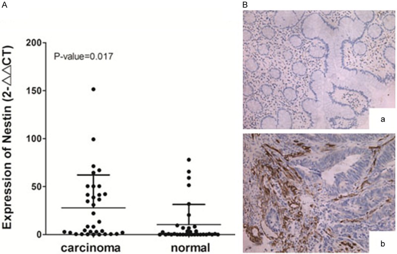
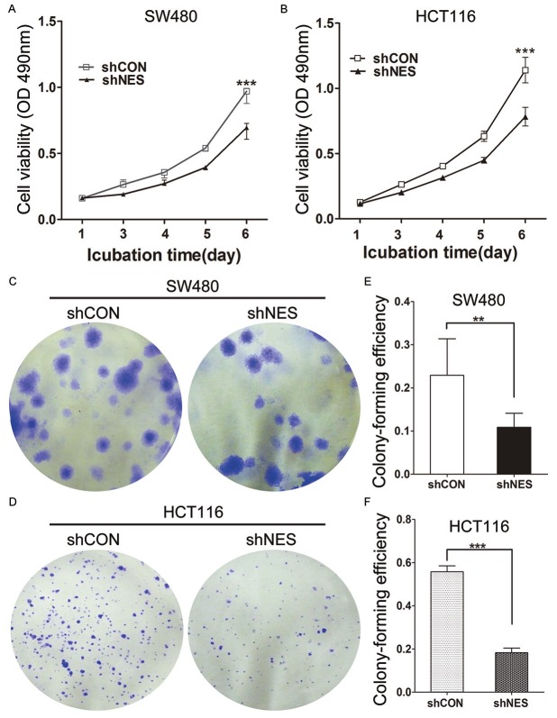
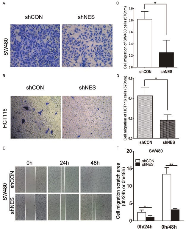
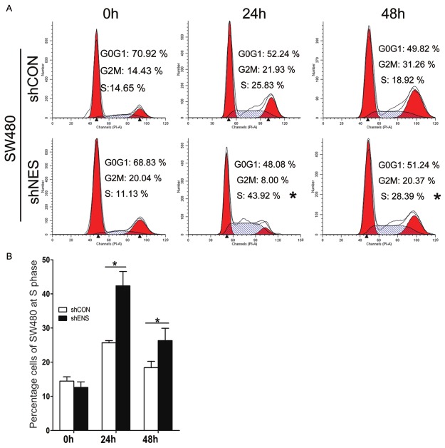
Nestin, a member of type VI intermediate filament protein family, is widely expressed in mammalian nervous tissue and stem/precursor cells of non-neuronal normal tissues. Nestin has also been investigated to determine possible tumor-promoting functions. However, whether Nestin is involved in colorectal cancer (CRC) cells remains unclear. In this report, Nestin expression was upregulated in stromal cells of human CRC tissues. Endogenous Nestin expression in CRC cell lines SW480 and HCT116 was knocked down by a lentivirus. MTT and colony formation assays revealed that Nestin deletion significantly inhibits the proliferation of CRC cell lines; flow cytometer analysis showed that Nestin deletion causes cell cycle arrest at S phase. Transwell chamber and wound healing scratch assays also revealed that Nestin deletion suppresses cell migration. Our findings indicated that Nestin plays an essential role in CRC progression; thus, Nestin can be applied as a therapeutic target of CRC.
Keywords: Nestin; colorectal cancer; metastasis.
Figures

Similar articles
-
Lentivirus-mediated knockdown of rhomboid domain containing 1 inhibits colorectal cancer cell growth.Mol Med Rep. 2015 Jul;12(1):377-81. doi: 10.3892/mmr.2015.3365. Epub 2015 Feb 17. Mol Med Rep. 2015. PMID: 25695376
-
[Abnormal expression of APRIL in colorectal cancer cells promotes tumor growth and metastasis].Zhonghua Zhong Liu Za Zhi. 2013 Apr;35(4):249-55. doi: 10.3760/cma.j.issn.0253-3766.2013.04.003. Zhonghua Zhong Liu Za Zhi. 2013. PMID: 23985251 Chinese.
-
Potential role of TRIM3 as a novel tumour suppressor in colorectal cancer (CRC) development.Scand J Gastroenterol. 2016;51(5):572-82. doi: 10.3109/00365521.2015.1124285. Epub 2015 Dec 22. Scand J Gastroenterol. 2016. PMID: 26691157
-
Chitinase 3-Like 1, Nestin, and Testin Proteins as Novel Biomarkers of Potential Clinical Use in Colorectal Cancer: A Review.Adv Exp Med Biol. 2020;1279:1-8. doi: 10.1007/5584_2020_506. Adv Exp Med Biol. 2020. PMID: 32170669 Review.
-
Nestin-expressing progenitor cells: function, identity and therapeutic implications.Cell Mol Life Sci. 2018 Jun;75(12):2177-2195. doi: 10.1007/s00018-018-2794-z. Epub 2018 Mar 14. Cell Mol Life Sci. 2018. PMID: 29541793 Free PMC article. Review.
Cited by
-
Glycine Promotes the Survival of a Subpopulation of Neural Stem Cells.Front Cell Dev Biol. 2018 Jul 11;6:68. doi: 10.3389/fcell.2018.00068. eCollection 2018. Front Cell Dev Biol. 2018. PMID: 30050902 Free PMC article.
-
Nestin Expression Is Associated with Relapses in Head and Neck Lesions.Diagnostics (Basel). 2021 Mar 24;11(4):583. doi: 10.3390/diagnostics11040583. Diagnostics (Basel). 2021. PMID: 33805026 Free PMC article.
-
Is There Such a Thing as a Genuine Cancer Stem Cell Marker? Perspectives from the Gut, the Brain and the Dental Pulp.Biology (Basel). 2020 Nov 27;9(12):426. doi: 10.3390/biology9120426. Biology (Basel). 2020. PMID: 33260962 Free PMC article. Review.
-
Nestin is a New Partner in Endothelial Colony Forming Cell Angiogenic Potential.Stem Cell Rev Rep. 2023 Oct;19(7):2541-2550. doi: 10.1007/s12015-023-10587-1. Epub 2023 Jul 15. Stem Cell Rev Rep. 2023. PMID: 37452965
-
Expression of Nestin associates with BRCA1 mutations, a basal-like phenotype and aggressive breast cancer.Sci Rep. 2017 Apr 24;7(1):1089. doi: 10.1038/s41598-017-00862-w. Sci Rep. 2017. PMID: 28439082 Free PMC article.
References
-
- Brenner H, Kloor M, Pox CP. Colorectal cancer. Lancet. 2014;383:1490–1502. - PubMed
-
- Jemal A, Siegel R, Ward E, Hao Y, Xu J, Murray T, Thun MJ. Cancer statistics, 2008. CA Cancer J Clin. 2008;58:71–96. - PubMed
-
- Cunningham D, Atkin W, Lenz HJ, Lynch HT, Minsky B, Nordlinger B, Starling N. Colorectal cancer. Lancet. 2010;375:1030–1047. - PubMed
-
- Enewold L, Horner MJ, Shriver CD, Zhu K. Socioeconomic disparities in colorectal cancer mortality in the United States, 1990-2007. J Community Health. 2014;39:760–766. - PubMed
-
- Andressen C, Stocker E, Klinz FJ, Lenka N, Hescheler J, Fleischmann B, Arnhold S, Addicks K. Nestin-specific green fluorescent protein expression in embryonic stem cell-derived neural precursor cells used for transplantation. Stem Cells. 2001;19:419–424. - PubMed
Publication types
MeSH terms
Substances
LinkOut - more resources
Full Text Sources
Medical