Cerebral cavernous malformations associated to meningioma: High penetrance in a novel family mutated in the PDCD10 gene
- PMID: 26246098
- PMCID: PMC4757286
- DOI: 10.1177/1971400915591688
Cerebral cavernous malformations associated to meningioma: High penetrance in a novel family mutated in the PDCD10 gene
Abstract
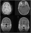
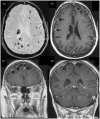
Multiple familial meningiomas occur in rare genetic syndromes, particularly neurofibromatosis type 2. The association of meningiomas and cerebral cavernous malformations (CCMs) has been reported in few patients in the medical literature. The purpose of our study is to corroborate a preferential association of CCMs and multiple meningiomas in subjects harbouring mutations in the PDCD10 gene (also known as CCM3). Three members of an Italian family affected by seizures underwent conventional brain Magnetic Resonance Imaging (MRI) with gadolinium contrast agent including gradient echo (GRE) imaging. The three CCM-causative genes were sequenced by Sanger method. Literature data reporting patients with coexistence of CCMs and meningiomas were reviewed. MRI demonstrated dural-based meningioma-like lesions associated to multiple parenchymal CCMs in all affected individuals. A disease-causative mutation in the PDCD10 gene (p.Gln112PhefsX13) was identified. Based on neuroradiological and molecular data as well as on literature review, we outline a consistent association between PDCD10 mutations and a syndrome of CCMs with multiple meningiomas. This condition should be considered in the differential diagnosis of multiple/familial meningioma syndromes. In case of multiple/familial meningioma the use of appropriate MRI technique may include GRE and/or susceptibility-weighted imaging (SWI) to rule out CCM. By contrast, proper post-gadolinium scans may aid defining dural lesions in CCM patients and are indicated in PDCD10-mutated individuals.
Keywords: Magnetic resonance imaging; PDCD10.; cerebral cavernous malformation; multiple meningioma.
© The Author(s) 2015.
Figures


Similar articles
-
PDCD10 gene mutations in multiple cerebral cavernous malformations.PLoS One. 2014 Oct 29;9(10):e110438. doi: 10.1371/journal.pone.0110438. eCollection 2014. PLoS One. 2014. PMID: 25354366 Free PMC article.
-
Description of Two Families with New Mutations in Familial Cerebral Cavernous Malformations Genes.J Stroke Cerebrovasc Dis. 2021 Dec;30(12):106130. doi: 10.1016/j.jstrokecerebrovasdis.2021.106130. Epub 2021 Sep 29. J Stroke Cerebrovasc Dis. 2021. PMID: 34597987
-
A single-center study on 140 patients with cerebral cavernous malformations: 28 new pathogenic variants and functional characterization of a PDCD10 large deletion.Hum Mutat. 2018 Dec;39(12):1885-1900. doi: 10.1002/humu.23629. Epub 2018 Sep 24. Hum Mutat. 2018. PMID: 30161288
-
Molecular diagnosis in cerebral cavernous malformations.Neurologia. 2017 Oct;32(8):540-545. doi: 10.1016/j.nrl.2015.07.001. Epub 2015 Aug 21. Neurologia. 2017. PMID: 26304651 Review. English, Spanish.
-
Cerebral cavernous malformations: from CCM genes to endothelial cell homeostasis.Trends Mol Med. 2013 May;19(5):302-8. doi: 10.1016/j.molmed.2013.02.004. Epub 2013 Mar 15. Trends Mol Med. 2013. PMID: 23506982 Review.
Cited by
-
The Dual Role of PDCD10 in Cancers: A Promising Therapeutic Target.Cancers (Basel). 2022 Dec 3;14(23):5986. doi: 10.3390/cancers14235986. Cancers (Basel). 2022. PMID: 36497468 Free PMC article. Review.
-
The multifaceted PDCD10/CCM3 gene.Genes Dis. 2020 Dec 30;8(6):798-813. doi: 10.1016/j.gendis.2020.12.008. eCollection 2021 Nov. Genes Dis. 2020. PMID: 34522709 Free PMC article. Review.
-
Endothelial cell clonal expansion in the development of cerebral cavernous malformations.Nat Commun. 2019 Jun 24;10(1):2761. doi: 10.1038/s41467-019-10707-x. Nat Commun. 2019. PMID: 31235698 Free PMC article.
-
Multiple meningiomas: Epidemiology, management, and outcomes.Neurooncol Adv. 2023 Jun 3;5(Suppl 1):i35-i48. doi: 10.1093/noajnl/vdac108. eCollection 2023 May. Neurooncol Adv. 2023. PMID: 37287575 Free PMC article.
-
Familial Multiple Cavernous Malformation Syndrome: MR Features in This Uncommon but Silent Threat.J Belg Soc Radiol. 2016 Mar 21;100(1):51. doi: 10.5334/jbr-btr.938. J Belg Soc Radiol. 2016. PMID: 30151459 Free PMC article.
References
-
- Riemenschneider MJ, Perry A, Reifenberger G. Histological classification and molecular genetics of meningiomas. Lancet Neurol 2006; 5: 1045–1054. - PubMed
-
- DeAngelis LM. Brain tumors. N Engl J Med 2001; 11; 344: 114–123. - PubMed
-
- Whittle IR, Smith C, Navoo P, et al. Meningiomas. Lancet 2004; 8; 363: 1535–1543. - PubMed
MeSH terms
Substances
LinkOut - more resources
Full Text Sources
Other Literature Sources
Research Materials

