

Structure and Function of p53-DNA Complexes with Inactivation and Rescue Mutations: A Molecular Dynamics Simulation Study
- PMID: 26244575
- PMCID: PMC4526489
- DOI: 10.1371/journal.pone.0134638
Structure and Function of p53-DNA Complexes with Inactivation and Rescue Mutations: A Molecular Dynamics Simulation Study
Abstract
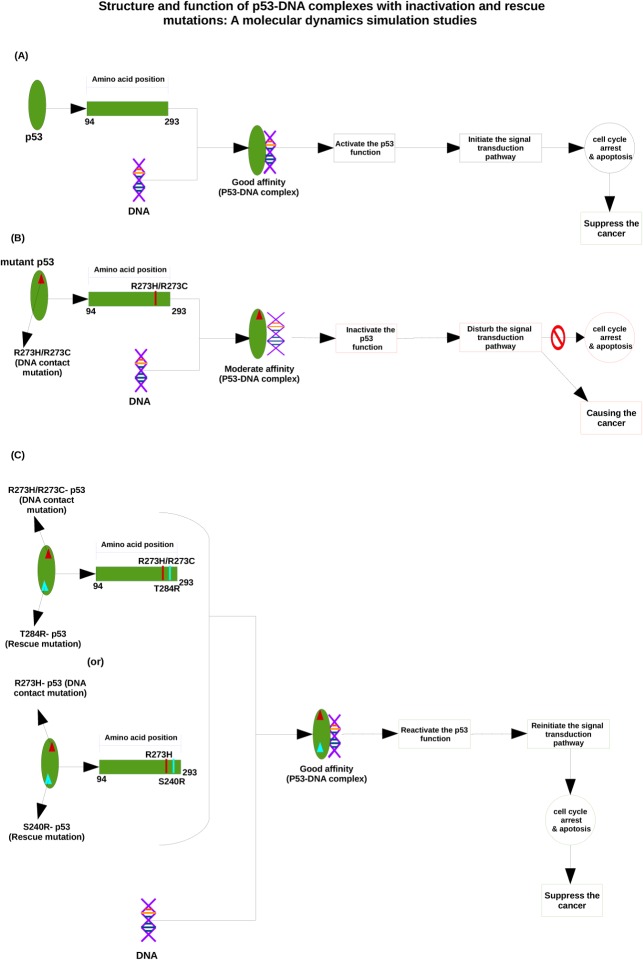
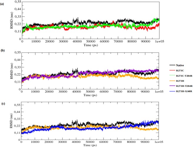
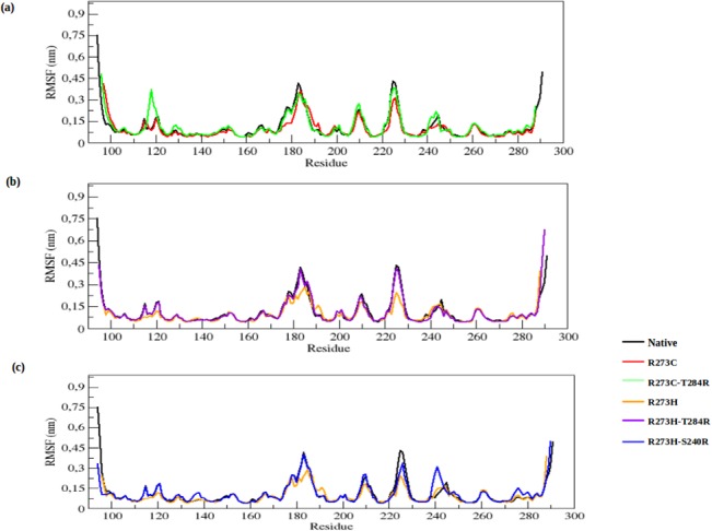
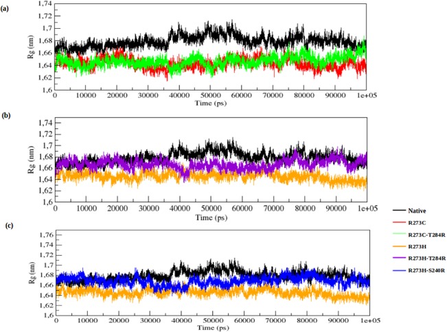
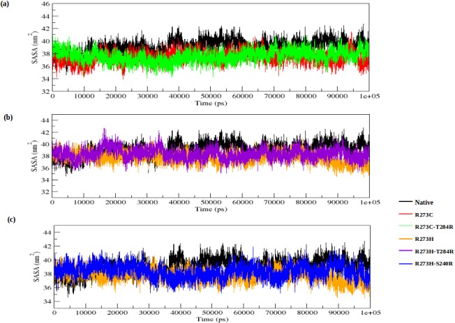
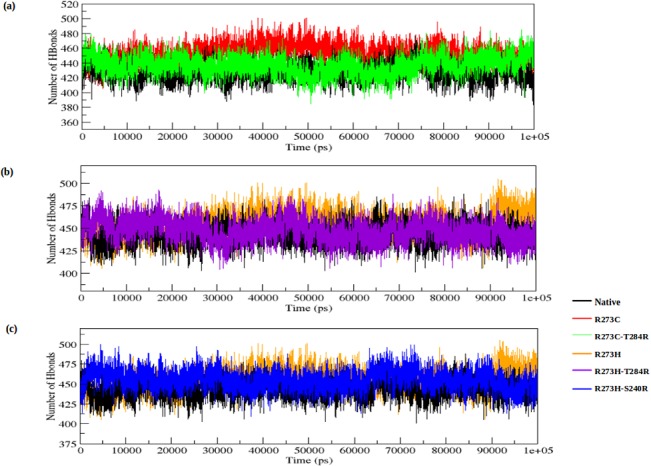
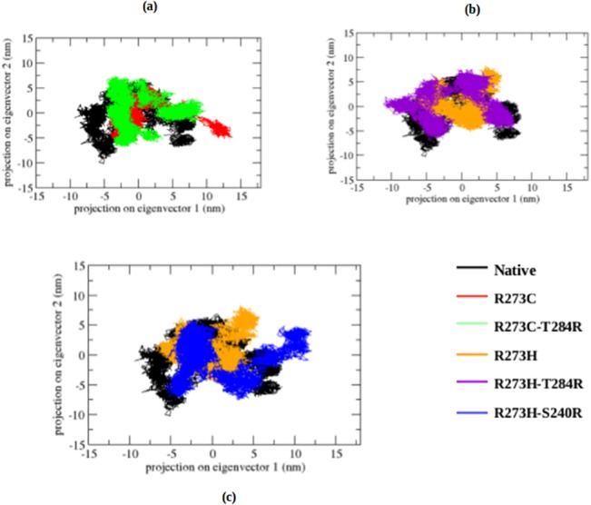
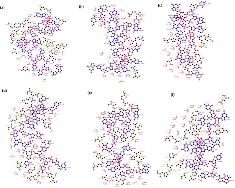
The tumor suppressor protein p53 can lose its function upon DNA-contact mutations (R273C and R273H) in the core DNA-binding domain. The activity can be restored by second-site suppressor or rescue mutations (R273C_T284R, R273H_T284R, and R273H_S240R). In this paper, we elucidate the structural and functional consequence of p53 proteins upon DNA-contact mutations and rescue mutations and the underlying mechanisms at the atomic level by means of molecular dynamics simulations. Furthermore, we also apply the docking approach to investigate the binding phenomena between the p53 protein and DNA upon DNA-contact mutations and rescue mutations. This study clearly illustrates that, due to DNA-contact mutants, the p53 structure loses its stability and becomes more rigid than the native protein. This structural loss might affect the p53-DNA interaction and leads to inhibition of the cancer suppression. Rescue mutants (R273C_T284R, R273H_T284R and R273H_S240R) can restore the functional activity of the p53 protein upon DNA-contact mutations and show a good interaction between the p53 protein and a DNA molecule, which may lead to reactivate the cancer suppression function. Understanding the effects of p53 cancer and rescue mutations at the molecular level will be helpful for designing drugs for p53 associated cancer diseases. These drugs should be designed so that they can help to inhibit the abnormal function of the p53 protein and to reactivate the p53 function (cell apoptosis) to treat human cancer.
Conflict of interest statement
Figures
Similar articles
-
Mutants TP53 p.R273H and p.R273C but not p.R273G enhance cancer cell malignancy.Hum Mutat. 2014 May;35(5):575-84. doi: 10.1002/humu.22528. Epub 2014 Apr 7. Hum Mutat. 2014. PMID: 24677579
-
Structural basis of restoring sequence-specific DNA binding and transactivation to mutant p53 by suppressor mutations.J Mol Biol. 2009 Jan 9;385(1):249-65. doi: 10.1016/j.jmb.2008.10.063. Epub 2008 Oct 30. J Mol Biol. 2009. PMID: 18996393
-
Ensemble-based computational approach discriminates functional activity of p53 cancer and rescue mutants.PLoS Comput Biol. 2011 Oct;7(10):e1002238. doi: 10.1371/journal.pcbi.1002238. Epub 2011 Oct 20. PLoS Comput Biol. 2011. PMID: 22028641 Free PMC article.
-
Structural and sequential context of p53: A review of experimental and theoretical evidence.Prog Biophys Mol Biol. 2015 Mar;117(2-3):250-263. doi: 10.1016/j.pbiomolbio.2014.12.002. Epub 2014 Dec 27. Prog Biophys Mol Biol. 2015. PMID: 25550083 Review.
-
Structure-function-rescue: the diverse nature of common p53 cancer mutants.Oncogene. 2007 Apr 2;26(15):2226-42. doi: 10.1038/sj.onc.1210291. Oncogene. 2007. PMID: 17401432 Review.
Cited by
-
Discovery of anti-MERS-CoV small covalent inhibitors through pharmacophore modeling, covalent docking and molecular dynamics simulation.J Mol Liq. 2021 May 15;330:115699. doi: 10.1016/j.molliq.2021.115699. Epub 2021 Feb 20. J Mol Liq. 2021. PMID: 33867606 Free PMC article.
-
The Challenges and Prospects of p53-Based Therapies in Ovarian Cancer.Biomolecules. 2023 Jan 12;13(1):159. doi: 10.3390/biom13010159. Biomolecules. 2023. PMID: 36671544 Free PMC article. Review.
-
Unravelling the anticancer potential of a square planar copper complex: toward non-platinum chemotherapy.RSC Adv. 2021 Dec 10;11(62):39349-39361. doi: 10.1039/d1ra06227a. eCollection 2021 Dec 6. RSC Adv. 2021. PMID: 35492449 Free PMC article.
-
Investigating pathogenic SNP of PKCι in HCV-induced hepatocellular carcinoma.Sci Rep. 2023 Aug 2;13(1):12504. doi: 10.1038/s41598-023-39804-0. Sci Rep. 2023. PMID: 37532886 Free PMC article.
-
The Structural Effect of FLT3 Mutations at 835th Position and Their Interaction with Acute Myeloid Leukemia Inhibitors: In Silico Approach.Int J Mol Sci. 2021 Jul 16;22(14):7602. doi: 10.3390/ijms22147602. Int J Mol Sci. 2021. PMID: 34299222 Free PMC article.
References
-
- Meulmeester E, Jochemsen AG. p53: a guide to apoptosis. Curr Cancer Drug Targets 2008;8: 87–97. - PubMed
-
- Lim YP, Lim TT, Chan YL, Song AC, Yeo BH, Vojtesek B, et al. The p53 knowledgebase: an integrated information resource for p53 research. Oncogene 2007;26: 1517–1521. - PubMed
-
- Gomez-Lazaro M, Fernandez-Gomez FJ, Jordán J. p53: twenty five years understanding the mechanism of genome protection. J PhysiolBiochem 2004;60: 287–307. - PubMed
-
- Sakaguchi K, Sakamoto H, Xie D, Erickson JW, Lewis MS, Anderson CW et al. Effect of phosphorylation on tetramerization of the tumor suppressor protein p53. J Protein Chem. 1997;16: 553–556. - PubMed
Publication types
MeSH terms
Substances
Grants and funding
LinkOut - more resources
Full Text Sources
Other Literature Sources
Research Materials
Miscellaneous
