. 2015 Oct 15;43(18):8790-800.
doi: 10.1093/nar/gkv764. Epub 2015 Jul 30.

The N-terminus of RPA large subunit and its spatial position are important for the 5'->3' resection of DNA double-strand breaks

Affiliations

The N-terminus of RPA large subunit and its spatial position are important for the 5'->3' resection of DNA double-strand breaks

Margaret Tammaro et al. Nucleic Acids Res. .

Abstract

The first step of homology-dependent repair of DNA double-strand breaks (DSBs) is the resection of the 5' strand to generate 3' ss-DNA. Of the two major nucleases responsible for resection, EXO1 has intrinsic 5'->3' directionality, but DNA2 does not. DNA2 acts with RecQ helicases such as the Werner syndrome protein (WRN) and the heterotrimeric eukaryotic ss-DNA binding protein RPA. We have found that the N-terminus of the RPA large subunit (RPA1N) interacts with both WRN and DNA2 and is essential for stimulating WRN's 3'->5' helicase activity and DNA2's 5'->3' ss-DNA exonuclease activity. A mutant RPA complex that lacks RPA1N is unable to support resection in Xenopus egg extracts and human cells. Furthermore, relocating RPA1N to the middle subunit but not to the small subunit causes severe defects in stimulating DNA2 and WRN and in supporting resection. Together, these findings suggest that RPA1N and its spatial position are critical for restricting the directionality of the WRN-DNA2 resection pathway.


Figures

Figure 1.
Figure 1.
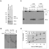
The N-terminal domain of RPA1 interacts with WRN and DNA2. (A) A gel showing the GST fusions of various parts of Xenopus RPA. The proteins were separated on a 12% SDS-PAGE gel and stained by Coomassie brilliant blue. The size markers are in kilodaltons. (B) A Western blot showing the interaction between Xenopus WRN and RPA1N. The various GST fusion proteins were incubated with the N-terminal 455 amino acids of WRN and then isolated by glutathione Sepharose beads. The proteins bound to beads were analyzed for WRN by Western blot. (C) A western blot showing the interaction between RPA1N (GST-1N) with WRN and DNA2 in Xenopus egg extracts. (D). A western blot showing the interaction between RPA1N (GST-1N) and DNA2.
Figure 2.
Figure 2.
RPA1N is required for the stimulation of WRN's helicase activity. (A) A gel showing recombinant Xenopus wild-type RPA and mutant RPA lacking RPA1N (1NΔ RPA). The proteins were separated by a 4–12% NUPAGE MOPS gel (Invitrogen, CA) and stained by Coomassie brilliant blue. (B) An agarose gel showing the binding activity of the wild-type and 1NΔ RPA to ss-m13 DNA. The binding reactions were separated by a 1% TAE-agarose gel and DNA was detected by staining with SYBR Gold. (C) Effect of wild-type RPA and 1NΔ RPA on the unwinding activity of WRN. The substrate was a 32P-labeled 56mer oligonucleotide annealed to m13 ss-DNA. After incubation at room temperature for 30 min, the reactions were separated by a 8% TAE-PAGE and DNA was detected by exposure of the dried gel to X-ray film. (D) Quantitaton of the unwinding activity in the presence of wild-type RPA or 1NΔ RPA. The percentages of the oligonucleotide dissociated were quantitated and the averages and standard deviations were calculated from three sets of data and plotted.
Figure 3.
Figure 3.
RPA1N is required for the stimulation of DNA2's 5′->3′ exonuclease activity. (A) Nuclease assay with a 32P-labeled ss-48mer oligonucleotide attached to magnetic beads via a biotinylated nucleotide at the 3′ end of the labeled strand. The substrate was incubated with DNA2 and wild-type or 1NΔ RPA (4 ng/μl) for the indicated times and the products were analyzed by a 8% TAE-PAGE. (B) The percentages of nucleotide products were quantitated and the averages and standard deviations were calculated from three sets of data and plotted.
Figure 4.
Figure 4.
RPA1N is required for resection in Xenopus egg extracts. The substrate for resection was a 5.7 kb linearized DNA with a ddC at the 3′ end and a 32P label immediately inside. The substrate was incubated with mock depleted or RPA depleted extracts supplemented with various RPA proteins or buffer. Samples taken at the indicated times were analyzed by a 1% TAE-agarose gel.
Figure 5.
Figure 5.
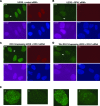
RPA1N is important for resection in cells. (A) Etoposide-induced RPA foci in U2OS cells treated with control siRNA. (B) Etoposide-induced RPA foci in U2OS cells treated with RPA1 siRNA. (C) Etoposide-induced RPA foci in U2OS cells expressing wild-type RPA1 treated with RPA1 siRNA. (D) Etoposide-induced RPA foci in U2OS cells expressing RPA1 that lacks the N-terminal domain treated with RPA1 siRNA. The RPA1 siRNA targets the 3′ UTR of the endogenous RPA1 gene, which is not present in the two ectopic RPA1 genes. Cells were treated with different siRNAs for 72 h and then with 250 μM etoposide for 2 h. They were co-stained for RPA, CenpF and EdU. EdU was added to the media 15 min before etoposide. (E) Close-ups of the nuclei indicated by arrows in (A)–(D).
Figure 6.
Figure 6.
The spatial location of RPA1N affects the stimulation of WRN's helicase activity. (A) A gel showing recombinant Xenopus wild-type RPA and two mutant RPAs. Proteins were separated by a 4–12% NUPAGE MES gel (Invitrogen, CA, USA) and stained by Coomassie brilliant blue. (B) An agarose gel showing the binding activity of the wild-type and mutant RPAs to ss-m13 DNA. (C) Effect of wild-type RPA and mutant RPAs on the unwinding activity of WRN. The substrate was a 32P-labeled 56mer oligonucleotide annealed to m13 ss-DNA. (D) Quantitaton of the unwinding activity in the presence of wild-type RPA or mutant RPAs. The percentages of the oligonucleotide dissociated were quantitated and the averages and standard deviations were calculated from three sets of data and plotted.
Figure 7.
Figure 7.
The spatial location of RPA1N is important for stimulating the ss-DNA exonuclease activity of DNA2. (A) Nuclease assay with a 32P-labeled ss-48mer oligonucleotide attached to magnetic beads via a biotinylated nucleotide at the 3′ end of the labeled strand. The substrate was incubated with DNA2 and wild-type or mutant RPAs (4 ng/μl) for the indicated times and the products were analyzed by a 8% TAE-PAGE. (B) The percentages of nucleotide products were quantitated and the averages and standard deviations were calculated from three sets of data and plotted. (C) Effect of wild-type and mutant RPAs on the degradation of a 5.7kb ss-DNA with a 32P label near the 3′ end in Xenopus egg extracts. RPA-depleted extracts were supplemented with buffer or various RPA proteins (20ng/μl). Samples were taken at the indicated times and analyzed by a 1% TAE-agarose gel.
Figure 8.
Figure 8.
The location of RPA1N is important for the resection of ds-DNA. The substrate for resection was a 5.7 kb linearized ds-DNA with a ddC at the 3′ end and a 32P label immediately inside. The substrate was incubated with mock depleted or RPA depleted extracts supplemented with various RPA proteins or buffer. Samples taken at the indicated times were analyzed by a 1% TAE-agarose gel electrophoresis.
Figure 9.
Figure 9.
Model of the position of RPA1N and stimulation of WRN's 3′->5′ helicase activity and DNA2's 5′->3′ exonuclease activity. (A) RPA1N in the wildtype complex is optimally positioned to interact with WRN at the ss/ds-DNA junction on the 3′ strand and with DNA2 approaching from the 5′ end. RPA binding to the rest of ss-DNA prevents the reannealing of the unwound strands and the endonuclytic cleavage by DNA2. (B) 1NΔ RPA can still bind to ss-DNA to protect it against endonuclytic cleavage by DNA2 and to facilitate unwinding by preventing reannealing of the single strands unwound by the low intrinsic helicase activity of WRN. (C) RPA1N in the 3–1N RPA complex is still positioned close enough to interact normally with WRN and DNA2 and stimulate their activities. (D) RPA1N in the 1N-2 RPA complex is shifted more drastically in space and is less effective to recruit WRN to the ss/ds-DNA junction. It is completely incapable of recruiting DNA2 to the 5′ end of ss-DNA.

Similar articles

Cited by

References

    1. Symington L.S. Role of RAD52 epistasis group genes in homologous recombination and double-strand break repair. Microbiol. Mol. Biol. Rev. 2002;66:630–670. - PMC - PubMed
    1. Nussenzweig A., Nussenzweig M.C. A backup DNA repair pathway moves to the forefront. Cell. 2007;131:223–225. - PubMed
    1. Ciccia A., Elledge S.J. The DNA damage response: making it safe to play with knives. Mol. Cell. 2010;40:179–204. - PMC - PubMed
    1. Symington L.S., Gautier J. Double-strand break end resection and repair pathway choice. Annu. Rev. Genet. 2011;45:247–271. - PubMed
    1. Toczylowski T., Yan H. Mechanistic analysis of a DNA end processing pathway mediated by the Xenopus Werner syndrome protein. J. Biol. Chem. 2006;281:33198–33205. - PubMed

Publication types

MeSH terms

LinkOut - more resources