. 2016 Jan;73(2):409-25.
doi: 10.1007/s00018-015-1999-7. Epub 2015 Jul 30.

The ecdysone receptor signalling regulates microvilli formation in follicular epithelial cells

Affiliations

The ecdysone receptor signalling regulates microvilli formation in follicular epithelial cells

Patrizia Romani et al. Cell Mol Life Sci. 2016 Jan.

Abstract

Epithelial morphogenesis contributes greatly to the development and homeostasis of the organs and body parts. Here, we analysed the consequences of impaired ecdysone receptor (EcR) signalling in the Drosophila follicular epithelium. Besides governing cell growth, the three EcR isoforms act redundantly in controlling follicle cell positioning. Flattening of the microvilli and an aberrant actin cytoskeleton arise from defective EcR signalling in follicle cells, and these defects impact on the organisation of the oocyte membrane. We found that this signalling governs a complex molecular network since its impairment affects key molecules as atypical protein kinase C and activated Moesin. Interestingly, the activity of the transcription factor Tramtrack69 isoform is required for microvilli and their actin core morphogenesis as well as for follicle cell positioning. In conclusion, our findings provide evidence of novel roles for EcR signalling and Tramtrack69 transcription factor in controlling stage-specific differentiation events that take place in the follicular epithelium.

Keywords: Cad99C; EcR dominant negative; Ecdysone signalling; Oogenesis.


Figures
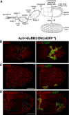
Fig. 1
Fig. 1
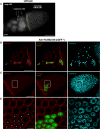
EcR signalling controls expression of the EcR gene during oogenesis. Schematic representation of a single ovariole containing a string of developing egg chambers of progressive age (a). The newly formed egg chamber emerges from the germarium, undergoes a continuous process of development and acquires morphological features that allow recognition of 14 different stages. In each egg chamber FCs form an epithelium that covers the nurse cells–oocyte complex. As oogenesis proceeds, complex differentiation events specify multiple subpopulation of FCs. Starting at stage 9 a group of anterior follicle cells, the border cells, delaminates from the epithelium and migrates through the nurse cells to reach the oocyte surface. As egg chamber grows, main body FCs change their shape from cuboidal to columnar and by stage 10 they cover the oocyte. The organisation of the stage 10B is illustrated in the schematic drawings of a sagittal plane through the centre of the egg chamber and of a surface view. At stage 10B few squamous FCs cover the nurse cells, the columnar main body FCs surround the oocyte and a group of anterior columnar FCs, namely the centripetally migrating FCs, starts to migrate at the interface between the oocyte and the nurse cells. The border cells after reaching the anterior region of the oocyte start to migrate dorsally. Anterior is to the left. The FC flp-out clones are marked by the expression of the GFP protein (green, dotted area in bd). Confocal microscopy analysis of stage 10B egg chambers dissected from hs-flp/Act>>Gal4; UAS-nGFP/+; UAS-EcRB2-F645A/+ females (bd) and stained with anti-EcRA (red in b), anti-EcRB1 (red in c) and anti-EcR common (red in d), antibodies. The stage 10B egg chambers are oriented with the anterior regions to the left. Scale bars bd 50 μm
Fig. 2
Fig. 2
Blocking EcR signalling causes mispositioning and reduced FC growth. Fluorescence microscopy analysis of a stage 10B egg chamber dissected from a wild-type female and stained with DAPI to show the positioning of the squamous FCs (arrows) and the main body FCs that are located in the anterior (left to yellow line) and posterior region (right to yellow line) of the egg chamber, respectively (a). The FC flp-out clones are marked by the expression of the GFP protein (green) (asterisks) (bc′). Confocal microscopy analysis of stage 10B egg chambers dissected from hs-flp/Act>>Gal4; UAS-nGFP/+; UAS-EcRB2-F645A/+ females and stained with anti-DE-Cad (red) ab and with To-Pro-3 nuclear dye (cyan). c′ Magnification of the boxed regions in c. The anterior region is to the left in all the panels. Scale bars ac 50 μm and c′ 10 μm
Fig. 3
Fig. 3
Blocking EcR signalling affects egg chamber development. Fluorescence microscopy analysis of ovarioles (a, b) and stage 10 egg chambers (a′, c) dissected from w/w; tub-Gal80 ts /+; tub-Gal4/+ control females (a, a′) and from w/w; tub-Gal80 ts /+; tub-Gal4/UAS-EcRB2-DN females (b, c) and stained with DAPI. The arrows in b indicate degenerating egg chambers that are absent in control ovarioles (a). The brackets in a′ and c indicate main body FCs. Fluorescence microscopy analysis of mid-oogenesis egg chambers dissected from w/w; Cy2-Gal4/UAS-nGFP; tub-Gal80 ts /+ (dg) and from w/w; Cy2-Gal4/UAS-nGFP; tub-Gal80 ts /UAS-EcRB2-DN (hk) females and stained with DAPI. The expression domain of the Cy2-Gal4 driver is marked by GFP (green in e, g and i, k). DAPI staining is in D and H (white). g, k Overlap of the optical images in e, f and in i, j, respectively. The arrows in dk mark the limit of the main body FCs. Anterior is up in all panels with the exception of c. Scale bars a, a′, b 100 μm, c 200 μm and dk 50 μm
Fig. 4
Fig. 4
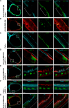
Blocking EcR signalling alters microvilli and the F-actin cytoskeleton. The FC flp-out clones expressing EcRB2-DN are marked by the expression of the GFP protein (a) (green). The expression domain of the Cy2-Gal4 driver is marked by GFP (green in b, c). Confocal microscopy analysis of a stage 10B egg chamber from hs-flp/Act>>Gal4; UAS-mGFP/+; UAS-EcRB2-DN/+ females (a). The microvilli are stained with anti-Cad99C ab (cyan) and actin with phalloidin (red). a From left to right show: (1) the merging of the three signals, (2) the magnification of the microvilli, and (3) of the actin staining alone, and (4) the magnified merging of the signals. The boxed region in the left panel marks a flp-out clone with flanking wild-type cells and indicates the magnified regions on the right. The brackets indicate the flp-out clone. The arrow shows the space between FCs and oocyte, the arrowhead shows the absence of this space. Confocal microscopy analysis of egg chambers dissected from w/w; Cy2-Gal4/UAS-nGFP; tub-Gal80 ts /UAS-EcRB2-DN (b) and from w/w; Cy2-Gal4/UAS-nGFP; tub-Gal80 ts /+ (c) females. The organisation and stainings of the panels in b and c are the same as in a. Asterisks indicate the alteration of the actin cytoskeleton. Anterior is up in all panels. Scale bars ac, left 50 μm and ac magnifications 5 μm
Fig. 5
Fig. 5
The morphogenesis of microvilli requires aPKC. Confocal microscopy analysis of a stage 10B egg chamber dissected from hs-flp/Act>>Gal4; UAS-nGFP/+; UAS-EcRB2-DN/+ stained with anti-aPKC ab (red) (a). The FC flp-out clone expressing EcRB2-DN is marked by the expression of the GFP protein (green). Confocal microscopy analysis of a stage 10B egg chamber dissected from hs-flp/Act>>Gal4; UAS-aPKC CA /+; +/+, stained with anti-Cad99C ab (cyan), anti-CD2 ab (green) and phalloidin (red) (b). The FC flp-out clone expressing aPKCCA is indicated by the absence of CD2 and is marked by the brackets. Confocal microscopy analysis of a stage 10B egg chamber dissected from hs-flp/Act>>Gal4; UAS-nGFP/+; UAS-aPKC-RNAi/+ female stained with anti-Cad99C ab (cyan) and phalloidin (red) (c). The FC flp-out clone expressing aPKC-RNAi is marked by the expression of the GFP protein (green, brackets). Confocal microscopy analysis of a stage 10B egg chamber dissected from hs-flp/Act>>Gal4; +/+; UAS-aPKC-RNAi/UAS-EcRB2-DN female stained with anti-Cad99C ab (cyan), anti-CD2 ab (green) and phalloidin (red) (d). The FC flp-out clone expressing EcRB2-DN and aPKC-RNAi is indicated by the absence of CD2 (brackets). Anterior is up in a, b, d and on the left in c. All boxed FC flp-out clones are magnified in the panels on the right. Scale bars ad, left 50 μm and ad magnifications 5 μm
Fig. 6
Fig. 6
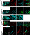
Blocking EcR signalling alters the organisation of the oocyte membrane. The organisation of ag from left to right is the same as in Fig. 4. Confocal microscopy analysis of a stage 10B egg chamber dissected from hs-flp/Act>>Gal4; +/+; UAS-EcRB2-DN/+ and stained with anti-CD2 ab (cyan) and Lectin dye (green) (a). The FC flp-out clone expressing EcRB2-DN is indicated by the absence of CD2 and is marked by the brackets. Confocal microscopy analysis of egg chambers dissected from w/w; Cy2-Gal4/+; tub-Gal80 ts /UAS-EcRB2-DN (b) and from w/w; Cy2-Gal4/+; tub-Gal80 ts /+ (c) females and stained with the Lectin dye (green) and phalloidin (red in b and cyan in c). The asterisks in b indicate protrusions of the actin cytoskeleton of the oocyte. Confocal microscopy analysis of a stage 10B egg chamber dissected from hs-flp/Act>>Gal4; UAS-mGFP/+; UAS-EcRB2-DN/+ female stained with anti-Cad99C (cyan) and anti-Yl (red) abs (d). The FC flp-out clone expressing EcRB2-DN is indicated by the GFP expression (green) and is marked by the brackets. Confocal microscopy analysis of egg chambers dissected from w/w; Cy2-Gal4/UAS-nGFP; tub-Gal80 ts /UAS-EcRB2-DN (e) and from w/w; Cy2-Gal4/UAS-nGFP; tub-Gal80 ts /+ (f) females stained with phalloidin (cyan) and anti-Yl ab (red). Confocal microscopy analysis of a stage 10B egg chamber dissected from hs-flp/Act>>Gal4; Flu-ph/+; UAS-EcRB2-DN/+ female stained with phalloidin (red), anti-CD2 ab (green), and anti-HA ab (cyan) (g). The absence of the CD2 indicates FCs expressing EcRB2-DN (brackets). Anterior is up in a, c, d and on the left in b, eg. Scale bars ag, left 50 μm and ag magnifications 5 μm
Fig. 7
Fig. 7
Blocking EcR signalling enhances levels of pMoe. The FC flp-out clones are marked by the expression of the GFP protein (green) (a, b, e) and by the absence of the CD2 marker (c, d). Confocal microscopy analysis of a stage 10B egg chamber dissected from hs-flp/Act>>Gal4; UAS-nGFP/+; UAS-EcRB2-DN/+ stained with anti-pMoe (red) and anti-Sip1 (cyan) ab (a). Confocal microscopy analysis of a stage 10B egg chamber dissected from hs-flp/Act>>Gal4; UAS-mGFP/+; UAS-EcRB2-DN/+ stained with anti-Slik ab (cyan) (b). Confocal microscopy analysis of a stage 10B egg chamber dissected from hs-flp/Act>>Gal4; UAS-aPKC CA /+; +/+ (c, d) stained with anti-CD2 (green in c, d), anti-Sip1 (cyan in c) and anti-pMoe (red) and anti-Slik (cyan) in d. Confocal microscopy analysis of a stage 10B egg chamber dissected from hs-flp/Act>>Gal4; UAS-mGFP/UAS-Moe CA-Myc females stained with anti-Cad99C ab (cyan) and phalloidin (red) (e). The dotted lines in ad mark the boundary between the wild-type and EcRB2-DN expressing cells (a, b) and between the wild-type and aPKCCA expressing cells (c, d). Anterior is on the left in panels ad, and up in e. Scale bars ae left 50 μm and ae magnifications 10 μm
Fig. 8
Fig. 8
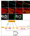
Morphogenesis of microvilli is altered in ttk 1e11 loss of function clones. Confocal microscopy analysis (a, b) and fluorescence microscopy analysis (c) of stage 10B egg chambers dissected from hs-flp, tub-Gal4, UAS-nGFP/+; +/+; FRT82B, tub-Gal80/FRT82B, ttk 1e11 stained with phalloidin (red) and anti-Cad99C ab (cyan in a), anti-aPKC ab (red in b) and DAPI (white in c). The ttk 1e11 FC clones are indicated by the presence of nGFP (green in ac). Scheme of the EcR signalling targets. p.m. plasma membrane (d). Anterior is up in a and on the left in b, c. Scale bars ac left panels 50 μm and a, b magnifications 10 μm

Similar articles

Cited by

References

    1. Yamanaka N, Rewitz KF, O’Connor MB. Ecdysone control of developmental transitions: lessons from Drosophila research. Annu Rev Entomol. 2013;58:497–516. doi: 10.1146/annurev-ento-120811-153608. - DOI - PMC - PubMed
    1. Schwedes CC, Carney GE. Ecdysone signaling in adult Drosophila melanogaster . J Insect Physiol. 2012;58(3):293–302. doi: 10.1016/j.jinsphys.2012.01.013. - DOI - PubMed
    1. Konig A, Yatsenko AS, Weiss M, Shcherbata HR. Ecdysteroids affect Drosophila ovarian stem cell niche formation and early germline differentiation. EMBO J. 2011;30(8):1549–1562. doi: 10.1038/emboj.2011.73. - DOI - PMC - PubMed
    1. Morris LX, Spradling AC. Steroid signaling within Drosophila ovarian epithelial cells sex-specifically modulates early germ cell development and meiotic entry. PLoS ONE. 2012;7(10):e46109. doi: 10.1371/journal.pone.0046109. - DOI - PMC - PubMed
    1. Bownes M. The roles of juvenile hormone, ecdysone and the ovary in the control of Drosophila vitellogenesis. J Insect Physiol. 1989;34:409–413. doi: 10.1016/0022-1910(89)90115-7. - DOI

Publication types

LinkOut - more resources