

Blood flow modulation of vascular dynamics
- PMID: 26218416
- PMCID: PMC4626080
- DOI: 10.1097/MOL.0000000000000218
Blood flow modulation of vascular dynamics
Abstract
Purpose of review: Blood flow is intimately linked with cardiovascular development, repair and dysfunction. The current review will build on the fluid mechanical principle underlying haemodynamic shear forces, mechanotransduction and metabolic effects.
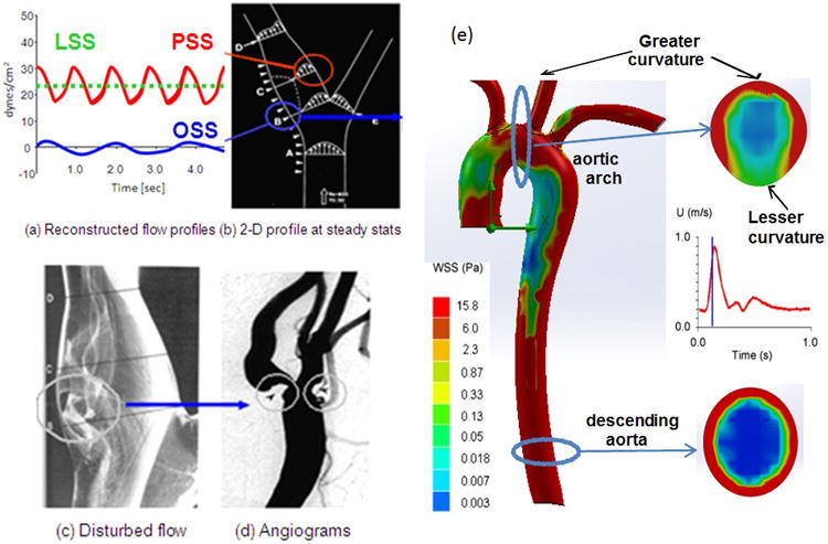
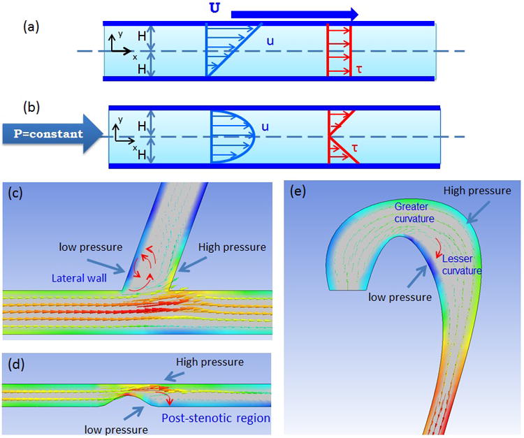
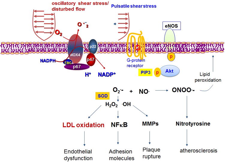
Recent findings: Pulsatile flow produces both time (∂τ/∂t) and spatial-varying shear stress (∂τ/∂x) to modulate vascular oxidative stress and inflammatory response with pathophysiological significance to atherosclerosis. The characteristics of haemodynamic shear forces, namely, steady laminar (∂τ/∂t = 0), pulsatile shear stress (PSS: unidirectional forward flow) and oscillatory shear stress (bidirectional with a near net 0 forward flow), modulate mechano-signal transduction to influence metabolic effects on vascular endothelial function. Atheroprotective PSS promotes antioxidant, anti-inflammatory and antithrombotic responses, whereas atherogenic oscillatory shear stress induces nicotinamide adenine dinucleotide phosphate oxidase-JNK signalling to increase mitochondrial superoxide production, protein degradation of manganese superoxide dismutase and post-translational protein modifications of LDL particles in the disturbed flow-exposed regions of vasculature. In the era of tissue regeneration, shear stress has been implicated in reactivation of developmental genes, namely, Wnt and Notch signalling, for vascular development and repair.
Summary: Blood flow imparts a dynamic continuum from vascular development to repair. Augmentation of PSS confers atheroprotection and reactivation of developmental signalling pathways for regeneration.
Conflict of interest statement
Figures
Similar articles
-
Shear-induced endothelial mechanotransduction: the interplay between reactive oxygen species (ROS) and nitric oxide (NO) and the pathophysiological implications.J Biomed Sci. 2014 Jan 13;21(1):3. doi: 10.1186/1423-0127-21-3. J Biomed Sci. 2014. PMID: 24410814 Free PMC article. Review.
-
MicroRNAs in flow-dependent vascular remodelling.Cardiovasc Res. 2013 Jul 15;99(2):294-303. doi: 10.1093/cvr/cvt096. Epub 2013 Apr 23. Cardiovasc Res. 2013. PMID: 23612583 Review.
-
Role of shear stress direction in endothelial mechanotransduction.Mol Cell Biomech. 2008 Mar;5(1):1-8. Mol Cell Biomech. 2008. PMID: 18524241
-
Flow detection and calcium signalling in vascular endothelial cells.Cardiovasc Res. 2013 Jul 15;99(2):260-8. doi: 10.1093/cvr/cvt084. Epub 2013 Apr 9. Cardiovasc Res. 2013. PMID: 23572234 Review.
-
The shear stress of it all: the cell membrane and mechanochemical transduction.Philos Trans R Soc Lond B Biol Sci. 2007 Aug 29;362(1484):1459-67. doi: 10.1098/rstb.2007.2128. Philos Trans R Soc Lond B Biol Sci. 2007. PMID: 17569643 Free PMC article. Review.
Cited by
-
Vascular dysfunction and pathology: focus on mechanical forces.Vasc Biol. 2021 Jun 9;3(1):R69-R75. doi: 10.1530/VB-21-0002. eCollection 2021. Vasc Biol. 2021. PMID: 34291191 Free PMC article. Review.
-
Endothelial mechanotransduction in cardiovascular development and regeneration: emerging approaches and animal models.Curr Top Membr. 2021;87:131-151. doi: 10.1016/bs.ctm.2021.07.002. Epub 2021 Oct 12. Curr Top Membr. 2021. PMID: 34696883 Free PMC article. Review.
-
It takes more than two to tango: mechanosignaling of the endothelial surface.Pflugers Arch. 2020 Apr;472(4):419-433. doi: 10.1007/s00424-020-02369-2. Epub 2020 Apr 1. Pflugers Arch. 2020. PMID: 32239285 Free PMC article. Review.
-
Primary Cilia and Atherosclerosis.Front Physiol. 2021 Feb 2;12:640774. doi: 10.3389/fphys.2021.640774. eCollection 2021. Front Physiol. 2021. PMID: 33633590 Free PMC article. Review.
-
Computational simulations of the 4D micro-circulatory network in zebrafish tail amputation and regeneration.J R Soc Interface. 2022 Feb;19(187):20210898. doi: 10.1098/rsif.2021.0898. Epub 2022 Feb 16. J R Soc Interface. 2022. PMID: 35167770 Free PMC article.
References
-
- Madamanchi NR, Vendrov A, Runge MS. Oxidative stress and vascular disease. Arteriosclerosis, thrombosis, and vascular biology. 2005;25:29–38. - PubMed
-
- Harrison D, Griendling KK, Landmesser U, et al. Role of oxidative stress in atherosclerosis. The American journal of cardiology. 2003;91:7A–11A. - PubMed
-
- Stone PH, Coskun AU, Kinlay S, et al. Effect of endothelial shear stress on the progression of coronary artery disease, vascular remodeling, and in-stent restenosis in humans: in vivo 6-month follow-up study. Circulation. 2003;108:438–444. - PubMed
